Revealing Sub-Surface Vibrational Modes by Atom-Resolved
Damping Force Spectroscopy
Abstract
We propose to use the damping signal of an oscillating cantilever in dynamic atomic force microscopy as a noninvasive tool to study the vibrational structure of the substrate. We present atomically resolved maps of damping in carbon nanotube peapods, capable of identifying the location and packing of enclosed Dy@C82 molecules as well as local excitations of vibrational modes inside nanotubes of different diameter. We elucidate the physical origin of damping in a microscopic model and provide quantitative interpretation of the observations by calculating the vibrational spectrum and damping of Dy@C82 inside nanotubes with different diameters using ab initio total energy and molecular dynamics calculations.
pacs:
81.05.Tp, 61.48.De, 68.37.Ps 68.35.JaDetecting surface and subsurface vibrations with atomic spatial resolution is a daunting endeavor. Reported observations of molecular vibrations by Inelastic Scanning Tunnelling Spectroscopy (IESTS) require an electrically conducting substrate Stipe et al. (1998). Atomic Force Microscopy (AFM) experiments involving ultrasonic oscillations of elastically indented samples Yamanaka et al. (1994); Kolosov et al. (1998) can be performed on electrically insulating systems, but yield subsurface images with nanoscale resolution at best. Building on the high spatial resolution and sensitivity of dynamic non-contact AFM Morita et al. (2002); García and Pérez (2002), we introduce Damping Force Spectroscopy (DFS) as a non-invasive tool to study subsurface structure and vibrational modes in complex molecular systems at the atomic scale. We have chosen carbon nanotube peapods Smith et al. (1998); Jorio et al. (2008); Britz and Khlobystov (2006) consisting of linear chains of fullerenes enclosed in single-wall carbon nanotubes (SWNTs) Kitaura and Shinohara (2006) as a prominent example of supramolecular compounds. Of particular interest are (M@Cn)@SWNT peapods containing M@Cn metallofullerenes, hollow cages of carbon atoms surrounding the metal atom M, known for their unusual electronic transport behavior Shimada et al. (2002).
Here, we demonstrate that monitoring the damping of an oscillating AFM tip provides invaluable information not only about topography, but also the subsurface vibrational modes that have not been observed before with atomic-scale spatial resolution. Our DFS studies of (Dy@C82)@SWNT indicate that the observed damping of the tip oscillation depends sensitively on its position and host tube diameter of (Dy@C82)@SWNT, in agreement with extensive molecular dynamics (MD) studies reported here. Results of our predictive calculations trace back the observed damping to the excitation of local vibrational modes by transferring energy from the oscillating AFM tip. This truly mechanical oscillator couples to the enclosing nanotube first and subsequently to the enclosed molecules, revealing their packing structure.
(Dy@C82)@SWNT peapods were prepared by encapsulating Dy@C82 metallofullerenes in open-ended SWNTs at a filling rate of %, as confirmed by high-resolution transmission electron microscopy and DFS EPAPS . The peapods were deposited at low coverage onto an insulating flat SiO2 surface of a Si substrate and observed by a home-built dynamic AFM EPAPS , shown schematically in Fig. 1(a). The AFM, equipped with a commercial Si cantilever (spring constant of 34.3 N/m, eigenfrequency of kHz) and Si tip (nominal tip radius of 20 Å), was operated at constant oscillation amplitude of Å under ultra-high vacuum ( Pa) at low temperature ( K).
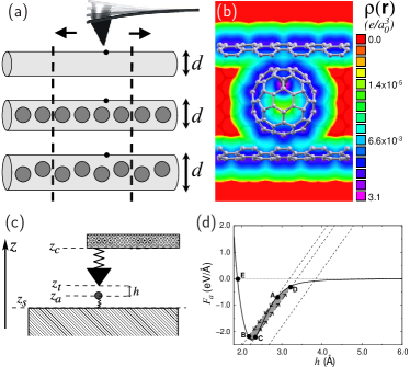
High-resolution AFM topography observations of SWNTs and peapods, illustrated in Fig. 1(a), are shown by contour plots in Fig. 2(a:1-4) EPAPS . Our topography results indicate that the diameter of the empty SWNT in Fig. 2(a:1) is Å. Short-range interatomic interactions, probed by the dynamic AFM, provide atomic-scale contrast in topography images Ashino et al. (2004). The observed atomic arrangement in this tube identifies its chiral index as Ashino and Wiesendanger (2006). Comparing these results to those for a peapod of the same diameter, presented in Fig. 2(a:2), we find the atomic-scale topography features to be amplified with respect to the empty SWNT. This contrast enhancement can likely be attributed to local stiffening of the nanotube wall next to the enclosed Dy@C82 molecules.
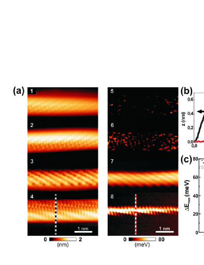
The optimum packing geometry of fullerenes in peapods is determined primarily by their diameter () and that of the enclosing nanotube () Britz and Khlobystov (2006). We expect an optimum snug fit at Å Yoon et al. (2005); Hodak and Girifalco (2001), with the center-to-center inter-fullerene distance Å, nearly independent of the nanotube diameter. With the diameter Å of Dy@C82, a Å wide SWNT should provide an optimum fit, with the encapsulated molecules separated by Å. The diameter Å of the narrowest peapod observed here, discussed in Fig. 2(a:4), is smaller than optimum. In this case, the enclosed molecules distort locally the nanotube wall Ashino et al. (2008), as seen in Fig. 2(a:4). We find the observed undulation period of Å to agree with the above estimated inter-fullerene distance .
More intriguing than the topography are our results for the spatial variation of the energy loss per cycle of the cantilever oscillation, defined as damping and monitored in DFS EPAPS . The damping data, shown in Fig. 2(a:5-8), have been recorded simultaneously with the topography data Morita et al. (2002); García and Pérez (2002). For better quantitative comparison, we present topography and damping data for the same system as line plots in Fig. 2(b). According to Fig. 2(a:5), almost no damping is observed in an empty SWNT. In comparison to these observations, Fig. 2(a:6) suggests that damping increases significantly in a peapod of the same diameter. Decreasing the diameter of the enclosing nanotube causes an increasingly snug fit of Dy@C82 in the peapod. Our data in Figs. 2(a:6-8) indicate that (i) increases, (ii) the atomic-scale features within are amplified, and (iii) helical undulations in grow in magnitude with decreasing diameter of the enclosing nanotube. In the case of a snug fit, atomic-scale features in show a similar or even larger contrast than topography features. Even though both are spatially correlated, we observe the strongest damping slightly off the center of the largest topographic protrusions on-top the encapsulated fullerenes.
The topography images in Figs. 2(a) and 2(b) show some lateral asymmetry, which may be due to Dy@C82 ordering within the peapod or an artifact EPAPS . While we have taken precautions to avoid artifacts caused by tip-induced nanotube displacement (we scanned in the axial direction) and feedback lag (we used a small scan velocity), we can not exclude an intrinsically asymmetric shape or doping profile of the AFM tip as the cause of the asymmetry. Irrespective of this finding, we conclude that monitoring the damping of the oscillating tip provides information superior to topography imaging and allows to discriminate between snug and loose packing of fullerenes inside the nanotube.
In Fig. 2(c) we plot the maximum energy loss per cycle observed during a DFS scan as a function of the host nanotube diameter . The data points in the diagram are presented for a and a nanotube with Å, a nanotube with Å, and and nanotubes with Å. Our results indicate a universal dependence of on the diameter only. From the narrowest to the widest nanotube, we find the value of to decrease from meV to meV and meV per cycle of the oscillating tip.
To uncover the origin of damping in DFS, we performed ab initio density functional calculations of the electronic structure, equilibrium geometry and elastic response of these systems, along with MD calculations of the dynamical coupling between the AFM tip and the peapod.
Our total energy calculations indicate that unlike the most stable C2 isomer of C82, the near-spherical isomer of Dy@C82 has a different C2v symmetry. This structure is energetically preferred by at least 0.5 eV over any other isomer and likely abounds in our samples. The total charge distribution of Dy@C82 inside the nanotube, providing optimum enclosure, is shown in Fig. 1(b).
The fundamental concept of an AFM imaging an elastic substrate is depicted in Fig. 1(c). The position of the tip apex atom depends on the position of the cantilever mount and the deflection of the elastic cantilever, represented by a spring. Similarly, an elastic substrate is represented by a different spring, allowing the position of the closest surface atom to differ from of the substrate. The force exerted on the tip by the substrate depends largely on the closest tip-substrate distance .
To elucidate the origin of damping in the dynamical AFM, we first consider the force exerted on a substrate carbon atom by an approaching AFM tip at height , shown by the solid line in Fig. 1(d). For a given position of the cantilever mount, the instantaneous position of the tip and the substrate atom depends not only on their mutual distance , which determines the nonlinear interaction force shown by the solid line, but also on the compensating forces of the strained cantilever and substrate, shown by the dotted lines, as well as the history. In the case of a soft cantilever or substrate, as the AFM tip approaches from far away, an instability occurs at point A, causing an abrupt decrease in the tip-substrate distance to point B. During the retraction cycle, a similar instability occurs at C, causing an abrupt increase in the tip-substrate distance to D. We note that even the closest-approach point B occurs in the non-contact regime, since is beyond the equilibrium tip-substrate separation , characterized by . The hysteresis corresponding to the shaded area in Fig. 1(d), delimited by A,B,C and D, represents the energy dissipation in the substrate during an ideal approach-retraction cycle.
To quantitatively analyze, how the energy loss of the oscillating tip depends on its position and the tube diameter, we performed a series of MD calculations EPAPS . We induced an initial perturbation by radially displacing a surface atom by 0.3 Å, corresponding to the distance in Fig. 1(d), and abruptly releasing it. The observed energy loss per cycle depends on the frequency of vibrational modes excited by the plucking process and the likelihood of exciting them. This energy is then dissipated efficiently owing to the high thermal conductivity of nanotube systems Berber et al. (2000). Since vibrations excited in the cantilever do not change with position, spatial contrast in is only due to the substrate.
In empty SWNTs, we found that plucking excites only the radial breathing mode (RBM) in the frequency range meV in the systems considered here. The energy needed to excite this mode is consistent with values seen in Fig. 2(a:5). We expect stronger damping in peapods due to additional modes introduced by the fullerenes. Ultra-soft vibrations of Dy within the C82 cage at meV are caused by the soft DyC82 interaction and the large mass of Dy. Since these modes are decoupled from the remaining modes, we will ignore the presence of Dy in the following discussion. Among the fullerene modes, librations about the center are the softest, followed by axial and off-axis motion of the center of mass. Harder fullerene modes include the RBM at meV, the quadrupolar deformation mode near meV, and higher multipolar modes. Especially in the case of a snug fit in the peapod, the nanotube and fullerene modes are strongly mixed.
For each of the nanotubes studied, we changed the axial position of the surface atom plucked from on-top a fullerene to in-between fullerenes, and also studied a position half-way between the two. Our simulations indicate that damping is strongest for the last position, due to the possibility of exciting librations and axial vibrations of the fullerene underneath. This off-center position, indicated in the inset of Fig. 3(a), agrees with the interpretation of our data in Fig. 2(a).
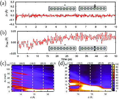
Our MD simulation results for C are presented in Figs. 3(a) and 3(b). The radial trajectory of the plucked atom, shown in Fig. 3(a), and that of the closest fullerene molecule, shown in Fig. 3(b), suggest that several modes are excited. To understand which modes dominate damping, we Fourier analyzed all trajectories in our study. The Fourier spectra of are shown in Fig. 3(c) and those of in Fig. 3(d) for zigzag nanotubes ranging from to . Data for intermediate nanotube diameters have been obtained by interpolation.
We consider the narrow C peapod to provide optimum coupling between the snugly fit fullerene and the enclosing nanotube. In this case, and its spatial modulation should be particularly strong. In contrast to an empty SWNT, the vibration spectrum of a surface atom in C is very rich, as seen in Fig. 3(c). Besides low-frequency modes with meV, it contains modes with equally spaced frequencies spanning the range meV. In the wider nanotube, the vibration spectrum is similar, but softer. In the and wider nanotubes, the fullerene-nanotube coupling is reduced drastically, as fullerenes may rearrange at little energy cost. As seen in Fig. 3(c), decreased fullerene-nanotube coupling causes a strong intensity drop of the high-frequency modes at meV. Especially in the narrow nanotubes, our results for the off-center plucking position in Fig. 3(c) differ from those of other simulations, where the plucked atom is on top of a fullerene. Since plucking on top a fullerene excites neither librations nor axial fullerene motion, energy dissipation in this plucking position is lower. Comparing the results for the two plucking positions explains the high spatial modulation of seen in Fig. 2(a:8).
The vibration spectrum of the center-of-mass motion of a C82 fullerene in the nanotube, shown in Fig. 3(d), is rather featureless and much softer than that in Fig. 3(c). The meV off-axis vibration mode, which dominates the spectrum, red shifts in wider nanotubes. Owing to the zigzag or helical arrangement of fullerenes in wider peapods, the off-axis vibrations of the fullerenes may couple to soft axial vibrations and librations, which appear in the Fourier spectrum of in Fig. 3(d). These important soft modes, which do not change , modify the spectrum indirectly by coupling anharmonically to off-axis modes. Especially in the wider nanotubes, we find the center-of-mass vibrations of C82 to be rather insensitive to the axial position of the plucked atom, thus contributing very little to the spatial modulation of .
In conclusion, Damping Force Spectroscopy can reveal geometrical packing and vibrational spectra of subsurface structures at the atomic scale. We expect that this technique will gain significant appeal for atomic-level investigations of three-dimensional supramolecular systems.
We thank Siegmar Roth and Dirk Obergfell for useful discussions and for sample preparation. We gratefully acknowledge financial support from the Deutsche Forschungsgemeinschaft and from the National Science Foundation under NSF-NSEC grant No. 425826 and NSF-NIRT grant No. ECS-0506309. Computational resources have been provided by the Michigan State University High Performance Computing Center.
References
- Stipe et al. (1998) B. C. Stipe, M. A. Rezaei, and W. Ho, Science 280, 1732 (1998).
- Yamanaka et al. (1994) K. Yamanaka, H. Ogiso, and O. Kolosov, Appl. Phys. Lett. 64, 178 (1994).
- Kolosov et al. (1998) O. V. Kolosov, M. R. Castell, C. D. Marsh, G. A. D. Briggs, T. I. Kamins, and R. S. Williams, Phys. Rev. Lett. 81, 1046 (1998).
- Morita et al. (2002) S. Morita, R. Wiesendanger, and E. Meyer, Noncontact Atomic Force Microscopy, NanoScience and Technology (Springer, Berlin, 2002).
- García and Pérez (2002) R. García and R. Pérez, Surf. Sci. Rep. 47, 197 (2002).
- Smith et al. (1998) B. W. Smith, M. Monthioux, and D. E. Luzzi, Nature 396, 323 (1998).
- Jorio et al. (2008) A. Jorio, G. Dresselhaus, and M. S. Dresselhaus, eds., Carbon Nanotubes: Advanced Topics in the Synthesis, Structure, Properties and Applications, Topics in Applied Physics, Vol. 111 (Springer, Berlin, 2008).
- Britz and Khlobystov (2006) D. A. Britz and A. N. Khlobystov, Chem. Soc. Rev. 35, 637 (2006).
- Kitaura and Shinohara (2006) R. Kitaura and H. Shinohara, Chem. Asian J. 1, 646 (2006).
- Shimada et al. (2002) T. Shimada, T. Okazaki, R. Taniguchi, T. Sugai, H. Shinohara, K. Suenaga, Y. Ohno, S. Mizuno, S. Kishimoto, and T. Mizutani, Appl. Phys. Lett. 81, 4067 (2002).
- (11) See EPAPS Document No. ### for experimental and computational details.
- Ashino et al. (2004) M. Ashino, A. Schwarz, T. Behnke, and R. Wiesendanger, Phys. Rev. Lett. 93, 136101 (2004).
- Ashino and Wiesendanger (2006) M. Ashino and R. Wiesendanger, Jpn. J. Appl. Phys. 45, 2286 (2006).
- Yoon et al. (2005) M. Yoon, S. Berber, and D. Tománek, Phys. Rev. B. 71, 155406 (2005).
- Hodak and Girifalco (2001) M. Hodak and L. A. Girifalco, Chem. Phys. Lett. 350, 405 (2001).
- Ashino et al. (2008) M. Ashino, D. Obergfell, M. Haluska, S. Yang, A. N. Khlobystov, S. Roth, and R. Wiesendanger, Nature Nanotech. 3, 337 (2008).
- Berber et al. (2000) S. Berber, Y.-K. Kwon, and D. Tománek, Phys. Rev. Lett. 84, 4613 (2000).