How perfect can graphene be?
Abstract
We have identified the cyclotron resonance response of purest graphene ever investigated, which can be found in nature on the surface of bulk graphite, in form of decoupled layers from the substrate material. Probing such flakes with Landau level spectroscopy in the THz range at very low magnetic fields, we demonstrate a superior electronic quality of these ultra low density layers ( cm-2) expressed by the carrier mobility in excess of 107 cm2/(V.s) or scattering time of ps. These parameters set new and surprisingly high limits for intrinsic properties of graphene and represent an important challenge for further developments of current graphene technologies.
pacs:
71.70.Di, 76.40.+b, 81.05.UwFabrication of graphene structures has triggered vast research efforts focused on the properties of two-dimensional systems with massless Dirac fermions. Nevertheless, further progress in exploring this quantum electrodynamics system in solid-state laboratories seems to be limited by insufficient electronic quality of manmade structures and the crucial question arises whether existing technologies have reached their limits or major advances are in principle possible. The substrate, and more general any surrounding medium, has been recently identified as a dominant source of extrinsic scattering mechanisms, which effectively degrade the electronic quality of currently available graphene samples J. Martin et al. (2008); Y.-W. Tan et al. (2007). Despite significant advances in technology, including the fabrication of suspended specimens K. I. Bolotin et al. (2008); X. Du et al. (2008), the realistic limits of the scattering time and mobility in graphene, achievable after elimination of major extrinsic scattering sources, remain an open issue. Experiments call for higher quality samples, which are almost certainly crucial for possible verification of interesting predictions concerning basic phenomena of quantum electrodynamics (e.g. Zitterbewegung) or observation of the effects of interactions between Dirac fermions, (resulting e.g. in the appearance of the fractional quantum Hall effect). Even more simple effects such as lifting the degeneracy of the spin and/or pseudo-spin degree of freedom in very high magnetic fields indicate the strong influence of the sample quality on the information that can be deduced from experiments D. A. Abanin et al. (2007); Z. Jiang et al. (2007a).
Recently, well-defined graphene flakes have been discovered in form of sheets, decoupled from, but still located on the surface of bulk graphite which naturally serves as a well-matched substrate for graphene Li et al. (2009). In this Letter, we report on response from these flakes in microwave magneto-absorption experiments and show that their Dirac-like electronic states are quantized into Landau levels (LLs) in magnetic fields down to 1 mT and at elevated temperatures up to 50 K. The deduced unprecedented quality of the studied electronic system sets surprisingly high limits for the intrinsic scattering time and the corresponding carrier mobility in graphene.
The cyclotron resonance has been measured in a high-frequency EPR setup in double-pass transmission configuration, using the magnetic-field-modulation technique. A flake of natural graphite was placed in the variable temperature insert of the superconducting solenoid and via quasi-optics exposed to the linearly polarized microwave radiation emitted by a Gun diode at frequencies 283.2 or 345 GHz (1.171 or 1.427 meV). The absorbed radiation has been followed by either heterodyne detection (283.2 GHz) or by the bolometer (345 GHz). To enhance the relatively weak response of graphene flakes, the modulation amplitude ( mT) had to be chosen close to the CR width, which broadens resonances observed at lower magnetic fields. All spectra have been corrected for the remanent field of the magnet.
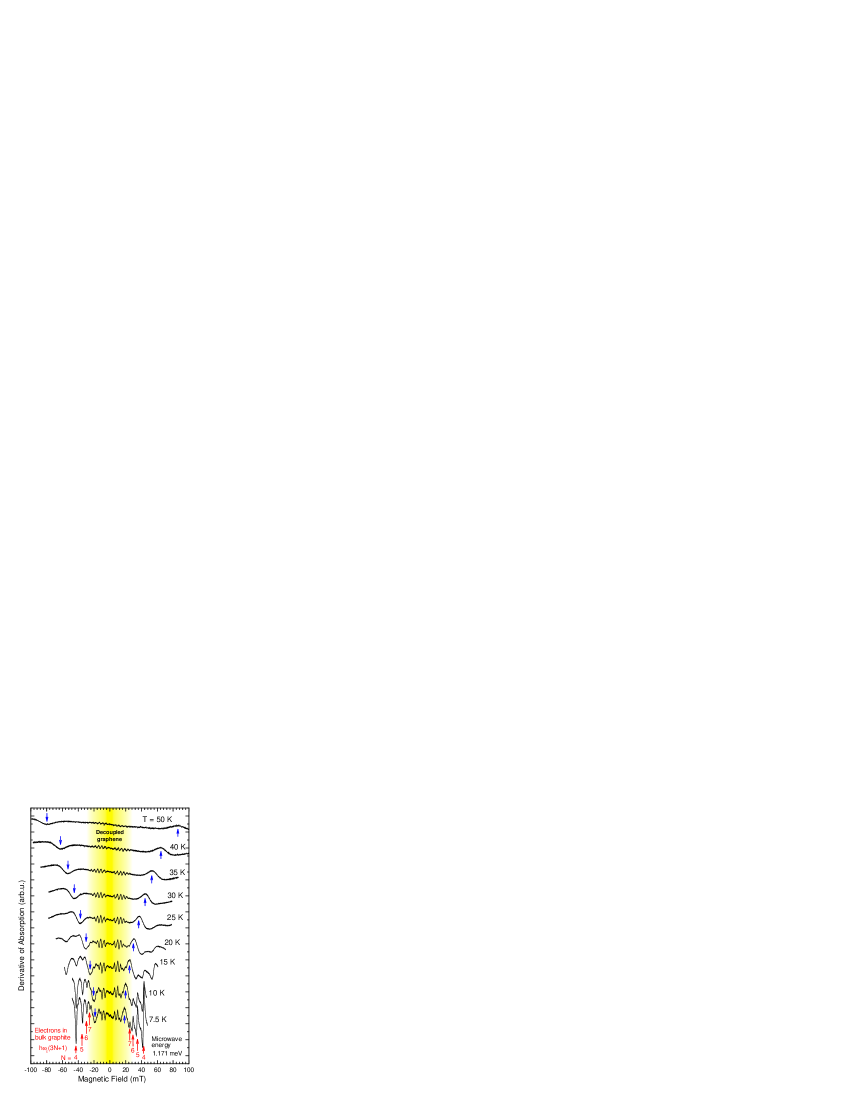
Our main experimental finding is illustrated in Fig. 1. The traces in this figure represent the magneto-absorption response of the natural graphite specimen at different temperatures, measured as a function of the magnetic field at fixed microwave frequency. They correspond to the derivative of the absorption strength with respect to the magnetic field since the field modulation technique has been applied. Strong at 7.5 K, but rapidly vanishing with temperature lines marked with red arrows in Fig. 1 can be easily recognized as cyclotron resonance (CR) harmonics of point electrons in bulk graphite. A possible origin of the temperature dependent transition marked with the blue arrow will be discussed later on. The features of primary interest, which we argue are due to decoupled graphene sheets on the graphite surface, are seen at very low fields, within the yellow highlighted area of Fig. 1.
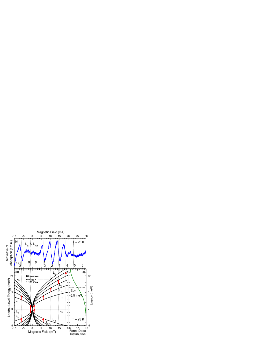
The interpretation of these low-field data is schematically illustrated in Fig. 2. The observed spectral lines (Fig. 2a) are assigned to cyclotron resonance transitions between adjacent LLs () with energies: K. S. Novoselov et al. (2005); Y. B. Zhang et al. (2005), characteristic of massless Dirac fermions in graphene sheets with an effective Fermi velocity . This velocity is the only adjustable parameter required to match the energies of the observed and calculated CR transitions. A best match is found for m.s-1 in fair agreement with values found in multilayer epitaxial graphene M. L. Sadowski et al. (2006); M. Orlita et al. (2008a) or exfoliated graphene on Si/SiO2 substrate Z. Jiang et al. (2007b); R. S. Deacon et al. (2007); Z. Li et al. (2008). As can be seen from Figs. 2b,c the multi-mode character of the measured spectra is directly related to thermal distribution of carriers among different LLs. The intensity of a given transition is proportional to the difference in thermal occupation of the involved LLs. Roughly speaking, the strongest transitions imply LLs in the vicinity of the Fermi level, which fixes EF at around 6-7 meV from the Dirac point.
To reproduce the experimental data, we assume the absorption strength is proportional to the longitudinal conductivity of the system:
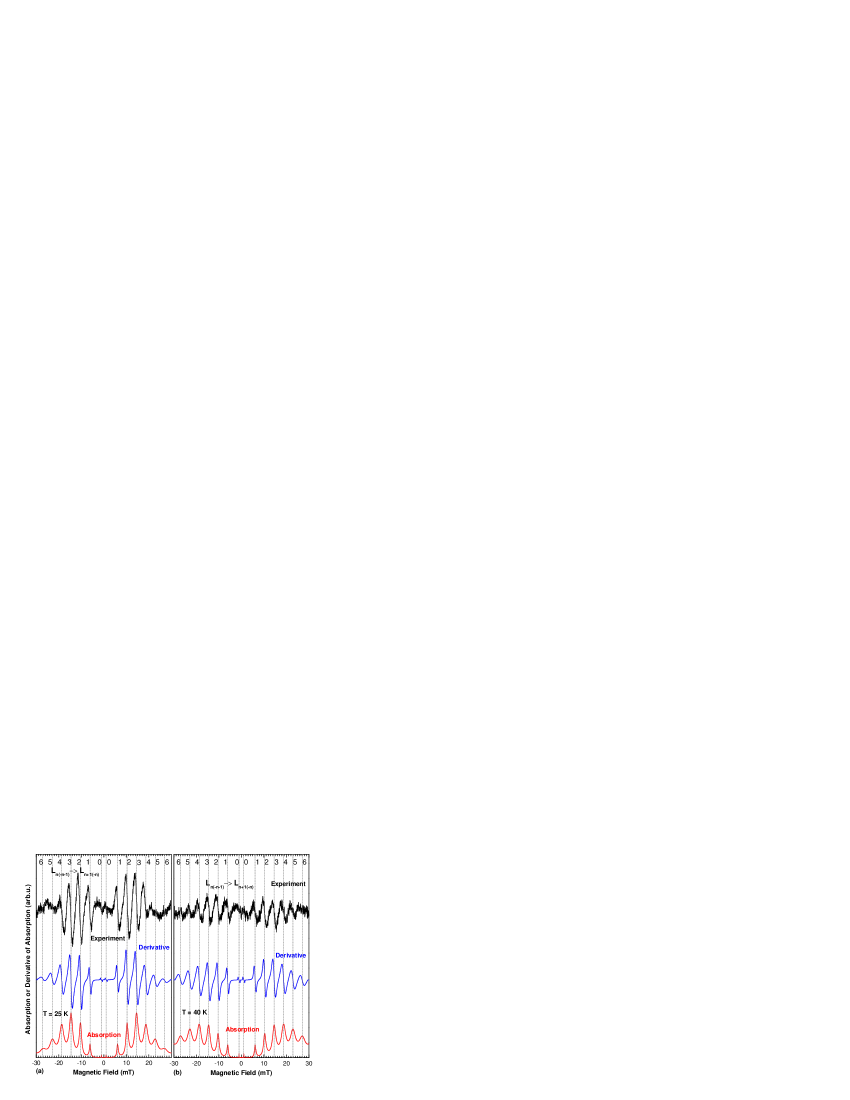
where is occupation of the -th LL and with for or equal 0 otherwise M. L. Sadowski et al. (2006); Gusynin et al. (2007). The calculated traces in Fig. 3 have been drawn taking eV for the line broadening, m.s-1 and meV. To directly simulate the measured traces, the derivative of the absorption with respect to the magnetic field has been calculated taking account of the field modulation mT used in the experiment. In spite of its simplicity, our model is more than in a qualitative agreement with our experimental data, see Fig. 3. The calculation fairly well reproduces the experimental trends: Multi-mode character of the spectra, the intensity distribution among the lines, as well as its evolution with temperature, and allows to estimate the characteristic broadening of the CR transitions.
Our modeling could be further improved but at the expenses of additional complexity which we want to avoid here. Assuming magnetic-field and/or LL index dependence of the broadening parameter and taking into account the possible fluctuation of the Fermi level within the ensemble of probed flakes would improve the agreement between experiment and theory, particularly at low temperatures. Comparing both, the measured and simulated traces, we are also more confident in the spectacular observation of the CR transition (involving the LL) at a magnetic field as low as 1 mT. Bearing in mind the small value of the extracted broadening parameter one may conclude that LL quantization should survive in studied graphene layers down to the field of T. Hence, the magnetic field of the Earth of T is no longer negligibly small. Instead, it can open an energy gap at the Dirac point up to meV, depending on the sample orientation.
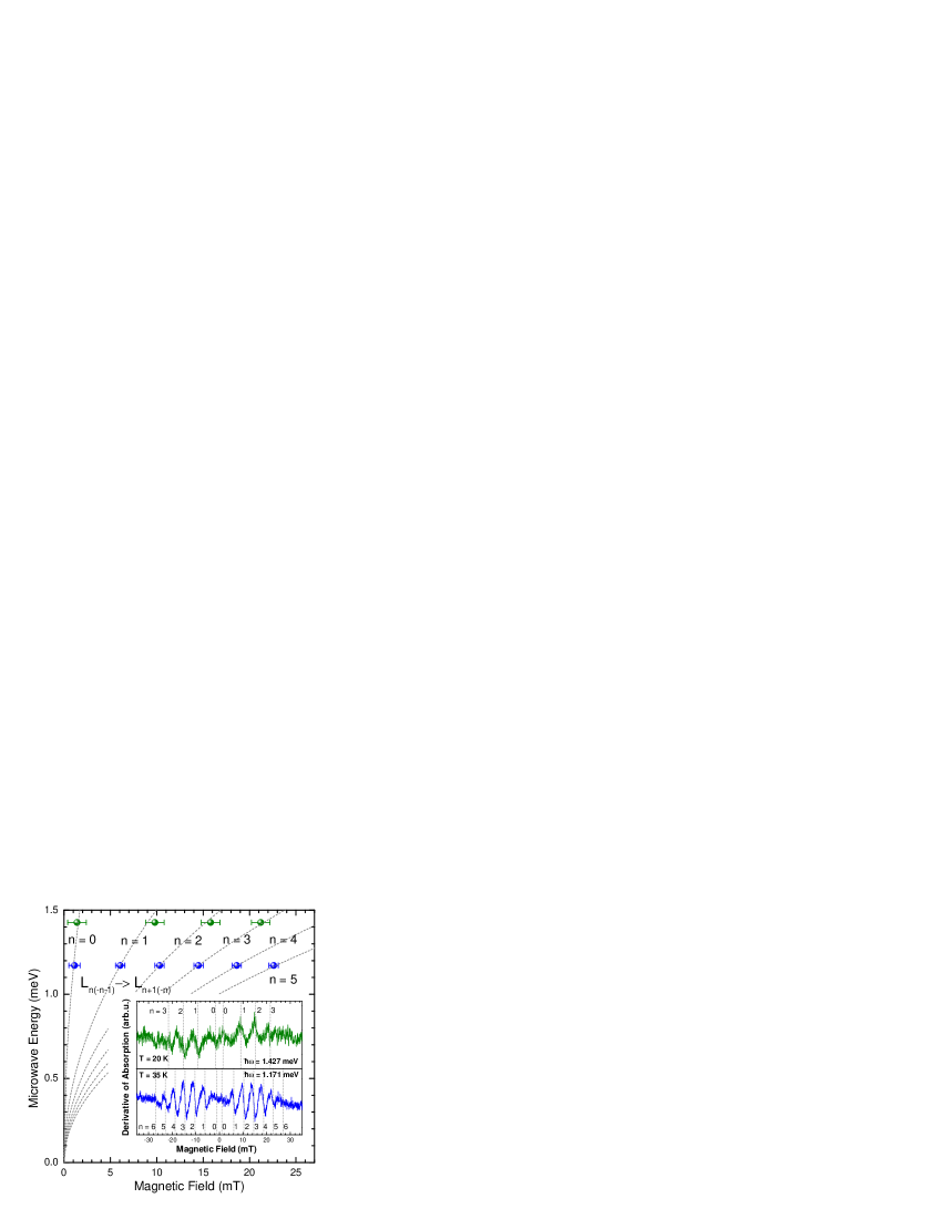
To crosscheck our interpretation, we have also measured the spectra using a different (higher) microwave energy meV, see Fig. 4. Despite the weaker sensitivity of the experimental setup at this frequency, we can clearly identify the same set of inter-LL transitions simply shifted to higher magnetic fields.
The finite Fermi energy meV, corresponding to a carrier density of cm-2, indicates that the probed layers are in thermodynamical contact with the surrounding material, which supplies these carriers. On the other hand, we find no signs of electrical coupling of these graphene layers to bulk graphite. Our experiments show that any possible energy gap opened due to this interaction at the Dirac point cannot exceed a few hundred eV. The absence of this gap convincingly confirms that we are indeed dealing with decoupled graphene and not with the point of bulk graphite, where Dirac-like fermions are also present M. Orlita et al. (2008b) but a (pseudo)gap of a few meV is expected A. Grüneis et al. (2008) and indeed observed Toy et al. (1977). The temperature evolution of the measured spectra is another important indication allowing us to discriminate between the graphene and bulk graphite contributions. No (or very weak) temperature broadening of CR transitions is expected for graphene S. V. Morozov et al. (2008); K. I. Bolotin et al. (2008); M. Orlita et al. (2008a), whereas the response of bulk graphite should follow the relatively strong decrease of the carrier scattering time, expressed by the average mobility, which reaches up to cm2/(V.s) at low temperatures, but falls down by one order of magnitude at K Brandt et al. (1988). Indeed, this behaviour is observed in Fig. 1. Whereas the CR harmonics of point electrons in bulk graphite Nozières (1958) seen in the spectra at mT, disappear very rapidly upon increasing , the graphene-like features survive and their intensity is simply following the vanishing difference in the occupation between the adjacent LLs. It is worth noticing that the graphene-like signal, although always substantially weaker then the response from bulk graphite, has been observed for a number of different specimens of natural graphite. Mechanical scratching of the sample surface and fast thermal cooling of the sample has been found to enhance the signal from decoupled graphene in comparison with bulk graphite, likely helping in detaching graphene sheets from the graphite crystal.
Since the well-defined LL quantization in our graphene flakes is observed down to mT, see Fig. 2, we obtain via the semi-classical LL quantization condition the carrier mobility cm2/(V.s), almost two orders of magnitude higher in comparison with suspended Y.-W. Tan et al. (2007); K. I. Bolotin et al. (2008); X. Du et al. (2008) or epitaxial graphene M. Orlita et al. (2008a). The LL broadening , obtained via comparison of our experiment with the simulated traces, allows us to estimate the scattering time ps (), which significantly exceeds those reported in any kind of manmade graphene samples, see e.g. Refs. Y.-W. Tan et al. (2007); K. I. Bolotin et al. (2008), and gives an independent estimation for the mobility cm2/(V.s) in good agreement with the estimate above. Even though we cannot verify this estimate by a direct electrical measurement, a near correspondence of the scattering time derived from CR measurements and transport scattering time was recently verified on samples with a significantly lower mobility Z. Jiang et al. (2007b). Moreover, the estimated mobility should not decrease with temperature, as no broadening of CRs is observed up to K, when CR intensities become comparable with the noise. This extremely high value of mobility combines two effects: the long scattering time and a very small effective mass . Remarkably, the same scattering time in a moderate density sample ( cm-2), would imply the mobility still remaining high, around cm2/(V.s), and comparable to best mobilities of two-dimensional electron gas in GaAs structures at these densities.
The model we used here to describe the magneto-absorption response of Dirac fermions in graphene is amazingly simple, based on a one-particle approximation, and it is perhaps surprising that it is so well applicable to simulate the experimental data, particularly in context of the outstanding quality of the electronic system studied. At first sight, the observation of collective excitations, due to, e.g., magneto-plasmons could be expected in our experiments and only preliminary theoretical work addresses the surprising approximate validity of Kohn’s theorem in graphene Bychkov and Martinez (2008); Asano and Ando (2009). Also size-confined magneto-plasmons are apparent in the microwave-absorption spectra of two-dimensional gas of massive electrons Allen et al. (1977), but apparently not seen in our experiments on graphene. This perhaps points out the qualitative difference in the plasmon behaviour in systems with quadratic and linear dispersion relations Hwang and Das Sarma (2007); Polini et al. (2009). On the other hand, we speculate that the transition, marked with blue arrows in Fig. 1 has a character of a collective excitation. It is characterized by a sensitivity to the magnetic field and by softening with temperature. The energy of this excitation can be deduced to scale roughly as , and this points towards its magnetic origin. For example, energy scaling is typical of a ferromagnetic resonance, see e.g. Ref. Liu and Furdyna, 2006. Nevertheless, the origin of the spectral feature marked by blue arrows in Fig. 1 remains an intriguing puzzle. We see this feature repeatedly on different samples, but at slightly different energies, each time however characteristically evolving with temperature. With its apparent magnetic characteristics one should seek its origin in impurities B. Uchoa et al. (2008), structural defects Yazyev and Helm (2007) or edge states Yang et al. (2008) in graphene, but possibly also on the surface of bulk graphite Červenka and Flipse (2009).
To conclude, graphene layers decoupled from bulk graphite have been probed in cyclotron resonance experiment, which offers an unambiguous evidence of extremely high carrier mobility in graphene exceeding cm2/(V.s). This measurement significantly shifts the limits of intrinsic mobility in graphene S. V. Morozov et al. (2008) and poses a quest for further development in the technology of graphene fabrication. Graphene samples with mobilities comparable to the best GaAs samples Hwang and Das Sarma (2008) thus seem to be achievable.
Acknowledgements.
P.N. and M.O. contributed equally to this work. Part of this work has been supported by EuroMagNET II under the EU contract, by the French-Czech Project Barrande No. 19535NF, by contract ANR-06-NANO-019 and by projects MSM0021620834 and KAN400100652.References
- J. Martin et al. (2008) J. Martin et al., Nature Phys. 4, 144 (2008).
- Y.-W. Tan et al. (2007) Y.-W. Tan et al., Phys. Rev. Lett. 99, 246803 (2007).
- K. I. Bolotin et al. (2008) K. I. Bolotin et al., Phys. Rev. Lett. 101, 096802 (2008).
- X. Du et al. (2008) X. Du et al., Nature Nanotech. 3, 491 (2008).
- D. A. Abanin et al. (2007) D. A. Abanin et al., Phys. Rev. Lett. 98, 196806 (2007).
- Z. Jiang et al. (2007a) Z. Jiang et al., Phys. Rev. Lett. 99, 106802 (2007a).
- Nozières (1958) P. Nozières, Phys. Rev. 109, 1510 (1958).
- Brandt et al. (1988) N. B. Brandt, S. M. Chudinov, and Y. G. Ponomarev, Semimetals 1: Graphite and its Compounds, vol. 20.1 of Modern Problems in Condensed Matter Sciences (North-Holland, Amsterdam, 1988).
- Li et al. (2009) G. Li, A. Luican, and E. Y. Andrei, Phys. Rev. Lett. 102, 176804 (2009).
- K. S. Novoselov et al. (2005) K. S. Novoselov et al., Nature 438, 197 (2005).
- Y. B. Zhang et al. (2005) Y. B. Zhang et al., Nature 438, 201 (2005).
- M. L. Sadowski et al. (2006) M. L. Sadowski et al., Phys. Rev. Lett. 97, 266405 (2006).
- M. Orlita et al. (2008a) M. Orlita et al., Phys. Rev. Lett. 101, 267601 (2008a).
- Z. Jiang et al. (2007b) Z. Jiang et al., Phys. Rev. Lett. 98, 197403 (2007b).
- R. S. Deacon et al. (2007) R. S. Deacon et al., Phys. Rev. B 76, 081406(R) (2007).
- Z. Li et al. (2008) Z. Li et al., Nature Phys. 4, 532 (2008).
- Gusynin et al. (2007) V. P. Gusynin, S. G. Sharapov, and J. P. Carbotte, Phys. Rev. Lett. 98, 157402 (2007).
- M. Orlita et al. (2008b) M. Orlita et al., Phys. Rev. Lett. 100, 136403 (2008b).
- A. Grüneis et al. (2008) A. Grüneis et al., Phys. Rev. B 78, 205425 (2008).
- Toy et al. (1977) W. W. Toy, M. S. Dresselhaus, and G. Dresselhaus, Phys. Rev. B 15, 4077 (1977).
- S. V. Morozov et al. (2008) S. V. Morozov et al., Phys. Rev. Lett. 100, 016602 (2008).
- Bychkov and Martinez (2008) Y. A. Bychkov and G. Martinez, Phys. Rev. B 77, 125417 (2008).
- Asano and Ando (2009) K. Asano and T. Ando, private communication (2009).
- Allen et al. (1977) S. J. Allen, D. C. Tsui, and R. A. Logan, Phys. Rev. Lett. 38, 980 (1977).
- Hwang and Das Sarma (2007) E. H. Hwang and S. Das Sarma, Phys. Rev. B 75, 205418 (2007).
- Polini et al. (2009) M. Polini, A. MacDonald, and G. Vignale, arxiv:0901.4528 (2009).
- Liu and Furdyna (2006) X. Liu and J. K. Furdyna, J. Phys.: Condens. Matter 18, 245 (2006).
- B. Uchoa et al. (2008) B. Uchoa et al., Phys. Rev. Lett. 101, 026805 (2008).
- Yazyev and Helm (2007) O. V. Yazyev and L. Helm, Phys. Rev. B 75, 125408 (2007).
- Yang et al. (2008) L. Yang, M. L. Cohen, and S. G. Louie, Phys. Rev. Lett. 101, 186401 (2008).
- Červenka and Flipse (2009) J. Červenka and C. F. J. Flipse, Nature Phys. p. to be published (2009).
- Hwang and Das Sarma (2008) E. H. Hwang and S. Das Sarma, Phys. Rev. B 77, 235437 (2008).