Intensity-field correlation of single-atom resonance fluorescence
Abstract
We report measurements of an intensity-field correlation function of the resonance fluorescence of a single trapped 138Ba+ ion. Detection of a photon prepares the atom in its ground state and we observe its subsequent evolution under interaction with a laser field of well defined phase. We record the regression of the resonance fluorescence source field. This provides a direct measurement of the field of the radiating dipole of a single atom and exhibits its strong non-classical behavior. In the setup an interference measurement is conditioned on the detection of a fluorescence photon.
pacs:
42.50.Ct, 32.80.-t, 37.10.TyResonance fluorescence of atoms, in particular individual atoms, has been the subject of quantum optical measurements for many years Mandel ; Loudon . For example, resonance fluorescence is routinely used as a tool to simply detect atoms, for spectroscopy purposes, for creating photons, including for single and twin-photon sources, and its quadrature components have been used to create non-classical states of light Loudon . In short, the observation of resonance fluorescence is a technology ubiquitous in experimental physics. While its features are well investigated and understood, for spectroscopy and in quantum optics, a direct observation of the time evolution of the source field at the single-atom, single-photon level has not been made.
The measurement of correlation functions, sensitive to the source-field, has been proposed in the seminal paper by W. Vogel Vogel . In a first and pioneering experiment, G. T. Foster et al. have been able to report a wave-particle correlation function of the field that emanates out of a cavity and corresponds on average to only a fraction of a photon excitation OroCar ; CarOro ; OroPRA . Correlation functions of the fields comprised of many photons have been observed with lasers and other light sources Mandel ; Loudon ; Twiss2 and were approached theoretically Laserphys . However, to the best of our knowledge, the time evolution of the field that corresponds to a single resonance fluorescence photon has not been recorded so far. Moreover, the only previous observation of the intensity-field correlation OroCar ; OroPRA made use of a strong local oscillator with photocurrent detection. The reported measurement employs a weak local oscillator and photon counting and thus operates in a different regime.
Motivation for this investigation is the development of a tool (i) to investigate and monitor the emission of a single-photon field and at a later stage (ii) to detect the influence of boundary conditions, such as walls, mirrors, other atoms and quite generally of a different (engineered) bath on the dynamics of the emission process. For all of these tasks it will be necessary to monitor the resonance fluorescence field and possibly even feed-back Eschner ; Bushev1 ; Bushev2 ; Uwe on the radiating dipole.
In this letter, we report on the measurement of a third-order correlation function of a single radiating atom, using standard photon counting techniques. Using a homodyne detection scheme, we record the resonance fluorescence field conditioned on the detection of an initial resonance fluorescence photon that prepares the atom in its ground state. Since the correlation function of two fields is termed and that of two intensities is termed Twiss , we accordingly coin the name for this third-order intensity-field correlation.
In this work the correlation measurement is triggered by detecting a fluorescence photon from a single trapped 138Ba+ ion, which projects the ion into its ground state. Stop events are obtained from a homodyne detector, where the fluorescence interferes with a local oscillator (LO) of well-controlled phase relative to the exciting laser. The experimental setup is interferometrically stabilized and the phase of the LO can be adjusted to anywhere within [0, 2]. The measurement is repeated many times for different phases of the LO- field, such that the integrated signal records the average conditional time evolution of the (fluctuating) amplitude of the electromagnetic wave that constitutes the emission of a single resonance fluorescence photon.
The schematic experimental setup and the level scheme of the 138Ba+ ion are shown in Fig. 1. A single Ba+ ion is loaded in a linear Paul trap using photo-ionization with laser light near 413 nm Rotter_diss . The ion is confined in the harmonic pseudo-potential of the trap with radial (axial) oscillation frequency 1.7 MHz ( 1 MHz). Micromotion is minimized using 3 pairs of dc electrodes. The ion is continuously laser-cooled by two narrow-band (laser linewidth of a few tens of kHz) linearly polarized tunable lasers at 493 nm (green) and 650 nm (red) exciting the S1/2–P1/2 and P1/2–D3/2 transitions, respectively. The green laser intensity is adjusted to give mostly elastically scattered photons Mandel . After Doppler cooling, the ion is left in a thermal motional state with a mean number of vibrational excitation . A weak magnetic field defines a quantization axis perpendicular to the laser polarization and vector. Including the Zeeman substates, the internal structure of the atom is described as an 8-level system with the lasers exciting transitions Toschek .
Resonance fluorescence is detected in channels aligned along the quantization axis in both directions. About 4 of the green fluorescence is collected with two custom-made lenses (HALO (LINOS), NA=0.4) placed about 1 cm from the trap center to the left and right side of the trap.

The left beam can either be sent to a PMT (PMT-start) or to a CCD camera. On the right hand side the fluorescence beam is collimated with a telescope and then mixed with the LO field on a mirror with 99 reflectivity. After coupling to a single mode optical fiber for mode matching, the interfering fields are detected at another PMT (PMT-stop) leaving a count rate of about 10 kcps for the fluorescence after the fiber. In both detection channels a quarter wave-plate and a Glan-Thompson polarizer select photons and filter out the transition. The phase of the interferometer is controlled with a Piezo mounted mirror in the LO path by monitoring the count rate of the homodyne signal. Thus the error in the phase of the LO is given by the shot noise of this signal and is estimated to about 10 degrees () and 24 degrees ( or ) for a typical integration time of 0.1 s. Phase locking by keeping the homodyne count rate constant is continuous with a time constant of several seconds and does not affect the contrast of our data within the limits set by the shot noise. Correlations between the PMT start and the PMT stop-counts are obtained by recording single photon arrival times with a Time Acquisition Card (Correlator) with up to 100 ps resolution.
For a theoretical analysis we consider a frame rotating at the (green) laser frequency, . Thus, the green source part of the radiated field by the ion reads
| (1) |
where is in the long-time limit after the exciting laser is turned on, represents a constant amplitude, and is the Pauli lowering operator from to , associated with a creation of a single photon. With the LO path blocked we measure the conventional normalized second order (intensity) correlation, . In terms of atomic operators it reads
| (2) |
Figure 2 depicts a measurement of this quantity. It exhibits the characteristic anti-bunching at short time, with a null rate of coincidences, (without background subtraction). Aside from this minor offset, it is well reproduced by our 8-level Bloch simulations. Fitting parameters are the laser powers and detunings. Thus, the is used for calibrating the laser settings for the measurement.

With the LO arm unblocked, we measure the homodyne signal conditioned on a photon emission from the ion, where the phase of the LO can be adjusted. We now write the detected fields in units of the square root of photon flux. represents the mean photon flux into the PMT-start, where is the product of the radiative decay rate and the overall collection and detection efficiency of the PMT-start. We similarly denote the fluorescence field at the PMT-stop by , where is the product of radiative decay rate and the respective collection and detection efficiency of a photon at the PMT-stop. Then representing the local oscillator field by the complex amplitude the field after the interferometer reads
| (3) |
and for positive we measure a total unnormalized second-order correlation
| (4) |
which expands out to
| (5) |
Here, is the intensity correlation function given by Eq. (2), and for the third-order correlation function at a given LO phase we write
| (6) |
For later convenience we define the LO phase relative to the asymptotic phase of the resonance fluorescence field, such that . The pre-factor in Eq. (5) is
| (7) |
while
| (8) |
is the visibility of the interference part in and
| (9) |
is the ratio of the florescence intensity to the total intensity at the PMT-stop.
According to Eq. (5), the expected correlation function consists of three parts. A -dependent part with visibility reveals the correlation due to the interference of the fluorescence with the LO. The remaining non-interfering part with weight consists of a nor- mal second-order correlation function (both start and stop counts from fluorescence photons) weighted by and a constant offset (stop counts from LO photons) weighted by . Normalizing by we obtain
| (10) |
This normalization is chosen such that at a phase of the LO, when vanishes asymptotically, yields an asymptotic value of 1.
Figure 3 shows the measured correlations between PMT-start and PMT-stop with the LO phase adjusted to and . Data are acquired after 30 minutes of accumulation for each curve and presented with a 1 ns resolution. The corresponding variance is obtained from shot noise, i.e. assuming Poisson statistics at all times . The solid curves show the theoretical prediction using Eq. (10) with a visibility and an intensity ratio . The measured correlations are well reproduced by superposition of the three contributions described by Eq. (10).
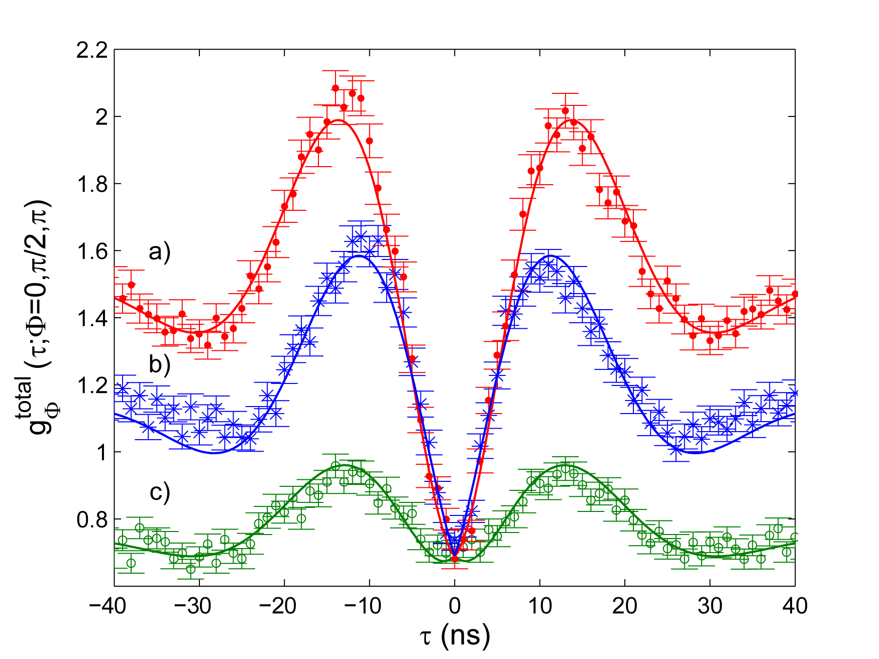
All curves contain a constant contribution and a scaled correlation. In addition, curve b), where the LO phase is set to , contains the imaginary part of the atomic polarization whose asymptotic contribution is zero. In contrast, curves a) and c), where the LO phase is set to 0 and , respectively, reveal the real part of the polarization which adds constructively or destructively to the other two contributions. All curves show the same coincidence rate at . Since both the contribution and the contribution are identically zero at , the measured coincidence rate at this point is solely determined by the offset of non- interfering LO photons at PMT-stop (and background counts). Calibration of the LO phase is obtained by looking for the maximum and minimum asymptotic values of and assigning to them the LO phases and , respectively.
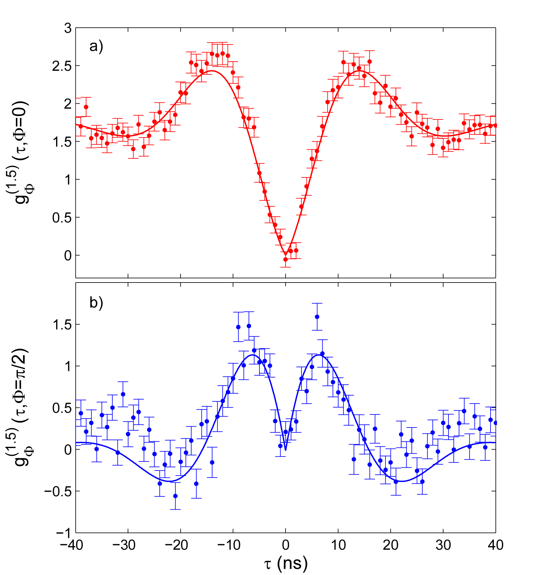
Determining the full complex intensity field correlation function requires its measurement for two orthogonal phases. We deduce it from the data in Fig. 3 and using Eq. (10) in the following way:
| (11) |
and
| (12) |
The result is shown in Fig. 4 together with the theoretical prediction of Eq. (10) (solid line). The data reveal the time evolution of the fluorescence field as it evolves from its initialization by an emitted photon into steady-state via damped Rabi oscillations. Comparing Fig. 4 a) with Fig. 2 we see that the grows linearly with around the point while the grows quadratically with showing clearly that the field rather than the intensity is measured.
The limitation for the visibility in the homodyne part of the setup is determined by the temporal overlap of two photon wave packets impinging at the mixing mirror. It is limited by the coherence time and the flux of the fluorescence photons with respect to the LO photons. Assuming elastically scattered photons, the coherence time is given by the 493 nm laser bandwidth with 50 Raab . The fluorescence count-rate of kcps is predetermined by the collected fraction of solid angle and the fiber-coupling efficiency. While the temporal overlap would benefit from a higher LO intensity (smaller ), the visibility of the interferometer would decrease and locking the interferometer would get more involved due to a larger shot-noise in the LO arm. Thus, optimizing the experiment resulted in a reduced visibility of and an intensity ratio of .
In summary, we have successfully measured an intensity-field correlation function of the resonance fluorescence from a single 138Ba+ ion. In our setup a photon detection from the ion starts the correlation measurement in a well defined state that evolves to steady-state via Rabi oscillations. The correlation function was obtained from the measured data points recorded with the LO being in and out-of phase with the fluorescence.
This measurement clearly shows the dynamical behavior of the atomic dipole. In principle, these measurements now allow for a detailed investigation of the fluctuating dipole and its non-classical statistics that leads to the fact that resonance fluorescence produces inherently squeezed light Mand . It is possible in principle to observe this effect using the third-order correlation function Vogel ; OroPRA . However, for this, the single atom must be only weakly excited which was not the case in the present experiment. Observation of the squeezing of the single-ion resonance fluorescence will be subject to further investigations. Finally, the current procedure will allow us to investigate the radiating dipole field under the influence of direct backaction Eschner and in the presence of boundary conditions Uwe ; Dubin and active feedback Bushev2 .
We thank L. Orozco for valuable discussions. This work has been partially supported by the Austrian Science Fund FWF (Project No. SFB F-015) and by the Institut fr Quanteninformation GmbH. J.E. acknowledges support by the European Commission (”EMALI”, MRTN-CT-2006-035369).
References
- (1) L. Mandel and E. Wolf, Optical Coherence and Quantum Optics (Cambridge University Press, New York, 1995).
- (2) R. Loudon, The Quantum Theory of Light, Clarendon Press, Oxford, 1983.
- (3) W. Vogel, Phys. Rev. Lett. 67, 2450 (1991).
- (4) H. J. Carmichael et al., Phys. Rev. Lett 85, 1855 (2000).
- (5) G. T. Foster et al., Phys. Rev. Lett 85, 3149 (2000).
- (6) G. T. Foster et al., Phys. Rev. A, 66, 033807 (2002).
- (7) R. Hanbury-Brown and R. Q. Twiss, Nature 178, 1046 (1956).
- (8) E. R. Marquina-Cruz and H. M. Castro-Beltran, Laser Physics 18, 157 (2008).
- (9) U. Dorner et al., Phys. Rev. A 66, 023816 (2002).
- (10) J. Eschner et al., Nature 413, 495 (2001).
- (11) P. Bushev et al., Phys. Rev. Lett. 92, 223602 (2004).
- (12) P. Bushev et al., Phys. Rev. Lett. 96, 043003 (2006).
- (13) R. Hanbury-Brown and R. Q. Twiss, Nature 177, 27 (1956).
- (14) Daniel Rotter, Dissertation, Innsbruck 2008.
- (15) M. Schubert et al., Phys. Rev. A 52, 2994 (1995).
- (16) Ch. Raab et al., Phys. Rev. Lett. 85, 538 (2000).
- (17) L. Mandel, Phys. Rev. Lett. 49, 136 (1982).
- (18) F. Dubin et al., Phys. Rev. Lett 98, 183003 (2007).