Knot-controlled ejection of a polymer from a virus capsid
Abstract
We present a numerical study of the effect of knotting on the ejection of flexible and semiflexible polymers from a spherical, virus-like capsid. The polymer ejection rate is primarily controlled by the knot, which moves to the hole in the capsid and then acts as a ratchet. Polymers with more complex knots eject more slowly and, for large knots, the knot type, and not the flexibility of the polymer, determines the rate of ejection. We discuss the relation of our results to the ejection of DNA from viral capsids and conjecture that this process has the biological advantage of unknotting the DNA before it enters a cell.
pacs:
87.15.-v, 82.35.Lr, 02.10.KnIcosahedral bacteriophages are viruses that infect bacteria. They typically consist of an almost spherical capsid head with dimensions of several tens of nanometers, on the order of the persistence length of DNA, and a narrow cylindrical tail with an internal diameter of only a few nanometers, through which the phage injects its DNA into bacteria Alberts et al. (1994). Remarkably long strands of DNA can be packed to almost crystalline densities inside the rigid capsid heads. For example, phage has a genome of length 16 microns squeezed into a spherical capsid with a diameter of just 58 nm. Internal pressures can thus be on the order tens of atmospheres. A number of viruses, such as as and 29 phages, exploit this pressure to force their DNA through their tail, and into their bacterial hosts.
Experiments, using fluorescent staining Mangenot et al. (2005) and light scattering de Frutos et al. (2005); Lof et al. (2007), have recently investigated DNA ejection from viral capsids. These show that the ejection rate can be affected by temperature Lof et al. (2007); de Frutos et al. (2005), the presence of binding proteins Lof et al. (2007), genome length Lof et al. (2007) and the concentration of salt or other ions de Frutos et al. (2005). Further work has shown that ejection can be suppressed, so that only a fraction of the genome is emitted, by adding polyethylene glycol to change the osmotic pressure of the solution surrounding the capsid Evilevitch et al. (2003). In addition, pauses, which may be correlated with position along the chain, are seen in the ejection of certain phages Mangenot et al. (2005). A number of the generic features of these experiments can be explained by treating the DNA as a simple model polymer which is driven from the capsid by the energetic and entropic penalty of close confinement Kindt et al. (2001); Tzlil et al. (2003); Inamdar et al. (2006); Ali et al. (2006, 2008).
In an intriguing set of experiments, Arsuaga et al. Arsuaga et al. (2002, 2005), directly extracted DNA from tailless bacteriophage mutants and showed that it was highly knotted, suggesting that this may also be the case inside the capsid. Moreover, recent simulations have provided further evidence for the prevalence of knots on a confined polymer: Micheletti et al. Micheletti et al. (2006) showed that the probability of knotting for a polymer contained in a sphere increases with the polymer length and the degree of confinement, in agreement with earlier work Michels and Wiegel (1986). Although the spectrum of knots seen in experiments was not exactly the same as those from simulations of a random polymer, a fact which presumably reflects the chiral nature of the DNA packing Arsuaga et al. (2005), the fact that DNA is highly knotted is remarkable. Knots can prevent the transcription of DNA by RNA polymerase, and cells have developed a number of active ways to control these entanglements, for example through the use of molecular machines like topoisomerases Alberts et al. (1994). It seems unlikely that viruses would use topoisomerases to unknot their DNA, so instead a different mechanism must be at work.
Motivated by these experiments, our aim in this letter is to study the effect of knots on the ejection of a viral genome from a phage capsid. The head-tail connector has a channel of diameter 2.3 nm in Kochan et al. (1984) and 1.7 nm in 29 Muller et al. (1997), compared with an interstrand spacing of about 2.5 nm for packed DNA Cerritelli et al. (1997); Hud and Downing (2001). Considering that for a knot to pass multiple strands must go through simultaneously, even simple knots in DNA are expected to be too large to fit. We assume that instead the DNA must be extruded by reptating through the knots. A biological advantage would be that the viral DNA would enter the bacteria unknotted.
Building on the success of previous modeling Ali et al. (2004, 2006, 2008) that reproduced generic effects seen in experiments, the approach we take is to represent the DNA by a simple bead-spring polymer. The polymer is initially confined to a spherical capsid and is coupled to a coarse-grained solvent. It is allowed to eject through a small hole, and the driving force is the pressure of the packed DNA in the capsid. We find that the knots control the rate at which the polymer can leave the capsid, with slower ejection rates observed for more complex knots. The knot acts as a ratchet, with the polymer being ejected as the knot reptates along it.
The coarse-grained polymer we consider comprises beads connected linearly by springs. The beads interact through the potential
| (1) | |||||
The first term in Eq. (1) is the FENE spring potential, the second a repulsive Lennard-Jones term representing an excluded volume interaction between the beads and the final term is a bending potential which can be used to control the persistence length of the polymer. We choose , and . We considered both flexible chains, , and semiflexible chains, , which corresponds to a persistence length of 10 beads, on the order of the diameter of the capsids, as is the case for real bacteriophages. The dynamics of the beads was simulated by using a velocity Verlet molecular dynamics algorithm. The viral capsid was modeled as a hard spherical shell by applying a force of magnitude to beads when their positions satisfied the inequality , where . In addition, we added a single hole small enough that only one bead at a time could pass through. Two sizes of capsid were considered: one with a radius of and a second with , which leads to a volume three times larger. The polymer was coupled to a solvent modeled using a stochastic rotation dynamics algorithm Malevanets and Kapral (1999). This provides a thermostat, which conserves momentum, and hence means that hydrodynamic interactions between polymer beads are included in the simulation. The capsid was permeable to the solvent, the physical case for phage capsids.
Two types of initial configuration, shown in Fig. 1, were considered. In both cases the knot was put in by hand in a tight configuration near the exit, its type confirmed using the Alexander polynomial. In the first case the remaining beads were positioned to form a spool Cerritelli et al. (1997). In the second they were initialized in a random configuration outside the capsid and the polymer was then packed by a motor pulling it through a second hole in the capsid, opposite to the first. After the packing was completed, the second hole was closed. In both cases the polymer was equilibrated (within its local minimum), with the first bead held in position just outside the capsid, before the start of the ejection.
Polymers of length 100 beads were used with the smaller capsid, and polymers of length 300 and 230 were used in the larger capsids for the random packing and the spooled packing respectively. All reported simulation results are averaged over at least 50 independent runs.
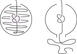
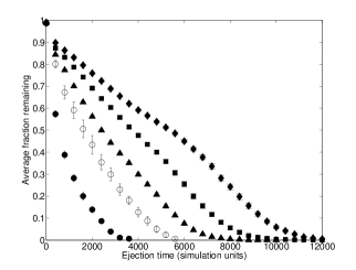
Fig. 2 shows typical results for the fraction of beads left in the capsid as a function of time from the release of the polymer. For this figure we used a small capsid and a random initial configuration but very similar results are found for other packings, initial conditions and capsid sizes. We compare results for unknotted chains to those with the knots , and . (Here we use the notation , where gives the minimal number of crossings in a projection of the knot onto a plane and is a standard way to distinguish between knots with the same number of crossings Orlandini and Whittington (2007).) Fig. 2 shows that there is a clear slowing of the rate of ejection when a knot is present. Moreover, the rate of ejection depends on the type of knot: the more complex the knot, the slower the rate of ejection.
At very early times the rates of ejection are similar and relatively high for both knotted and unknotted polymers. This corresponds to the knot being tightened and pushed to the capsid entrance as any free beads between the knot and the exit are ejected. The knot is too large to escape, so once it has moved to the hole it is held there by the excess pressure inside the capsid. Now the polymer has to reptate through the knot before any monomers are free to allow further ejection. Essentially the knot is acting as a ratchet. If it diffuses a small distance into the capsid, this frees a length of polymer between the knot and the capsid entrance. The knot is then quickly pushed back towards the entrance by the driving force and the free section of polymer is ejected. Near the end of the ejection, when only about beads remain in the capsid, the ejection speeds up and there is a shoulder in the curves in Fig. 2. This corresponds to the knot becoming undone.
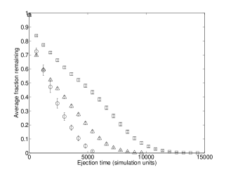
To confirm the effect of the knot on the dynamics, Fig. 3 compares similar ejection curves for a semiflexible polymer for length for three different initial conditions (i) unknotted, (ii) with a knot initially at the the polymer mid-point (iii) with a knot initially at capsid exit. For case (ii) the polymer initially ejects with the same speed as the unknotted polymer (i), but, once the knot has reached the capsid exit, the rate becomes similar to case (iii).
Simulations for longer polymers in a capsid of radius 4.36 showed the same generic behavior. Some small differences were that the final unknotting occurred with a somewhat larger number of beads left in the capsid because there was more free space, and that the rates sometimes showed a slight decrease with time, most likely because the pressure decrease has an effect on knot dynamics. For the randomly packed polymer we sometimes also observed initial jamming, with large variations in ejection time between runs. This occurred because the knot was initially trapped in a very tight configuration. However once ejection did start, the rate was independent of the initial jamming time.

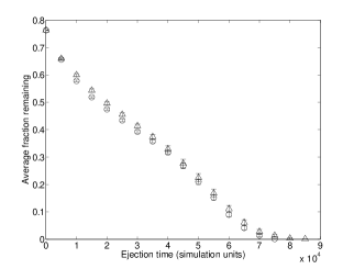
A quantitative comparison of ejection rates is shown in Fig. 4. Results are presented for both flexible and semiflexible polymers for knots with up to 6 crossings for chains in the small capsid, and for knots with up to 12 crossing for chains in large capsids. The results plotted are for the knots of type . Other knots, such as and , were considered. The results were very similar to the knots with the same C. Average rates were obtained by measuring the time taken for between 75% to 35% of the beads to be expelled. This protocol was chosen to avoid any early time jamming and the late time unravelling of the knot. Small changes in the cut-offs gave no qualitative changes in the results.
Fig. 4 shows the trend of decreasing ejection rate with increasing knot complexity. For the unknotted case, there are clear differences between the different kinds of polymers and capsids, but for more complex knots, the rates converge and seem to be almost completely determined by the knot type. This suggests that, for tight, confined knots, the rate of reptation of a knot may be independent of its flexibility. A similar independence of knot properties - the minimum length-to-radius ratio required for a tube forming a knot Buck and Rawdon (2004) and the diffusion constant Huang and Makarov (2007) - from flexibility has been reported.
Finally, note that the ejection rates show a weak oscillation as a function of knot size. The plausible explanation for this is that the knots we consider here with even and odd crossings belong to families with different topologies. Knots , and are torus knots and , and are even twist knots. It is reasonable that there is a weak dependence of the reptation rate along the chain on the knot topology. We have observed a similar oscillation of the polymer diffusion coefficients of knots on unconfined polymers under tension Matthews et al. .
Our model of a bacteriophage is highly simplified. For example, we ignored the tail. We checked that this does not have an important effect on the qualitative behavior by simulating a capsid with a tail attached. Perhaps more importantly, to make simulation feasible, our model of DNA inevitably ignores much chemical detail and our polymer diameter to persistence length ratio is much larger than in real DNA. Moreover, viral DNA is typically much longer than the polymers we study. However, we argue that the generic effects we observe: that the ejection rate is dominated by the rate of knot reptation, and that the knot is unravelled as it emerges from the capsid, are basic properties of knotted polymers moving through a narrow exit hole, and should therefore be robust to the inclusion of these details. Knots in phages may initially be loose but we expect that they will be tightened as the DNA is forced through the capsid entrance.
Experiments suggest that there could be multiple knots in the DNA in a viral capsid Arsuaga et al. (2002, 2005). To investigate the effect of multiple knotting we ran 50 simulations in each of which three knots, chosen randomly from , , , and were placed on the chain, near to the entrance of a larger capsid. Fig. 5 compares the averaged ejection curve to that for a single knot, showing that the knots stack up at the capsid entrance and behave like a single prime knot with a similar number of crossings. In the longer DNA in a viral capsid one might expect to see multiple knots which would sequentially collect at the exit. Thus the knot length would increase with time, which is expected to slow the ejection rate. It will be interesting to see if any sign of this effect can be seen experimentally.
To summarize, we have used simulations to determine the effect of knotting on the ejection of flexible and semiflexible polymers from a spherical capsid. We find that the ejection rate is controlled primarily by the knot, not the pressure of confinement. The knot moves to the hole in the capsid and then acts as a ratchet. Polymers with more complex knots eject more slowly. For tightly confined knots the flexibility of the polymer is not key in determining the rate of ejection.
Repeating packing experiments Smith et al. (2001) with knotted DNA would shed light on the dynamics of knots on polymer chains, and it is also of interest to ask whether knots can affect the motion of biomolecules as they traverse nanopores Dekker (2007).
Acknowledgments: We thank Issam Ali, Enzo Orlandini, Davide Marenduzzo and Cristan Micheletti for helpful discussions.
References
- Alberts et al. (1994) B. Alberts, D. Bray, J. Lewis, M. Raff, K. Roberts, and J. Watson, Molecular Biology of the Cell (Garland Publishing, New York, 1994).
- Mangenot et al. (2005) S. Mangenot, M. Hochrein, J. Radler, and L. Letellier, Curr. Biol. 15, 430 (2005).
- de Frutos et al. (2005) M. de Frutos, L. Letellier, and E. Raspaud, Biophys. J. 88, 1364 (2005).
- Lof et al. (2007) D. Lof, K. Schillen, B. Jonsson, and A. Evilevitch, J. Mol. Biol. 368, 55 (2007).
- Evilevitch et al. (2003) A. Evilevitch, L. Lavelle, C. M. Knobler, E. Raspaud, and W. M. Gelbart, Proc. Nat. Acad. Sci. U.S.A. 100, 9292 (2003).
- Kindt et al. (2001) J. Kindt, S. Tzlil, A. Ben-Shaul, and W. M. Gelbart, Proc. Nat. Acad. Sci. U.S.A. 98, 13671 (2001).
- Tzlil et al. (2003) S. Tzlil, J. T. Kindt, W. M. Gelbart, and A. Ben-Shaul, Biophys. J. 84, 1616 (2003).
- Inamdar et al. (2006) M. M. Inamdar, W. M. Gelbart, and R. Phillips, Biophys. J. 91, 411 (2006).
- Ali et al. (2006) I. Ali, D. Marenduzzo, and J. M. Yeomans, Phys. Rev. Lett. 96, 208102 (2006).
- Ali et al. (2008) I. Ali, D. Marenduzzo, and J. M. Yeomans, Biophys. J. 94, 4159 (2008).
- Arsuaga et al. (2002) J. Arsuaga, M. Vazquez, S. Trigueros, D. W. Sumners, and J. Roca, Proc. Nat. Acad. Sci. U.S.A. 99, 5373 (2002).
- Arsuaga et al. (2005) J. Arsuaga, M. Vazquez, P. McGuirk, S. Trigueros, D. W. Sumners, and J. Roca, Proc. Nat. Acad. Sci. U.S.A. 102, 9165 (2005).
- Micheletti et al. (2006) C. Micheletti, D. Marenduzzo, E. Orlandini, and D. W. Sumners, J. Chem. Phys. 124, 064903 (2006).
- Michels and Wiegel (1986) J. P. J. Michels and F. W. Wiegel, Proc. R. Soc. London, Ser. A 403, 269 (1986).
- Kochan et al. (1984) J. Kochan, J. L. Carrascosa, and H. Murialdo, J. Mol. Biol. 174, 433 (1984).
- Muller et al. (1997) D. J. Muller, A. Engel, J. L. Carrascosa, and M. Velez, EMBO J. 16, 2547 (1997).
- Cerritelli et al. (1997) M. E. Cerritelli, N. Cheng, A. H. Rosenberg, C. E. McPherson, F. P. Booy, and A. C. Steven, Cell 91, 271 (1997).
- Hud and Downing (2001) N. V. Hud and K. H. Downing, Proc. Nat. Acad. Sci. U.S.A. 98, 14925 (2001).
- Ali et al. (2004) I. Ali, D. Marenduzzo, and J. M. Yeomans, J. Chem. Phys. 121, 8635 (2004).
- Malevanets and Kapral (1999) A. Malevanets and R. Kapral, J. Chem. Phys. 110, 8605 (1999).
- Orlandini and Whittington (2007) E. Orlandini and S. G. Whittington, Rev. Mod. Phys. 79, 611 (2007).
- Buck and Rawdon (2004) G. Buck and E. J. Rawdon, Phys. Rev. E 70, 011803 (2004).
- Huang and Makarov (2007) L. Huang and D. E. Makarov, J. Phys. Chem. A 111, 10338 (2007).
- (24) R. Matthews, A. A. Louis, and J. M. Yeomans, unpublished results.
- Smith et al. (2001) D. E. Smith, S. J. Tans, S. B. Smith, S. Grimes, D. L. Anderson, and C. Bustamante, Nature 413, 748 (2001).
- Dekker (2007) C. Dekker, Nat. Nanotechnol. 2, 209 (2007).