Charge transport-mediated recruitment of DNA repair enzymes
Abstract
Damaged or mismatched bases in DNA can be repaired by Base Excision Repair (BER) enzymes that replace the defective base. Although the detailed molecular structures of many BER enzymes are known, how they colocalize to lesions remains unclear. One hypothesis involves charge transport (CT) along DNA [Yavin, et al., PNAS, 102, 3546, (2005)]. In this CT mechanism, electrons are released by recently adsorbed BER enzymes and travel along the DNA. The electrons can scatter (by heterogeneities along the DNA) back to the enzyme, destabilizing and knocking it off the DNA, or, they can be absorbed by nearby lesions and guanine radicals. We develop a stochastic model to describe the electron dynamics, and compute probabilities of electron capture by guanine radicals and repair enzymes. We also calculate first passage times of electron return, and ensemble-average these results over guanine radical distributions. Our statistical results provide the rules that enable us to perform implicit-electron Monte-Carlo simulations of repair enzyme binding and redistribution near lesions. When lesions are electron absorbing, we show that the CT mechanism suppresses wasteful buildup of enzymes along intact portions of the DNA, maximizing enzyme concentration near lesions.
pacs:
87.15.H,82.39.Pj,05.10.Gg,05.40.-aI Introduction
The genomes of all living organisms are constantly under attack by mutagenic agents such as reactive oxygen species and ionizing radiation. Such processes can damage bases giving rise to localized lesions in the DNA Bruner et al. (2000); Nash et al. (1996) that can lead to harmful mutations and diseases such as cancer. For example, guanine residues can be oxidized, generating a radical called 7,8-dihydro-8-oxoguanine, or oxoG for short. Unlike the non-oxidized form, this radical can pair with both cytosine and adenine, ultimately giving rise to GC TA transversion mutations Bruner et al. (2000) upon multiple replications. Lesions can also arise through alkylation, hydration and deamination.Bruner et al. (2000)
One defense mechanism against these mutation processes is the Base Excision Repair (BER) pathway. BER enzymes recognize and undo damage to DNA by adsorbing onto the sugar-phosphate backbone, locating the lesion and excising it. The biomechanical functions of repair enzymes have been well established and their 3D structures are known in great detail.Parikh et al. (1997) There are four main types of BER enzyme: DNA glycosylases, AP-endonucleases, DNA polymerases and DNA ligases. Each of these enzymes has a different role in the BER family. For example, DNA glycosylases initiate the repair pathway, detecting and recognizing distinct forms of DNA damage while the endonucleases are responsible for cleaving the sugar-phosphate backbone. Together, these enzymes maintain the overall integrity of DNA, generally ensuring that miscoded proteins are kept to a minimum.
The problem of how a BER enzyme locates a lesion on DNA is a specific example of how enzymes find localized targets. The DNA of E. coli contains about base pairs. If we assume that BER enzymes find lesions through a pure 1D diffusive “sliding” process with diffusion constant , the search time is roughly . Estimating to be , the value for a human DNA glycosylase, Blainey et al. (2006) we obtain a search time of about days, much longer than even the reproductive period of E. coli. Therefore, it is likely that other mechanisms are responsible for DNA target location.
In 1970, Riggs et al. Riggs et al. (1970a, b) measured the association rate of the LacI repressor protein to its target on DNA to be about . This was puzzling because the theoretical upper limit for the association rate of a LacI enzyme diffusing in 3D is predicted (via the Debye-Smoluchowski formula) to be about 2 orders of magnitude less. This fundamental biophysical problem was studied in the seminal work of von Hippel and co-workers Berg et al. (1981); Winter et al. (1989); von Hippel and Berg (1989); Berg and von Hippel (1987) and the “faster-than-diffusion” search of targets on DNA has received recent attention. Mirny (2008); Wunderlich and Mirny (2008); Klenin et al. (2006); Slutsky and Mirny (2004); Hu et al. (2006); Cherstvy et al. (2008) Facilitated diffusion is one mechanism von Hippel and Berg (1989); Slutsky and Mirny (2004); Wunderlich and Mirny (2008); Halford and Marko (2004) proposed to explain the accelerated search. Instead of diffusing directly to their target, the searching enzymes can spend part of their time attached to the DNA and perform a 1D random walk along part of the strand. If the enzyme is able to spend 50% of its time on the DNA and 50% of its time diffusing in 3D, and the diffusion constants in 1D and 3D are comparable, the association rate is predicted to increase by as much as 100, Slutsky and Mirny (2004) bringing it in line with the experiments in Riggs et al. Riggs et al. (1970a, b) However, other authors have shown that (i) typical enzymes are highly associated with DNA, spending over 99.999% of their time on the strand Wunderlich and Mirny (2008) and (ii) the diffusion constant in 1D can be 1000 times smaller than in 3D,Wang et al. (2006) resulting in a negligible reduction of the search time. Hence, facilitated diffusion in its basic form is not adequate to explain the fast reaction rates observed. Extensions to the facilitated diffusion theory can incorporate finite enzyme concentrations, Cherstvy et al. (2008) “antenna” effects resulting from the conformation of the DNA, Hu et al. (2006) fast intersegment transfers of the protein, Slutsky and Mirny (2004) specific/non-specific protein-DNA interactions, Slutsky and Mirny (2004) and directed DNA sliding.Loverdo et al. (2008)
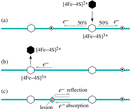
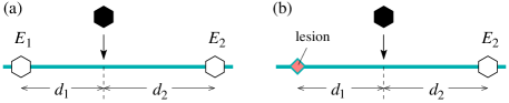
Although BER enzymes may colocalize to lesions by exploiting the facilitated diffusion mechanisms cited above, other mechanisms are likely required for efficient and timely recruitment to lesions. A charge-transport (CT) mechanism has been recently proposed as a possible basis for efficient scanning by MutY, a type of DNA glycosylase.Yavin et al. (2005); Boon et al. (2003) MutY is known to contain an iron-sulfur cluster which plays a key role in the CT mechanism. The cluster can take one of two forms: [4Fe-4S]2+ and [4Fe-4S]3+. When MutY is in solution, the cluster is in the state and is resistant to oxidation. However, upon binding to DNA, the cluster potential is shifted, making the state more accessible. The result is that after binding, MutY-[4Fe-4S]2+ is easily oxidized and releases an electron along the DNA, as shown in Fig 1(a). It should be noted that the state of MutY has a binding affinity that is about 4 orders of magnitude larger than that of the state. Boal et al. (2005) Therefore MutY-[4Fe-4S]2+ spends most of its time in solution whereas MutY-[4Fe-4S]3+ exists primarily adsorbed onto DNA.
Although controversial about 15 years ago, long range electron transport in DNA is now a well accepted phenomenon.Giese (2002); Schuster (2000) Experiments indicate that charge transport can occur over (about 12 base pairs) in less than a nanosecond Turro and Barton (1998); Murphy et al. (1993) and the influence of DNA strand crossovers on CT is generally small. Giese (2002) Although electron dynamics along DNA is in general very complicated, some aspects of the process are now understood. For example, both guanine and adenine can act as carriers of positive charge; in analogy with semiconductors, oxidized DNA can transport charge via the transfer of holes from base to base.
Quantifying how BER enzymes adsorb to DNA and how they are recruited to lesions has so far been restricted to simple scaling arguments.Eriksen (2005) In this paper, in order to explore the implications of DNA target selection solely by CT, we assume that adsorbed MutY BER enzymes do not slide along the DNA. However, upon first attachment to DNA, the enzyme will emit an electron that propagates along the strand in a random direction and its cluster will go from the [4Fe-4S]2+ to the [4Fe-4S]3+ state. Should this electron become absorbed by another MutY-[4Fe-4S]3+ enzyme further along the DNA, the form is reduced and desorbs (Fig 1(b)). If the electron back-scatters and returns to the original MutY, it self-desorbs. Although the model proposed in this paper is intended to specifically describe the colocalization and redistribution of MutY through the redox reaction of its iron-sulfur cluster, many BER enzymes, in fact, contain such a cluster, e.g. endonuclease III. Therefore, we think that our model may be more general and could also describe the binding kinetics of other enzymes.
Since unbiased stochastic motion in 1D always leads to return of the electron, Redner (2001) in the absence of any other electron absorbers on the DNA, a MutY BER enzyme that is deposited will eventually self-desorb with probability 1. However, BER enzymes can be recruited to DNA by preexisting electron absorbers. These are typically guanine radicals (“oxoG”) and other lesions, indicated in Fig. 1(c) by circumscribed dots and filled diamonds, respectively. It has been suggested that oxoG plays an important role in the seeding of MutY onto DNA.Yavin et al. (2005) The oxoG radicals, like adsorbed enzymes, are able to absorb electrons, preventing them from returning and desorbing BER enzymes that originally released them. Therefore, the oxoG radical in Fig. 1(a) can absorb one left-moving electron and prevent it from back-scattering and desorbing the right-most enzyme. Upon reduction, oxoG radicals convert to normal guanine bases, no longer absorb electrons, and no longer take part in the CT mechanism.
Other lesions do not simply annihilate by absorbing electrons; rather, they require the physical presence of BER enzymes to excise them. These lesions may recruit smaller, more abundant proteins from solution that permit multiple electron absorption. Another possibility is that the lesions reflect electrons. Both cases are shown in Fig. 1(c). Therefore, our basic model consists of right and left-moving electrons, guanine radicals, oxidized and reduced forms of BER enzymes, and lesions on the DNA strand. Newly adsorbed BER enzymes instantly release electrons (right or left-moving), while oxoG radicals, lesions, and oxidized BER enzymes absorb electrons and prevent their passage.
In this paper, we model the adsorption, desorption and redistribution of repair enzymes using the redox mechanism shown in Fig. 1. We first derive some exact results in the absence of any lesions; in particular, enzyme adsorption probabilities and the time taken for returning electrons to induce enzyme desorption. These results enable us to define rules for Monte-Carlo simulations of the dynamics of multiple enzymes. For electron absorbing lesions, simulations show that if enzymes are deposited onto a DNA at a rate that is slow compared to the electron dynamics, the distance between a lesion and the closest enzyme scales as for large , while total number of enzymes adsorbed between two lesions scales as . However, because of the CT mechanism, this accumulation is not uniform along the DNA and the maximum enzyme density always occurs at lesions. Hence for electron-absorbing lesions, the CT mechanism concentrates enzymes to damaged bases in DNA, consistent with the qualitative predictions in Yavin, et al. Yavin et al. (2005) and Boon, et al. Boon et al. (2003)
The outline of this paper is as follows. In the next section, we develop a model for the electron dynamics based on the stochastic Broadwell model. Bicout (1997); Broadwell (1964a, b); Christlieb et al. (2004) Pairs of guanine radicals, BER enzymes or lesions define the boundary of a segment (a “gap”) over which an electron can propagate. Section III contains our results. In Section III.1, we we derive enzyme sticking probabilities and the time taken for returning electrons to desorb the enzymes that originally emitted them. In particular, we derive the MutY desorption rate in terms of the electron scattering (flip rate) and the electron speed. In Section III.2, we perform implicit-electron Monte-Carlo simulations to study the redistribution and accumulation of enzymes between two fixed lesions on the DNA. Finally, in Section IV, we discuss facilitated recruitment of enzymes to lesions in the context of the CT hypothesis, as well as the biological advantages and disadvantages of the proposed CT mechanism.
II Stochastic Charge Transport Model
II.1 One-sided Broadwell problem
| Symbol | Definition | Units |
|---|---|---|
| Probability density of rightward electron | 1/L | |
| Probability density of leftward electron | 1/L | |
| Position along DNA | L | |
| Position of electron release | L | |
| Time | T | |
| Density of oxoG guanine radicals on DNA | 1/L | |
| Distance between two oxoGs/enzymes | L | |
| electron flip rate | 1/T | |
| Electron speed | L/T | |
| Electron decay rate | 1/T | |
| Deposition rate of enzymes | 1/(L T) |
| Symbol | Math defn. | Descriptive definition |
|---|---|---|
| Rightward/ Leftward electron probability density | ||
| Coordinate along DNA | ||
| Position of electron release | ||
| Time | ||
| “Gap size” : distance between two oxoGs/enzymes/lesions | ||
| Electron flip rate | ||
| Electron decay rate | ||
| - | Position of enzyme adsorption | |
| - | Enzyme-lesion/enzyme-enzyme distance (see Fig. 6) |
In analogy with Bicout’s analysis for the unrelated problem of microtubule growth dynamics,Bicout (1997) we now present similar equations for the dynamics of electrons associated with repair enzymes. Consider Fig. 2(a): oxoG guanine radicals with density are distributed randomly along an infinite strand of DNA. A single repair enzyme initially attaches to the DNA at a random position, in between two electron absorbing oxoGs. The enzyme immediately emits an electron along the DNA to the left or right with equal probability. The electron can only move with speed , in the positive or negative directions, executing random flips between the two directions with rates . Furthermore, emitted electrons can be annihilated with rate through nonspecific interactions with random electron absorbers diffusing in the bulk.
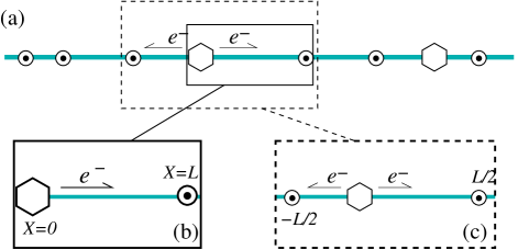
In general, two steps are required for a MutY enzyme to bind to DNA. First, when MutY-[4Fe-4S]2+ is in contact with the DNA, it has to undergo oxidation by releasing an electron. The oxidized form of the enzyme binds more strongly to DNA. Second, the released electron must be absorbed by some particle other than the enzyme (an oxoG, an already adsorbed MutY or a lesion) to prevent it from returning and reducing the enzyme. This allows the enzyme binding to become “permanent”. Therefore the net binding probability depends on (i) the probability of electron release by MutY-[4Fe-4S]2+ (when in contact with the DNA) and (ii) how far neighboring electron absorbers are from the adsorbed MutY. In this paper, we assume that when enzymes adsorb onto the DNA, they always oxidize, releasing an electron with probability 1; in Fig. 2(b), the electron is released to the right with probability 1 and in Fig. 2(c), the electron is released to the left or right with probability 1/2. In principle, an enzyme can attach to and then immediately detach from the DNA without releasing its electron, but assuming the electron release rate is large, we neglect this process. The adsorption probabilities we derive later in this section will depend only on the gap size and the parameters for electron motion.
Finally, we assume that the DNA is immersed in an infinite reservoir of enzymes which is kept at a fixed chemical potential. The rate of deposition of enzymes onto the DNA is assumed to be constant. A deposited enzyme can either adsorb by having its released electron captured by neighboring electron absorbers or it can desorb due to its electron returning.
To build our full solution, we first derive exact analytical expressions for the “one-sided” problem shown in Fig. 2(b) which consists of an enzyme at and a guanine radical at . At time , an electron is emitted from a position (subsequently, we will take the limit ) with speed in the positive -direction. For the one-sided problem, the electron is emitted only to the right. The probability that the electron is at a position between and at time , and moving to the right with velocity is denoted . Similarly, denotes the probability density of an electron moving with speed in the negative -direction. The electron can flip directions by scattering from inhomogeneities and thermally excited conformational variations along the DNA.D’Orsogna and Rudnick (2002); Bruinsma et al. (2000) We model this flipping process as a spatially homogeneous process occurring with constant rate , independent of any structure along the DNA such as base pair sequence.
The evolution equations for the probability densities are
| (1) |
where . Eqs. (1) describe the probability density of electrons being advected to the right and left. The flipping of the electron motion is represented through and couples the equations for and . Furthermore, the densities decay in time with an annihilation rate . Electrons can be annihilated by being absorbed by other proteins (besides BER enzymes) in solution. If these proteins adsorb onto the DNA, absorb an electron and desorb back into solution, an electron is permanently removed from the DNA.
The boundary conditions and initial conditions are
| (2) | |||||
| (3) | |||||
| (4) |
The boundary conditions (2) arise because the enzyme at and the oxoG at (see Fig. 2(b)) are both perfect electron absorbers. When , the initial condition (3) reflects the fact that an electron is released to the right from the enzyme at . Initially, there are no leftward traveling electrons in Fig. 2(b), justifying Eq. (4). All variable and parameters are listed in Tables 2.
We now define dimensionless independent variables through the guanine radical density and the rightward electron travel time :
| (5) |
so that Eqs. (1) can be written in the form
| (6) |
where and
| (7) |
and . In Eq. (7),
| (8) |
is the dimensionless flipping rate and electron decay rate. The boundary and initial conditions (2), (3), (4) become
| (9) |
where . In the physical problem, an electron is released from the enzyme as soon as it initially attaches to the DNA. Therefore, we solve Eqs. (6) with (7) and (9) taking the limit (for details, see Appendix A). The dimensionless variables are tabulated and defined in Table 2. Henceforth all of our results and analyses will be presented for .
II.2 Two-sided Broadwell problem
Now consider the two-sided problem depicted in Fig. 2(c). A repair enzyme lands at position between two oxoG guanine radicals that are a distance apart. The solution to the full problem can be found by splitting it into two subproblems and using our results from Section II.1. Instead of solving for the densities on , we can solve for separately on (with the enzyme initially deposited at and the guanine radical at ), on (with the enzyme at and the guanine radical at ) and combine the results. The enzyme desorption and adsorption probabilities (10) extend straightforwardly:
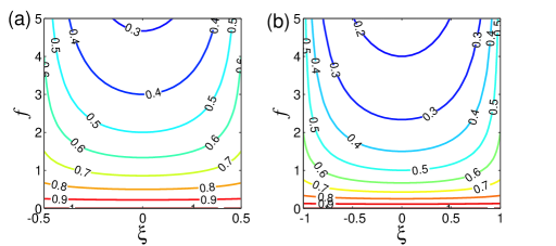
| (11) |
A plot of the sticking probability for different values of and for two different gap sizes is shown in Fig. 3. For a fixed gap size, and sufficiently large (corresponding to a diffusive electron motion), permanent BER enzyme adsorption is less likely to occur near the center of the gap because absorption of the electron by guanine radicals is less likely to occur. The permanent adsorption or sticking probability is more uniform when is small (corresponding to a ballistic electron motion): whether the oxoG radical is close or far away from the enzyme makes little difference to the adsorption probability. Finally, for fixed , increasing the gap size decreases the adsorption probability because guanine annihilation by the electron is less likely to occur. The diffusive and ballistic behaviors of the Broadwell model are derived in Appendix B.
| Symbol | Descriptive definition | See Eq. |
|---|---|---|
| Enzyme adsorption probability | (11) | |
| Enzyme desorption probability | (11) | |
| Enzyme adsorption prob. averaged over landing position | (12) | |
| Enzyme desorption prob. averaged over landing position | (20) | |
| Enzyme adsorption prob. averaged over landing posn. and gap size | (14,16) | |
| Random variable for conditional return time of electron | (17) | |
| Mean conditional return time (MCRT) of an electron | (18) | |
| MCRT of an electron averaged over landing position | (19) | |
| MCRT of an electron averaged over landing posn. and gap size | (21) |
III Results and Discussion
III.1 Statistics of repair enzymes away from lesions
In this section, we present and discuss deposition statistics that are valid far away from lesions. First, using Eq. (11), we average over the landing position to calculate mean sticking/adsorption probabilities of repair enzymes that are deposited between two guanine radicals that are a distance apart. The inter-radical distances (“gaps”) in DNA will, in general, be randomly distributed. Therefore we ensemble-average our results over the distribution that is expected to obey. Second, we find the mean return times of electrons, i.e. the time taken for a deposited enzyme to be desorbed by its own electron, providing it desorbs. Again, our results are ensemble-averaged over randomly distributed gap sizes. The quantities we shall compute and analyze in this section are listed in Table 3.
All the results presented are for adiabatic depositions. A deposition is adiabatic if the inter-deposition time is much larger than the time scale of the electron dynamics. In other words, for every enzyme deposited, its released electron completes its motion before the deposition of the next enzyme. At any given time, there is at most one traveling electron on the DNA. For details, see Appendix C.
III.1.1 Repair enzyme sticking probability
One quantity of interest is the probability that any given repair enzyme that lands on the DNA will not be kicked off by its own electron, and will remain adsorbed. Enzyme sticking relies on efficient capture of the released electron by neighboring electron absorbers (guanine radicals and adsorbed enzymes). Intuitively, one would expect that a greater density of absorbers with smaller gaps would result in a more efficient capture of enzymes.
For a single repair enzyme deposited onto the DNA, landing at a position (see Fig. 2(c)) inside a gap of length , centered about , the probability of it remaining on the DNA is given by in Eq. (11). This quantity can be averaged over all possible deposition positions within the gap to obtain
| (12) |
This result is plotted in Fig. 4 (dashed line). Eq. (12) gives the sticking probability of a repair enzyme newly deposited between two electron absorbers separated by , uniformly averaged over its deposition position within the gap.
We now average over the gap length distribution to compute the sticking probability for deposited enzymes that land anywhere along the entire DNA strand. For an infinite, lesion free DNA, depositing an enzyme will, in general, change the local guanine and enzyme distribution. Hence, the sticking probabilities will also change with each successive deposition, making the calculation difficult in the context of the Broadwell model. However it is possible to calculate the sticking probability for a given gap distribution. In special cases where this distribution is known or simple to calculate, we can compute the efficiency of enzyme recruitment onto the DNA.
Consider the case of a DNA with a discrete distribution of gaps . Suppose that on the DNA, a fraction of the gaps have size . Now consider many realizations of a single enzyme deposited onto this DNA. The fraction of enzymes that lands in gaps of size is and the fraction of these that stays adsorbed, using Eq. (12), is
| (13) |
The fraction of enzymes that stays adsorbed (in any gap) is obtained by summing over . In the continuum limit, , , where is the continuous gap length and is the probability distribution function (PDF) for . We obtain
| (14) |
Note that , the result that one might expect by naively averaging Eq. (12) over the gap distribution.
Since depends on the number of enzymes deposited, it is time dependent. In principle, one could calculate how changes as enzymes are adiabatically deposited. The corresponding evolution of the sticking probability is then given by Eq. (14). One possible way of finding how evolves is to use a mean field theory for the particle distributions, but we leave this as the subject of a future investigation.
In the special case where one enzyme is deposited onto a DNA that only has guanine radicals, we can calculate and hence explicitly. If the guanine radicals have a number density , then the gap lengths, on average, are , which corresponds to a unit dimensionless gap size (see Eq. (5)). Hence and the dimensionless gap sizes, , are exponentially distributed (see Appendix D) according to
| (15) |
so we set . Substituting this result into Eq. (14), we obtain
| (16) |
where is the exponential integral. This analytic result is plotted in Fig. 4 (solid line) and is confirmed by Monte-Carlo simulations (circles). The sticking probability increases when either the electron-absorber density increases, the electron velocity increases or the flip rate decreases.
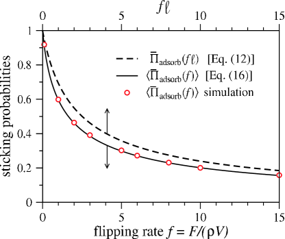
Equation (16) is valid only when the number of enzymes that have stuck is much less than the initial number of oxoG radicals. In this limit, the distribution of gap lengths will remain approximately exponential. For the human genome of base pairs, there are approximately oxoGs present at any given time.Helbock et al. (1998) In this case, we expect that Eq. (16) should be fairly accurate for about the first dozen depositions.
Note that (Eq. (12)) with gives the enzyme sticking probability inside an inter-radical gap of unit length, whereas (Eq. (16)) gives the enzyme sticking probability averaged over exponentially distributed inter-radical gaps lengths, but with unit mean. Intuitively, one would expect the boundaries defining the smaller gaps to be more efficient at sequestering electrons than those associated with larger gaps. However, the enhanced electron trapping by smaller gaps, leading to otherwise increased sticking probabilities is compensated by a higher deposition flux into larger gaps (large gaps collect more enzymes than small gaps). The net result of averaging over exponentially distributed gap sizes is for the larger gaps to dominate and lower the overall gap-averaged sticking probability. This is shown in Fig. 4 where for all values of , when .
III.1.2 Mean conditional return time of electrons
We now find the mean time that a BER enzyme stays on the DNA after its initial deposition, conditioned on its own electron returning and knocking the enzyme off. This quantity allows us to estimate a rate of desorption that can be used in more coarse-grained, higher level descriptions of the CT mechanism.
Consider depositing an enzyme into a gap of size at a position satisfying . The probability that the electron (“”) returns in a time , given that it returns is,
| (17) | |||||
In Eq. (17), is the leftward electron density at position at time given that the electron was released from at (see Fig. 1(b) for the case). This density comes from solving Eqs. (6) and (7) along with the conditions (9).
The mean conditional electron return time can then be computed from
| (18) |
Using Eq. (17), in Eq. (18) can be found in terms of the Laplace-transformed density which is given in Eq. (26) of Appendix A. Upon averaging over the initial landing positions , we obtain
| (19) |
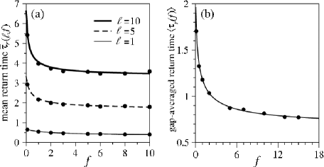
We plot , and validate Eq. (19) using MC simulations in Fig. 5(a).
Finally, we further ensemble-average over gap lengths . Consider many realizations of the deposition of a single enzyme onto an infinite DNA with oxoGs whose gaps are exponentially distributed. The average time that the enzyme stays adsorbed, given that its electron eventually returns to knock it off, is . The calculation of is similar to that of described in Section III.1.1, but modified to account for the fact that the number of enzymes that self-desorb (i.e., the number of return times that are finite) depends on . If an enzyme is deposited into a gap of size , the probability of self-desorbing after a finite time is given by (see Eq. (12))
| (20) |
Therefore, the required expression for is
| (21) |
In the numerator of Eq. (21), is the fraction of deposited enzymes that (i) land in a gap that has a length between and and (ii) eventually self-desorb after finite time. In the denominator, is the fraction of deposited enzymes that self-desorb after a finite time. The result (21) is confirmed by simulation data in Fig. 5(b).
Equation (21) was derived by considering the deposition of a single enzyme onto an infinite DNA with exponentially distributed gap lengths. However, as is the case with Eq. (16), it is also approximately true for a small number of depositions onto a finite DNA: providing the number of oxoGs annihilated is small compared to the total number of oxoGs, the distribution of gap lengths is still approximately exponential. Hence, for a given deposition rate of enzymes per unit length onto an infinite DNA, Eq. (21) will hold approximately for times such that the fraction of oxoGs annihilated is small.
Given a deposition rate of enzymes (per unit length), we can estimate a desorption rate (per unit length) from Eq. (21). If desorption were a Poisson process, then the desorption rate, , is found from the inverse of the mean unbinding time of the repair enzyme. Although the desorption process in our model depends on the dynamics of electron charge transport (rendering it to be non-Poisson), the inverse of the ensemble averaged conditional return time of an electron , is nonetheless a reasonable definition for the detachment rate . We expect this value of to be accurate, as long as the fraction of oxoGs annihilated by repair enzymes is small. The probabilities and times relevant to electron dynamics are summarized in Table 3.
III.2 Colocalization of enzymes to lesions
We now consider a permanent lesion on the DNA (one that does not annihilate upon absorption of an electron). Such a lesion may be bound to other enzymes and cofactors so that it can act as a sink for multiple electrons, or it can reflect electrons. In this section, we consider lesions that can either absorb or reflect electrons, as shown in Fig. 1(c). We are primarily interested in the average number of depositions required for a repair enzyme to be adsorbed within a certain (small) distance from the lesion.
For the sequential deposition of many enzymes onto a DNA populated with guanine radicals and lesions, the evolution of enzyme and guanine densities is not amenable to exact analytical solution. Therefore, our approach will be to track enzyme-lesion distances and enzyme concentrations on the DNA by performing Monte-Carlo simulations.
Each simulation consists of a series of adiabatic depositions. A deposition is simply the spontaneous appearance of a MutY-[4Fe-4S]3+ enzyme at a randomly chosen position along the DNA. Note that a deposition is an attempted adsorption: it can result either in the enzyme sticking to the DNA, or desorbing from it. In our simulations, the number of enzymes on the DNA can grow without bound. We do not model the bulk dynamics for MutY-[4Fe-4S]2+ enzymes in solution.
In our model, each enzyme that is deposited releases an electron along the DNA. However, rather than performing time-consuming, explicit simulations of a Broadwell process, we exploit our analytic results to implicitly account for the electrons. The rules for enzyme desorption and adsorption come from the probabilities and found in Eqs. (11). Specifically, consider the deposition of an enzyme, , between two already adsorbed enzymes, and (see Fig. 6(a)). Let the distance from to be , . Then the probability of adsorbing and knocking off is and the probability of self-desorbing is . In the case where an enzyme is deposited between a lesion and an adsorbed enzyme (see Fig. 6(b)), the adsorption and desorption probabilities have to be modified. If is replaced by an electron-reflecting lesion, the probability of permanently adsorbing without displacing is zero. The probability of adsorbing and knocking off is and the probability of self-desorption is .
If is replaced by an electron-absorbing lesion, the probability of permanently adsorbing without displacing is , the probability of adsorbing and knocking off is and the probability of self-desorption is . These probabilities are summarized in Tables 5 and 5.
| Event: | self-desorbs | adsorbs, desorbs | adsorbs, desorbs |
|---|---|---|---|
| Probability: |
| Event: | self-desorbs | adsorbs, stays adsorbed | adsorbs, desorbs |
|---|---|---|---|
| Probability: (reflecting lesion) | |||
| Probability: (absorbing lesion) |
MC simulations were performed on a periodic domain of size containing a single lesion, which is equivalent to a single finite domain with length and lesions at and . We start our simulations with no adsorbed BER enzyme (MutY), but with a unit density of guanine radicals (oxoG) whose gaps follow an exponential distribution (see Eq. (15)). When a single enzyme is deposited randomly on , the positions of the two particles (either oxoGs, lesions or already adsorbed enzymes) on either side are recorded and and are calculated (see Fig. 6). Using the probabilities in Tables 5 and 5, the outcome of this deposition event is determined: either the newly deposited enzyme adsorbs, or it desorbs due to its electron returning. Note that if an adsorption occurs, exactly one of three other events also has to occur: (i) a neighboring enzyme is reduced and desorbs (ii) a neighboring oxoG is annihilated or (iii) an electron is absorbed by a neighboring lesion.
Figure 7 shows density profiles obtained from our MC simulations. In Fig. 7(a), the depletion of guanine radicals is greater away from lesions: a guanine radical that is close to a lesion can, essentially, only be annihilated from one side. Near , the probability of oxoGs being annihilated from the left by a rightward-moving electron is very small. Similarly, near , the probability that oxoGs are annihilated from the right by leftward-moving electrons is also very small.
Figure 7(b) shows that electron reflecting lesions eventually prevent the build up of enzymes near lesions. The presence of an electron-reflecting lesion increases the local self-desorption rate. Note that the enzyme self-desorption probability is always greater in Fig. 6(b) than it is in Fig. 6(a) – when the lesion is electron reflecting. Therefore, near a reflecting lesion, the recruitment of enzymes by guanine radicals has to compete with this increased self-desorption rate. Although the density near the lesion increases with time, for a fixed time, its value is always smaller than the bulk value. Another way to understand the enzyme depletion is through a particle conservation argument. Since the total number of guanine radicals and BER enzymes is conserved, an increase in oxoG density near the boundaries must correspond to a decrease in the enzyme density.
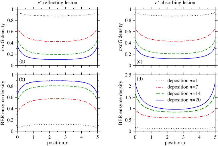
Figures 7(c) and 7(d) show density profiles near electron-absorbing lesions. The oxoG densities in Fig. 7(c) remain essentially unchanged from those surrounded by electron-reflecting lesions (Fig. 7(a)). As shown in Figs. 7(b) and 7(d), the BER enzyme density profiles are also similar for a small number of depositions, away from lesions. On the other hand, 7(d) also shows that for larger deposition numbers, the BER enzyme density near electron-absorbing lesions increases markedly.
The total number of particles on the DNA strand can be found by integrating the densities from to . For example, in Fig. 7(b), the solid curve representing the enzyme density after one attempted deposition takes the value over most of the domain and decreases slightly near the lesions. Therefore the (average) number of enzymes that remain adsorbed after one attempted deposition is approximately . This is in excellent agreement with the solid curve in Fig. 4 and Eq. (16) for since
Figure 7 only shows the densities up to 20 deposition attempts. When the number of depositions is much greater than 20, all of the enzyme-seeding guanine radicals are annihilated. In the absence of any electron absorbers on the DNA, there can be no net increase in enzyme number, and the enzyme density in Fig. 7(b) eventually saturates to unity everywhere in the domain, identical to the initial oxoG density. Each guanine radical is eventually replaced by a BER enzyme, so the long-time BER enzyme density mimics the initial oxoG density.
In contrast, when the lesions are electron absorbing, there are always two permanent electron absorbers in the system. In this case, the number of enzymes can grow without bound, even when all the oxoGs are depleted.
Figure 8(a) shows how enzymes converge to electron absorbing lesions located at and . At any given time, we label the enzymes on the DNA according to their position so that . Both the number of enzymes on the DNA, , and their positions, , are functions of , the number of (attempted) depositions that have occurred. We plot the quantities , and as functions of deposition number in Fig. 8(a). When fewer than 3 enzymes are adsorbed on the DNA, we define , . From our simulations, we find the scaling
| (22) |
in the large limit. For a BER enzyme to successfully excise a lesion, we assume that it has to be within a few base pairs of it. We set the physical enzyme-lesion distance , where is the width of a base pair which we take to be nm, and estimate . Approximately 1 in 40,000 guanine bases are guanine radicals, Helbock et al. (1998) so , and the number of attempted depositions required for the closest sticking enzyme to be within 5 base pairs of the lesion is . If each deposition takes at least 0.0005 seconds, 111 For E. Coli, the maximum deposition rate can be obtained by assuming a nucleoid radius of approximately m. Upon assuming a MutY diffusivity of cm2/s, the Debye-Smoluchowski estimate is M-1s-1 For MutY concentration of , the average time between depositions is . this amounts to a total (minimum) search time of about 50 minutes. Although this is a significant reduction compared to the original 1D sliding search time discussed in the Introduction, it is likely that MutY locates lesions even more quickly through a combination of the CT mechanism and facilitated diffusion along the DNA strand.
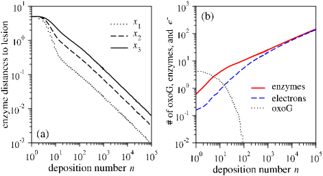
The solid curve in Fig. 8(b) shows the total number of enzymes on the DNA as a function of the deposition number when the lesions at and are electron absorbing. Upon depletion of the guanine radicals (shown by the dotted curve dropping to ), the enzyme number increases as . The dashed curve in Fig. 8(b) shows the number of electrons absorbed by the lesion. Initially, this is less than the enzyme number since enzymes adsorb mainly by oxoG annihilation. However, as all the radicals are used up, the dashed curve asymptotes to the curve for the enzyme total, indicating that the net increase of enzymes on the DNA is due primarily to lesion-induced colocalization.
Given that the enzyme-lesion distance scales as for electron absorbing lesions, one can directly show that the number of enzymes on the DNA scales as through a simple argument. If the enzyme-lesion distance is it takes attempts before an added enzyme lands closest to the lesion. When is large, the enzyme-lesion distance is small and the electron released by the newly deposited enzyme will be absorbed. For every depositions, on average, one permanent adsorption occurs. Hence, for every depositions, adsorptions occur.
While the convergence of CT enzymes towards lesions scales as , the convergence of passive enzymes (those that simply adsorb onto DNA without emitting electrons) scales as . The faster convergence of passive enzymes 222 While passive enzymes converge more quickly to enzymes when measured in terms of the deposition number , CT repair enzymes converge more quickly when measured in terms of the number of adsorptions . In fact, the enzyme-lesion distance for repair enzymes scales as compared to for passive enzymes with (every deposition results in an adsorption). is a consequence of linearly increasing the passive enzyme density on the DNA. The CT mechanism on the other hand, prevents the recruitment of large numbers of BER enzyme on the DNA at any given time with the total number scaling as for large . Hence, although BER enzymes only colocalize near lesions (note the maxima in the enzyme density occur at the lesions in Fig. 7(d)), and the CT mechanism suppresses the wasteful build-up of enzymes in undamaged parts of the DNA.
IV Summary and Conclusions
We developed a mathematical model for a proposed charge-transport mediated mechanism of Base Excision Repair (BER) enzyme colocalization to DNA lesions. Enzymes adsorb and desorb through a charge transport (CT) mechanism Yavin et al. (2005); Boon et al. (2003) which we model using a stochastic Broadwell process. Our main finding is that the CT mechanism concentrates repair enzymes at lesions provided the lesions are electron absorbing.
We first calculated enzyme sticking probabilities and self-desorption rates in the absence of lesions. Our results for an infinite, lesion free DNA, populated with guanine radicals, are summarized in Figs. 4 (which predicts the enzyme sticking probability) and 5 (which predicts the electron’s mean conditional return time). For the deposition of a single enzyme onto an infinite DNA, the results are exact; for a given deposition rate per unit length, we expect the results to hold approximately providing the fraction of guanine radicals (oxoGs) annihilated is small. We also explored how enzymes colocalize to lesions using Monte-Carlo simulations. Enzymes were adiabatically deposited onto a circular DNA with a single lesion. We found that electron-absorbing lesions colocalize CT enzymes, and while electron-reflecting lesions do not (Fig. 7).
Simple faciliated diffusion is often unable to account for the fast search times observed in certain DNA-protein reactions. Riggs et al. (1970a, b) Cherstvy et al. Cherstvy et al. (2008) state that under realistic conditions, facilitated diffusion cannot occur and propose that acceleration is achieved through the collective behavior of proteins. In the context of target search by enzymes, the CT mechanism complements facilitated diffusion models. Slutsky and Mirny (2004); von Hippel and Berg (1989); Hu et al. (2006) The CT-mediated mechanism is one such example of collective behavior. Instead of basing the enzyme search problem on the time for a single protein to find its target, the CT mechanism relies on a collective build-up of enzyme density at the lesion. Hence, issues important in facilitated diffusion theories, such as the starting point of the enzyme relative to the lesion and the length of the DNA become irrelevant in the CT mechanism.
In the case where targets (lesions) are electron absorbing, we find that the maximum enzyme density always occurs at the permanent lesions and furthermore that the CT mechanism maintains a low density of enzymes far from lesions to suppress oxoGs, which are another form (albeit less permanent) of DNA damage. In fact, after an initial transient where all oxoGs are annihilated, the density of enzymes for most of the DNA will be of the order of the oxoG density, which is very low (about 1 in 160,000 base pairs). Subsequent enzyme depositions will colocalize only near the lesion. Our results show that although (attempted) depositions are required for the concentration to build up to a sufficient level at the lesions in order for them to be excised, the number of enzymes actually adsorbed on the DNA is much less, at . Although this is a significant reduction, it is still greater than the copy number of MutY (), so it is likely that the effects of 1D diffusion of MutY are important. Blainey et al. (2006)
When considering the collective behavior of enzymes, one important constraint is that the number of BER enzymes available to participate in the search mechanism is fixed. The copy number for MutY, in particular, is about 20, Bai and Lu (2007) placing a bound on the total number of enzymes that can be successfully adsorbed on the DNA strand. Thus, the CT search mechanism is effective only if the number of oxoGs is not significantly greater than . Although guanine radicals absorb electrons, thereby seeding the adsorption of BER enzymes, too many radicals can deplete the reservoir of BER enzyme before they significantly concentrate to the lesions.
Although in our model, there are two modes of enzyme recruitment – oxoG-mediated and lesion-mediated (when the lesion is electron absorbing) – it is the latter that colocalizes enzymes to lesions. We re-emphasize that the initial recruitment by guanine radicals can only increase the enzyme density to a level that is of the order of the initial radical density. This density is far too low to ensure reliable excision of the lesion. However, upon subsequent depositions, enzymes rapidly colocalize and the accumulation is more focused.
Although our simple model successfully predicts colocalization of CT BER enzymes to electron-absorbing DNA lesions, it neglects many potentially important aspects. For example, BER enzymes are not point particles but have a finite size of about 10-15 base pairs. Random adsorption of finite sized particles has been studiedD’Orsogna and Chou (2005) and could be used to enhance our current model. We also neglected the sliding of BER enzymes on DNA. Inclusion of finite size effects and enzyme sliding into our model is likely to decrease the search time to a lesion. The effect of other proteins on the DNA, besides BER enzymes, is also important. These proteins could physically prevent the adsorption of BER enzymes, absorb electrons emitted by BER enzymes or shield the lesion from electrons (or possibly all three). We currently do not know the effect of molecular crowding on the CT model, but this topic is discussed by Li et al. Li et al. (2008) One possible approach to studying these more subtle attributes is to develop and analyze them within coarse-grained, mass-action type models, in conjunction with Monte-Carlo simulations.
Acknowledgements.
This work was supported by grants from the NSF (DMS-0349195) and the NIH (K25 AI41935). The authors thank J. Genereux, A. K. Boal and J. K. Barton for helpful discussions.Appendix A Solution of the one-sided Broadwell problem
Taking the Laplace transform of Eq. (6), we obtain
| (23) |
where , and
| (24) |
The solution to Eq. (23), can be found in two separate regions and and matching the solutions with the appropriate jump conditions derived from integrating Eq. (23) over an infinitessimal segment centered about :
| (25) |
The general solution of Eq. (23), , can be expressed in the form
| (26) |
where , are given by
| (27) |
Appendix B Limiting cases of the Broadwell model
Upon eliminating from Eqs. (1), satisfies
| (29) |
Similarly, eliminating from Eqs. (1) gives Eq. (29) but with replaced with . Upon neglecting electron decay, , and Eq. (29) simplifies to
| (30) |
where we have used the nondimensionalization (5) and the non-dimensional flip rate . When , we neglect the first term in Eq. (30) to obtain a diffusion equation with diffusivity . When , we neglect the second term to obtain a wave equation with unit wave speed. These limits correspond to a diffusive and ballistic electron motion respectively.
Appendix C Adiabatic approximation
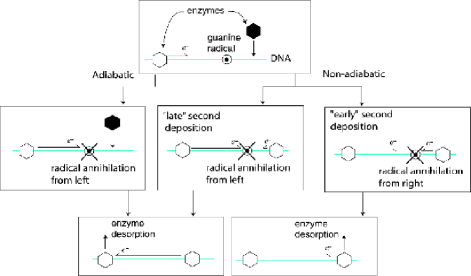
Since our stochastic analysis does not account for electron-electron interactions, we assume “adiabatic” deposition of BER enzymes. An adiabatic deposition of enzymes occurs when each enzyme is deposited sufficiently slowly so that the emitted electron completes its motion before the deposition of the next enzyme. At any given time, there is at most one traveling electron on the DNA.
Consider Figure 9: two enzymes are deposited on either side of a guanine radical with the left enzyme further away. For this example, assume that the electrons are always emitted toward the radical. In an adiabatic deposition, the deposition of the right enzyme occurs after the oxoG is annihilated. The final configuration consists of an adsorbed right enzyme and a desorbed left enzyme. In a non-adiabatic deposition, the right enzyme can be deposited before the annihilation of the oxoG. The final enzyme configuration depends critically on the time between the first and second depositions. If this time is long (a “late” second deposition), the oxoG is annihilated by the rightward electron and the final configuration is identical to the adiabatic case. If the inter-deposition time is short (an “early” second deposition), the leftward electron can annihilate the oxoG first and the final configuration corresponds to an adsorbed left enzyme and a desorbed right enzyme.
For a deposition to be adiabatic, the electron dynamics must be much faster than that of enzyme depositions:
| (31) |
where is the density of guanine radicals and is an intrinsic enzyme deposition rate per unit length of DNA. Thus, the adiabatic limit arises when and , with fixed (to keep the overall probabilities unchanged in Eq. (11)). Note that can still be small in an adiabatic deposition, as is the case in Fig. 9.
Appendix D Guanine gap distribution
Consider a lattice made up of sites on which guanine radicals can randomly appear at a rate of radicals per unit time , per lattice site. Each lattice site can hold at most one guanine radical. The size of the gap between two guanine radicals is the number of empty sites between them. Let denote the total number of gaps of size (measured in lattice sites) at time . Then obeys D’Orsogna and Chou (2005)
| (32) |
We will take the continuum limit of Eq. (32) when the number of sites becomes infinite, the guanine radicals become points on a line and the gap length becomes a continuous random variable, taking any value between 0 and . We aim to calculate the probability distribution function (PDF) of the gap length given a fixed average density of guanine radicals .
Let be the total length of the lattice and be the width of a single lattice site so that . Furthermore, the time taken for guanines to appear on the lattice is , where and .
Now we define dimensionless variables , and where
| (33) | |||||
| (34) | |||||
| (35) |
Note that and that for large , is approximately the total number of gaps at time ; hence in Eq. (35) is the fraction of gaps that have size at time .
The desired continuum limit is now obtained by taking , so that in Eq. (33) becomes a continuous variable ranging from to , and : the number of radicals that appear and the DNA length become infinite in such a way that stays a constant. When these limits are taken, becomes the probability of finding a gap of length at time and . Upon setting , we obtain the integro-differential equation
| (36) |
The Laplace transform in of Eq. (36) is
| (37) |
where and we have used the initial condition . Differentiating Eq. (37) with respect to gives
| (38) |
which is solved by , To determine the integration constant , we take the limit of Eq. (36) to obtain
| (39) |
where the last equality arises from the normalization of . The Laplace transform of Eq. (39) gives . Hence, resulting in and . Therefore, if is the non- dimensionlized gap length at , we find
| (40) |
References
- Bruner et al. (2000) S. D. Bruner, D. P. G. Norman, and G. L. Verdine, Nature 403, 859 (2000).
- Nash et al. (1996) H. M. Nash, S. D. Bruner, O. D. Schärer, T. Kawate, T. A. Addona, E. Spooner, W. S. Lane, and G. L. Verdine, Current biology 6, 968 (1996).
- Parikh et al. (1997) S. S. Parikh, C. D. Mol, and J. A. Tainer, Structure 5, 1543 (1997).
- Yavin et al. (2005) E. Yavin, A. K. Boal, E. D. A. Stemp, E. M. Boon, A. L. Livingston, V. L. O’Shea, S. S. David, and J. K. Barton, Proceedings of the National Academy of Science 102, 3546 (2005).
- Boon et al. (2003) E. M. Boon, A. L. Livingston, M. H. Chmiel, S. S. David, and J. K. Barton, Proceedings of the National Academy of Science 100, 12543 (2003).
- Blainey et al. (2006) P. C. Blainey, A. M. van Oijen, A. Banerjee, G. L. Verdine, and X. S. Xie, Proceedings of the National Academy of Science 103, 5752 (2006).
- Riggs et al. (1970a) A. D. Riggs, S. Bourgeois, and M. Cohn, J. Mol. Biol. 53, 401 (1970a).
- Riggs et al. (1970b) A. D. Riggs, H. Suzuki, and S. Bourgeois, J. Mol. Biol. 53, 401 (1970b).
- Berg et al. (1981) O. G. Berg, R. B. Winter, and P. H. von Hippel, Biochemistry 20, 6929 (1981).
- Winter et al. (1989) R. B. Winter, O. G. Berg, and P. H. von Hippel, Biochemistry 20, 6961 (1989).
- von Hippel and Berg (1989) P. H. von Hippel and O. G. Berg, J. Biol. Chem. 264, 675 (1989).
- Berg and von Hippel (1987) O. G. Berg and P. H. von Hippel, J. Mol. Biol. 193, 723 (1987).
- Mirny (2008) L. A. Mirny, Nature Physics 4, 93 (2008).
- Wunderlich and Mirny (2008) Z. Wunderlich and L. A. Mirny, Nucleic Acids Research 36, 3570 (2008).
- Klenin et al. (2006) K. Klenin, H. Merlitz, J. Longowski, and C.-X. Wu, Phys. Rev. Lett. 96, 018104 (2006).
- Slutsky and Mirny (2004) M. Slutsky and L. A. Mirny, Biophys. J. 87, 4021 (2004).
- Hu et al. (2006) T. Hu, A. Y. Grosberg, and B. I. Shklovskii, Biophys. J. 80, 2731 (2006).
- Cherstvy et al. (2008) A. G. Cherstvy, A. B. Kolomeisky, and A. A. Kornyshev, J. Phys. Chem. 112, 4741 (2008).
- Halford and Marko (2004) S. E. Halford and J. F. Marko, Nucleic Acids Res. 32, 3040 (2004).
- Wang et al. (2006) Y. M. Wang, R. H. Austin, and E. C. Cox, Phys. Rev. Lett. 97, 048302 (2006).
- Loverdo et al. (2008) C. Loverdo, O. Bénichou, M. Moreau, and R. Voituriez, Nature Physics 4, 134 (2008).
- Boal et al. (2005) A. K. Boal, E. Yavin, O. A. Lukianova, V. L. O’Shea, S. S. David, and J. K. Barton, Biochemistry 44, 8397 (2005).
- Giese (2002) B. Giese, Annu. Rev. Biochem. 71, 51 (2002).
- Schuster (2000) G. B. Schuster, Acc. Chem. Res. 33, 253 (2000).
- Turro and Barton (1998) N. J. Turro and J. K. Barton, J. Biol. Inorg. Chem. 3, 201 (1998).
- Murphy et al. (1993) C. J. Murphy, M. R. Arkin, Y. Jenkins, N. D. Ghatlia, S. H. Bossman, N. J. Turro, and J. K. Barton, Science 262, 1025 (1993).
- Eriksen (2005) K. A. Eriksen, Theoretical Biology and Medical Modelling 2, 15 (2005).
- Redner (2001) S. Redner, A guide to first-passage processes (Cambridge University Press, 2001).
- Bicout (1997) D. J. Bicout, Phys. Rev. E 56, 6656 (1997).
- Broadwell (1964a) J. E. Broadwell, Phys. Fluids 7, 1243 (1964a).
- Broadwell (1964b) J. E. Broadwell, J. Fluid Mech. 19, 401 (1964b).
- Christlieb et al. (2004) A. J. Christlieb, J. A. Rossmanith, and P. Smereka, Comm. Math. Sci. 2, 443 (2004).
- D’Orsogna and Rudnick (2002) M. R. D’Orsogna and J. Rudnick, Phys. Rev. E 66, 041804 (2002).
- Bruinsma et al. (2000) R. Bruinsma, G. Gruner, M. R. D’Orsogna, and J. Rudnick, Phys. Rev. Lett. 85, 4393 (2000).
- Helbock et al. (1998) H. J. Helbock, K. B. Beckman, M. K. Shigenaga, P. B. Walter, A. A. Woodall, H. C. Yeo, and B. N. Ames, Proc. Natl. Acad. Sci. USA 95, 288 (1998).
- Bai and Lu (2007) H. Bai and A.-L. Lu, J. Bacteriol. 189, 902 (2007).
- D’Orsogna and Chou (2005) M. R. D’Orsogna and T. Chou, J. Phys. A 38, 531 (2005).
- Li et al. (2008) G.-W. Li, O. G. Berg, and J. Elf, arXiv:0809.1063 (2008).