Rationale for enhancement factor in single molecule Raman spectroscopy
Abstract
We extend the Purcell’s original idea [Phys. Rev. 69, 682 (1946)] on modification of photon spontaneous emission rate to modification of photon spontaneous scattering rate. We find the interplay of local incident field enhancement and local density of photon states enhancement in close proximity to a silver nanoparticle may result in up to -fold rise of Raman scattering cross-section. Thus single molecule Raman detection is found to be explained by consistent quantum electrodynamic description without any chemical mechanism involved. A model of the so-called “hot points” in surface enhanced spectroscopy has been elaborated as local areas with high Q-factor at incident and scattered (emitted) light frequencies. For verification of the model we consider further experiments including transient Raman experiments to clarify incident field enhancement and scanning near-field optical mapping of local density of photon states.
pacs:
42.50.-p, 33.20.FbSince the discovery of molecular scattering of light with individual signatures of specific bondings in 1928 ref1 , vibrational spectroscopy has become the routine analytical tool in molecular physics and chemistry. Discovery of the giant enhanced Raman signals promoted by nanotextured metal surfaces and metal nanoparticles ref2 stimulated search for extreme Raman spectroscopy sensitivity and has resulted in pioneering works ref3 ; ref4 reported on single molecule Raman signatures. In spite of challenging experimental records, a consistent theory explaining up to enhancement factors documented has not been developed to date and the observation of single molecule Raman signals remains unexplained. Typically, local incident field enhancement factor ref6 is considered as the major contribution to SERS signals giving factors up to ref6 for most favorable combination of a metal nanobody shape, a molecule location and incident light frequency. Further enhancement factors are searched for among chemical mechanisms ref6 . Notably, the theory is essentially reduced to classical electromagnetism with no quantum electrodynamics (QED) involved.
A few years ago one the authors ref7 highlighted yet another enhancement factor, namely local density of photon states effect on Raman scattering rate in mesoscopic structures including metal nanobodies.Indeed, Raman scattering rate (number of scattered photons per second) can be written as the product of three terms
| (1) |
where is the incident light frequency, is the scattered light frequency, is incident light intensity and is the density of photon states (photon DOS). In this presentation, the three enhancement factors become apparent, i.e. local incident field enhancement (the first term), chemical enhancement (the second term) and density of photon states enhancement (the third term). Notably this expression holds equally for Raman or Mandelstam-Brillouin scattering (), for resonance (Rayleigh) scattering () and for spontaneous emission of photons. Such general formulation of spontaneous emission and scattering of photons dates back to the very first Dirac’s paper on quantum electrodynamics ref8 . While contribution of photon DOS redistribution is well recognized and examined for spontaneous emission in mesoscopic structures (e.g. ref9 -ref16 and refs therein), its contribution to modified scattering of photons has not been systematically recognized. The first calculation of local DOS contribution to SERS for the case of a metal cylinder ref17 did offer an optimistic value of which in fact should be taken as strong overestimate since non-radiative contribution to decay rate has been involved into consideration which does not contribute to photon emission and scattering rate.
In this paper, we report on simultaneous consideration of incident field enhancement and local density of photon states enhancement near a metal particle with prolate spheroidal shape as a reasonable primary model for single molecule Raman spectroscopy. Joint action of these two factors at the same point of space is found to offer up to -fold enhancement of Raman scattering rate. To the best of our knowledge this is the first evidence that consistent theory of single molecule Raman spectroscopy and comprehensive description of so-called “hot points” in surface enhanced spectroscopies can be constructed without necessarily involvement of chemical mechanisms but with consistent QED consideration.
We start from the early expression for Raman scattering probability proposed by G. Placzek ref18
| (2) |
where is the incident photon number, is the scattered photon number, is the matrix element of the transition under consideration, is the speed of light in vacuum. The first term in the square brackets describes stimulated scattering whereas the second term corresponds to spontaneous scattering. Notably, the second term is photon DOS in vacuum for a given polarization within a unit solid angle. To get full probability of scattering into angle and 2 polarizations the factor should be added to arrive at full photon DOS in vacuum
| (3) |
Therefore spontaneous Raman scattering rate in vacuum reads
| (4) |
For spontaneous scattering rate near a metal nanobody, the vacuum density of photon states (3) should be replaced by the local density of states (LDOS).
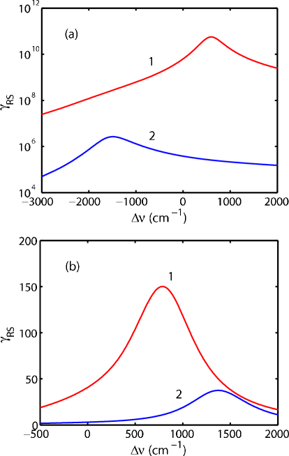
To calculate incident field enhancement of Raman scattering cross-section of a molecule near a nanoparticle normalized to that for the same molecule in vacuum we use the well-known approach based on the so-called electromagnetic theory of giant Raman scattering ref19 . Within this theory we can consider a molecule with polarizability located in z-axis of the Cartesian co-ordinates near a prolate spheroid ref20 . If is considered as the function of molecule position with other parameters fixed then the function will have maximum at certain point , where is a larger semi-axis of spheroid (spheroid is stretched along z-axis). Generally, can dramatically change with minor change in . For example, shift in by 1 Å can result in 2-3 orders of the magnitude change in . For typical values of = 10 for silver nanoparticles with 50-100 nm size one has values of 1-2 Å. Fig. 1 presents as a function of relative frequency shift for a molecule located near a silver prolate spheroidal nanoparticle with =80 nm and aspect ratio 8/5, and near silver nanosphere of the same volume as spheroid at the point . Dielectric permittivity of silver from Ref. ref21 has been used in calculations. The position of maximal values of Raman cross-section is defined approximately by position of maximal absolute value of nanoparticle’s polarizability. For selected silver nanoparticles it corresponds to 383.5 nm and 347.8 nm for spheroid, and 354.9 nm for sphere. This defines selection of an incident light wavelength chosen in the presented figure. For normal orientation of induced dipole moment of a molecule the enhancement factor readily reaches . In case of tangential orientation of induced dipole moment such values are less than . Normal orientation of induced dipole moment implies whereas tangential one means that or axes, where is an electric field of an incident light.
In spite of impressive factor from local incident field enhancement it is well lower than observed -fold single molecule Raman signal ref3 . In what follows we show that further enhancement can be understood if photon LDOS near a metal nanobody is properly taken into account as is seen from Eq. (4). Photon LDOS near a nanobody normalized with respect to that in free space formally coincides with the enhancement factor radiative rate gains near a nanobody with respect to rate in free space. This statement can be proved based on the fluctuation-dissipation theorem ref22 and is proposed as reasonable operational approach to the very definition of local density of photon states ref23 . With modified LDOS Raman scattering cross-section reads
| (5) |
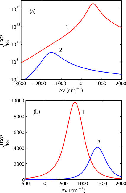
Calculation of LDOS has been made based on solution of a quasi-steady-state problem for a dipole electromagnetic field source near a nanoellipsoid in accordance with our previous works ref28 . In a quasi-steady-state approximation the full rate of spontaneous decay can be partitioned into two components. The first one is the radiative part related to photon emission. The second one is the non-radiative part related to dissipation of energy to a metal nanobody. The radiative part is found as the ratio of emmitted power by a dipole source near a nanobody to power of the same source in free space. Unlike ref17 only radiative part of the full decay rate has been accounted for since it is the radiative part of the full atomic decay rate which is determined by LDOS. The non-radiative part corresponds to Joule losses (heating of a nanobody) ref24 . In Fig. 2 calculated Raman cross-section is plotted with Eq. (5) taken into account as function of spectral shift for the same molecule and nanoparticle parameters and their relative displacement as in Fig. 1. One can see in addition to local incident field enhancement, local density of states enhancement provide factors of the several orders of the magnitude. Notably, maximal enhancement occurs for detuning from the incident field enhancement which is typical Raman shift for common organic molecules. Optimal combination of the two enhancement factors can result in more than -fold enhancement factors for proper displacement of a molecule near a nanoparticle and for proper orientation of its dipole moment with respect to incident field and a spheroid axis (Fig. 2a). Such values inherent in certain “hot points” make Raman detection of single molecules plausible. Enhancement is essentially frequency dependent which qualitatively corresponds to experiments but often is attributed to chemical factors.
The above consideration is valid not only for molecular spectroscopy but for all versions of vibrational spectroscopies, e.g. it can be applied for single quantum dot vibrational spectroscopy ref25 . It is also valid for Mandelstam-Brillouin scattering as well as for Rayleigh scattering. The latter has been discussed in our previous paper ref26 and gains additional argumentation in the context of the recent report on enhanced hyper-Rayleigh scattering in metal-dielectric nanostructures ref27 .
We believe that simultaneous action of incident field enhancement and local density of photon states enhancement does provide a reasonable rationale for single molecule Raman spectroscopy. The results presented in Fig. 2 are considered as a first step towards extensive theory for single molecule Raman detection. Triaxial ellipsoidal nanoparticles are expected to offer even higher field enhancement and LDOS enhancement factors ref28 . Furthermore, coupled metal nanoparticles which have been found to exhibit higher efficiency in Raman scattering enhancement ref29 have also been proven theoretically to possess superior local field ref29 and LDOS ref30 enhancement in the spacing between spheres and are believed their SERS efficiency can be described by simultaneous incident field and LDOS enhancements.
The proposed model sheds light on the so-called “hot points” as such places on a nanotextured metal surface or near metal nanobodies where simultaneous spatial redistribution of electromagnetic field occurs both at the frequency of the incident radiation and at the frequency of scattered radiation . The first effect is the so-called field enhancement factor whereas the second is local density of states enhancement. Enhancement of photon LDOS starting from the pioneering paper by E.M. Purcell ref9 can be interpreted as development of the certain Q-factor in the space region where a test emitter (atom or other quantum system) is placed. Since Q-factor implies a system is capable to accumulate energy (then Q value equals to the ratio of energy accumulated in the system to the portion of energy the system looses in a single oscillation period), formation of high local density of states areas in many instances can be treated as development of multiple microcavities at the scattered frequency over nanotextured metal surface. From the other side, such microcavities promote electromagnetic wave tunneling including light leakage towards the surface in near-field optical microscopy. Therefore surface mapping of high LDOS areas can be performed by scanning near-field microscopy as has been proposed in Ref. ref31 but to the best of the authors’ knowledge has never been applied to SERS-active structures.
Local field enhancement for incident light can not be interpreted as surface redistribution of incident light, i.e. as light “microfocusing” as anticipated by many authors. Since SERS is considered within linear light-matter interaction (contrary to e.g. surface enhanced second harmonic generation) the total Raman signal harvesting from a piece of area containing statistically large number of molecules will be the same independently of surface redistribution of light intensity because total incident light intensity integrated over the piece of area remains the same. Within the framework of linear light-matter interaction, Raman signal enhancement by means of incident field enhancement can only be understood in terms of high local Q-factors for incident light, i.e. in terms of light accumulation near the surface rather than light redistribution over the surface. Q-fold rise up of light intensity then occurs near hot points as it happens in microcavities and interferometers. However, accumulation of light energy needs certain time. Therefore huge Raman signals can develop only after certain time which is necessary for transient processes to finish resulting in steady increase of incident light intensity near hot point. Transient SERS experiments are therefore to be performed to clarify Q-factor effects in hot points formation.
Local DOS enhancement in a sense accounts for concentration of electromagnetic field at . This statement unambiguously implies probe, non-existing field ref32 . However, concentration of real field by many authors was anticipated to offer enhancement factor ref6 by analogy to factor for input light intensity. That anticipation is by no means justified because enhancement occurs only in the close subwavelength-scale vicinity of a nanobody and can not contribute to light harvesting in typical far field experiments. Concentration of really emitted light can actually contribute to SERS but only as induced Raman scattering [ term in Eq. (2)].
In conclusion, a rationale has been proposed for more than -fold enhancement factors in Raman spectroscopy in terms of local field enhancement and local density of photon states enhancement in the same point but at different frequencies, a model of the so-called “hot points” has been elaborated as local areas with high Q-factor at incident and scattered light frequencies and further experiments towards verification of the model have been outlined. The proposed consideration extends the original Purcell’s idea on strong modification of photon spontaneous emission rate to modification of spontaneous photon scattering rates.
The work has been supported by the EU NoE “PHOREMOST”, National Research Program “Crystalline and Molecular Structures” and by Belarusian National Basic Research Foundation.
References
- (1) C.V. Raman and K.S. Krishnan, Nature 121, 501 (1928).
- (2) Surface Enhanced Raman Scattering, edited by R.K. Chang and T.E. Furtak (Plenum, New York, 1981).
- (3) S. Nie and S.R. Emory, Science 275, 1102 (1997).
- (4) K. Kneipp, Y. Wang, H. Kneipp, L.T. Perelman, I. Itzkan, R.R. Dasari, and M.S. Feld, Phys. Rev. Lett. 78, 1667 (1997).
- (5) Surface-Enhanced Raman Scattering, K. Kneipp, M. Moskovits, H. Kneipp (Eds.) (Springer-Verlag, Berlin, 2006).
- (6) S.V. Gaponenko, Phys. Rev. B 65, 140303(R) (2002).
- (7) P.A.M. Dirac, Proc. Royal Soc. London, Ser. A 114, 243 (1927).
- (8) E.M. Purcell, Phys. Rev. 69, 682 (1946).
- (9) V.P. Bykov, Radiation of Atoms in a Resonant Environment (World Scientific, Singapore, 1993).
- (10) E. Yablonovitch, Phys. Rev. Lett. 58, 2059 (1987).
- (11) E.P. Petrov, V.N. Bogomolov, I.I. Kalosha, and S.V. Gaponenko, Phys. Rev. Lett. 81, 77 (1998).
- (12) P. Anger, P. Bharadwaj, and L. Novotny, Phys. Rev. Lett 96, 113002 (2006).
- (13) I.V. Bondarev, G. Ya. Slepyan, and S.A. Maksimenko, Phys. Rev. Lett 89, 115504 (2002).
- (14) D.V. Guzatov and V.V. Klimov, Phys. Rev. A 75, 052901 (2007).
- (15) D.S. Mogilevtsev and S.Ya. Kilin, Quantum Optics Methods of Structured Reservoirs (Belarusian Science, Minsk, 2007).
- (16) V.S. Zuev, A.V. Frantsesson, J. Gao and J.G. Eden. J. Chem. Phys. 122, 214726 (2005).
- (17) G. Placzek, in Handbuch der Radiologie, vol. 6, part 2, E. Marx (Ed.) [in German] (Akademische Verlagsgellschaft, Leipzig, 1934), p. 205.
- (18) M.I. Stockman, in Ref. 6, p. 47.
- (19) J. Gersten and A. Nitzan, in Ref. 2, p. 89.
- (20) P.B. Johnson, R.W. Christy, Phys. Rev. B 6, 4370 (1972).
- (21) S.M. Barnett, R. Loudon, Phys. Rev. Lett. 77, 2444 (1996).
- (22) G. D’Aguanno, N. Mattiucci, M. Centini, M. Scalora, and M.J. Bloemer, Phys. Rev. E 69, 057601 (2004).
- (23) D.V. Guzatov and V.V. Klimov, Chem. Phys. Lett. 412, 341 (2005).
- (24) R.R. Chance, A. Prock, and R. Silbey, Adv. Chem. Phys. 37, 1 (1978).
- (25) S.V. Gaponenko, Optical Properties of Semiconductor Nanocrystals (Cambridge University Press, Cambridge, 2005).
- (26) A.A. Lutich, S.V. Gaponenko, N.V. Gaponenko, I.S. Molchan, V.A. Sokol, and V. Parkhutik, Nano Letters 4, 1755 (2004).
- (27) E.M. Kim, S.S. Elovikov, T.V. Murzina, A.A. Nikulin, O.A. Aktsipetrov, M.A. Bader, and G. Marowsky, Phys. Rev. Lett. 95, 227402 (2005).
- (28) A.M. Michaels, J. Jiang, and L. Brus, J. Phys. Chem. B 104, 11965 (2000).
- (29) V.V. Klimov and D.V. Guzatov, Phys. Rev. B 75, 024303 (2007).
- (30) C. Chicanne, T. David, R. Quidant, J.C. Weeber, Y. Lacroute, E. Bourillot, A. Dereux, G. Colas des Francs, and C. Girard, Phys. Rev. Lett. 88, 097402 (2002).
- (31) S.V. Zhukovsky and S.V. Gaponenko, Phys. Rev. E 77, 046602 (2008).