Possible mechanisms for initiating macroscopic left-right asymmetry in developing organisms
Abstract
How might systematic left-right (L/R) asymmetry of the body plan originate in multicellular animals (and plants)? Somehow, the microscopic handedness of biological molecules must be brought up to macroscopic scales. Basic symmetry principles suggest that the usual “biological” mechanisms – diffusion and gene regulation – are insufficient to implement the “right-hand rule” defining a third body axis from the other two. Instead, on the cellular level, “physical” mechanisms (forces and collective dynamic states) are needed involving the long stiff fibers of the cytoskeleton. I discuss some possible scenarios; only in the case of vertebrate internal organs is the answer currently known (and even that is in dispute).
Keywords:
Cytoskeleton, motor proteins, actin, chirality, handedness:
PACS numbers: 87.19.l, 87.16.Ka, 87.16.Nn, 87.17.Ee1 Introduction
The anatomy of most animals – and many plants – breaks left/right symmetry, the same way for all or most individuals (though this doesn’t necessarily have any functional significance). The key processes at the cell or organism level – diffusion, regulation of gene expression, perhaps elasticity – don’t distinguish left from right, so how can the developing organism “learn” this wolpert-brown ; wood-review ; levin-LR-review ; wood-review-2005 ?
Of course, the proteins (or other biological molecules) that constitute the cells are handed; 111 It should be emphasized I am not interested here in the original prebiotic symmetry-breaking which determined the molecules’ handedness frank53 ; avertisov-molecules ; garcia-bellido . but how can this information be brought to the macroscopic scale? Any mechanism for that, I suggest, involves physics to an unusual extent: forces and motions acting on the stiff semi-macroscopic polymers that make up each cell’s “cytoskeleton”. Furthermore, I argue that just from a priori, basic symmetries, the possible mechanisms are strongly constrained. That is why this topic seemed appropriate for a conference in honor of Landau, even though he would probably have considered the entirety of biological physics to be “pathological”.
One motivation to pursue L/R specification is “cleaner” palmer than most development instabilities in that (i) it emerges out of a functionally symmetric state (if not, the original L/R specification just was earlier). Maybe, e.g., the differentiation of brain regions emerges in a similar fashion, but the way it goes can be explained by the morphological asymmetries of neighboring tissues; (ii) it is binary – the minimum of information; (iii) mutant embryos have unambiguous visual signatures. Perhaps for these reasons, L/R experiments have surged over the past decade.
The rest of the paper begins with an introduction in which I list examples of L/R asymmetry in model organisms, lay down key facts and assumptions, and classify the mechanism. The next four sections go on to tell four stories of L/R asymmetry (of which only the first is experimentally settled): cilia driving fluid flow in vertebrates (Sec. 2), a hypothetical mechanism wherein screw processive molecular motors transport signaling molecules (Sec. 3), shearing actin arrays causing a twist in cell division as in molluscs (Sec. 4), and finally rotating microtubule arrays in plants leading to a macroscopic twining as in vines (Sec. 5).
1.1 Examples
The first example is the internal organs of vertebrates cooke-review ; hirokawa-review : heart, lungs, etc. are located asymmetrically. In humans, mcmanus-RHLH the frequency of mirror-reversal is . Common model species are mouse, chick, Xenopus frog, or zebrafish.
A second example is the human brain: sun-brain-review : of course, right-hand dominance is a side effect of left-brain dominance. The congenital reversal frequency mcmanus-RHLH is , and surprisingly is independent of the handedness of internal organs 222 See references in mcmanus-RHLH , pp. 5-6; cooke-review , Part IV; and levin-LR-review , Sec. 10. This independence is also confirmed in frogs malashichev-review . so it probably has a different mechanism. (While some anatomical and functional brain asymmetries are found in other animals, the relation to human brain asymmetry malashichev-review ; halpern-LR ; malashichev-book is still unclear.)
Thirdly, even the humble C. elegans nematode “worm” is L/R asymmetric wood-review ; wood-review-2005 ; wood-orig ; poole-hobert ; hobert-brain-review — a creature so small that all cells are numbered by embryologists (they develop in an identical, and known, pattern), and every embryo repeats exactly the same sequence of divisions. Here, the gut twists, and a certain chemosensing neuron has functional L/R asymmetry. (Flies also have twists in their guts and genitals speder-drosophila-orig ; hozumi-drosophila-orig ; speder-noselli-review0 .)
As a fourth and final animal example, mollusc shells — say snails — are well known to coil right-handed (reversal frequency ). This has functional consequences in that mating is awkward between snails of different handedness, snail-species and a snake has evolved asymmetric jaws snail-snakes to better crush right-handed snails.
There is a parallel story of handedness in plants (Sec. 5) — certainly in climbing plants, whose roots and shoots both spiral with a species-dependent handedness. 333 Note that “phyllotactic spirals” in plants, responsible for Fibonacci numbers in leaf placement, in pine cones and sunfloweers — do not have a fixed handedness. (An interesting symmetry corollary — this would be obvious to Landau! — is that roots growing against a vertical hard surface can deviate by a “Hall angle” from heading straight downwards, and they do.)
Since this is biology, we do not expect universal answers; if a conjectured mechanism turns out to be wrong for one mentioned example, it might be valid for another!
1.2 Question and starting assumptions
An embryo can develop two axes by spontaneous symmetry breaking: (i) the anterior/posterior (A/P) axis, i.e. head/tail — call this the axis; (ii) the dorsal/ventral (D/V) axis,i.e. back/front — call this the axis. It is well understood that a combination of reaction and diffusion by chemical signals can generate this sort of pattern formation. (Here “reaction” includes regulation of DNA transcription and translation to proteins.) Note that in practice the symmetry breaking is often biased externally, e.g. by the point sperm entered egg.
A third () axis could certainly form, normal to the others, by a spontaneous symmetry breaking. But the “right-hand rule”
| (1) |
must be ensured: how can it be done? The key claim of this paper is that symmetry requires that each of the three symbols on the right-hand side of (1) has a specific physical (biological) correlate entering the mechanism. That is, there must be two kinds of preexisting polarization representing the and , plus some functionally chiral element representing the “” in (1).
I mention two important assumptions. First, there is a spontaneous symmetry breaking: some robust mechanism ensures an asymmetric outcome, but that by itself would produce an equal mixture of L and R organisms. (This is evidenced experimentally, in some cases, in mutants that have such a randomization.) The uniform outcome is due to an additional small biasing field (exponentially small in the system size), just as a tiny magnetic field decides the magnetization sense of an Ising magnet cooled through its Curie temperature. The practical significance is that our mechanism need only produce a weak bias (let’s arbitrarily aim for ), since the assumed symmetry breaking amplifies it enormously. 444 Symmetry breaking may also explain the functional reason for a bias, when there is no social reason for organisms to all have the same handedness. In the absence of a bias field, occasionally an organism would be a mixture of “left” and “right” type domains, which would be a congenital defect.
Second, I also assume that L/R asymmetry stems from the microscopic chirality of molecules (under genetic control), and not e.g. an asymmetry produced in the egg by a right-handed mother. (This is easily refuted by the inheritance patterns; and wouldn’t such a mechanism have error rate?)
Finally, since we seek the earliest L/R asymmetry, it follows (tautologically) that whatever caused it must have been L/R symmetric. 555Since all the molecules are asymmetric, more carefully we should say the cause is functionally L/R symmetric; just what that means will be clarified by examining the specific scenarios worked out in later sections.
1.3 Classifying mechanisms
There are three useful categories.
1.3.1 Two levels of any mechanism
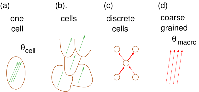
Any explanation of L/R specification really requires two stories, one at the cellular level and one at the collective level. The cell level story starts from proteins — that’s where the cytoskeleton fibers come in — and goes to properties of the whole cell, or the interaction of one cell and a neighbor. The collective story starts from the cell behavior and explains how this specifies a left and a right side in the whole embryo. For each of the following sections, I will indicate both levels; sometimes one or the other is rather trivial, but there are always the two levels. The symmetry principle applies at each level.
1.3.2 Two styles in development
In embryology, wolpert-text there are two general “styles”, applying to different animal phyla. An “early” style applies to molluscs and C. elegans: as cells first divide, each gets a determining label, schematically like a binary string. All its descendents retain that string, while possibly adding bits that refine the specification of cell type in the mature animal. Thus, cell fates are fixed early. On the other hand, vertebrates and insects have a “late” style: through many cell divisions the cells are unspecified, then fates are undetermined by pattern formation within a multicellular embryo. Evidently, an essentially cell-level L/R mechanism suffices for early-style creatures, whereas a collective L/R mechanism seems to be needed in late-style creatures.
1.3.3 Two ways to represent L and R
There are two ways that “leftness” might be represented in an embryo (Fig. 1). One is called “positional information”: wolpert-text it means some chemical has a concentration which is roughly a function of (say) ; three such chemicals can specify all coordinates. By sensing the concentrations of all three, a cell learns its position within the body and hence which organ it should become. (To do that, it does not necessarily need to know which direction is left, or posterior, etc.)
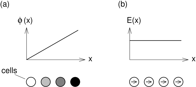
The alternate representation is “polarization” of cells, so that each “knows” which direction is left, but not where it sits along the L/R axis. We could write this as a local vector . Evidently the relation of polarization and positional information is . Wolpert’s pioneering paper wolpert-brown about the L/R mechanism as a symmetry problem envisaged a “polarization” representation. I believe that was meant only as a thought experiment, rather than a literal proposal for the mechanism; “positional information” seems in many cases easier to generate, as well as being the information ultimately needed to “inform” a developing tissue as to its fate.
It is not trivial to convert one kind of L/R information to the other. Indeed, given , a eukaryotic cell is big enough and sophisticated enough to sense a concentration gradient between one side and the other, and develop a polarizations in response, to differentiate (in the calculus sense!). But the reverse construction — integratiing — cannot be done locally. To generate an imbalance in the concentration, some chemical it must get actively transported through the organism with a bias along .
1.4 Cartoon of the cytoskeleton
The mechanism is not of the usual “biological” type. That would mean transport (by diffusion or otherwise) of signaling molecules and reactions. But transport alone won’t suffice for L/R, since diffusion doesn’t distinguish handedness. All possible mechanisms seem to involve actual forces or torques exerted by molecular motors, which somehow structure the cytoskeleton made of stiff, semi-macroscopic fibers (that the motors run along).
The cytoskeleton is the framework in each cell of a eukaryotic (higher) organism, built from long, stiff, directed, and helical macro-molecules (which I will call “fibers”); there are specific kinds of motor molecules for each kind of fiber. cytoskeleton . The two kinds of fiber are (i) microtubules (mt), with dynein or kinesin motors on them; (ii) actin fibers, with myosin motors on them. Each family of motors contains numerous subvarieties, used by the cell for special purposes. In particular, myosin V is the main myosin variety that moves along a fiber for long distances (“is processive”); other varieties, e.g. myosin II used in muscles, only makes contractions. We note that, despite the microscopic differences between mt and actin, when abstracted to the model level they may look very similar: i.e., a physics approach may uncover (even quantify) a kind of universality.
The two directions along the fibers are not symmetry-equivalent – the growth direction is called the fiber’s “polarity”. A given kind of motor (literally) walks in a fixed sense (which we assume to be the sense for this paper). It transports “cargoes”, which are typically chemicals in vesicles (small membrane bags attached to the motor by linking protein(s): see Fig. 2(a)). Occasionally (by thermal fluctuation) a motor falls off its fiber, and diffuses till it reattaches to the same or another fiber
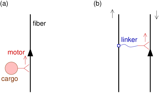
In place of a cargo, a motor can be linked somehow to another fiber [see Fig. 2(b)] so as to drive the motion of one relative to the other e.g. in cell division. Fibers form networks with many crosslinks, which are often dynamic, i.e. fibers are constantly appearing, growing, shrinking, and vanishing, producing a dynamic steady state describable by statistical mechanics.
2 Nodal flow mechanism
Here I merely review the only well-understood mechanism, a late-stage type mechanism acting in vertebrates hirokawa-review .
2.1 Cell-level story
We start with an approximately flat embryo that has already formed A/P and D/V axes. The following sentence describes the key and sufficient cause: watch carefully to see where each element of (1) gets mentioned. On its ventral side, near the “node” (a key place in a developing vertebrate embryo), are special cells with cilia — moving tails that stick out (“”) from surface, but tilted (“”) towards the embryo’s posterior end. PNAS-nodecilia . These cilia move circularly (), — clockwise, looking down — unlike regular cilia (which move back and forth).
2.1.1 Root of L/R asymmetry
So, why do the node cilia move circularly? This is plausible from their structure: each cilium has 9 pairs of microtubules that run its length, and each pair is linked by dynein molecules with their head-to-foot direction oriented clockwise. The crucial L/R event is that a ring of special protein molecules assembles in the cell membrane. This serves as the template to start the microtubule pairs with dynein linkages, and the whole structure apparently grows outward ring after ring by stacking each component onto its own kind in the previous ring. In the end, then, the L/R asymmetry is not due to the inherent helicity of the microtubules, but rather the handedness of the templating complex. Plenty of physics remains to be worked out in this system, namely applying elastic theory and fluid dynamics to show why the given structure executes circularly polarized motion, and in which sense brokaw-chiral .
2.2 Collective level (and alternate story)
The array of cilia just described is sufficient to break L/R symmetry: symmetry allows it to drive a fluid flow L to R across the embryo (as observed). If a signaling chemical is released, the flow carries it to the L side, where it can bias the symmetry breaking. The key check is that you reverse the flow externally and get out reversed embryos — in mice.
But the relative importance of early versus late mechanisms may depend on the kind of vertebrate: early L/R asymmetries were clearly seen in Xenopus frogs levin-ionflow-review , due to a distinct L/R mechanism. Possibly before the first cell division, preexisting (maternal) chemicals – in particular ion transporter proteins – are getting asymmetrically distributed in the egg; plausibly the L/R asymmetry comes via some kind of actin/myosin mechanism like the one discussed in Sec. 4, below levin-aw . The collective level of this mechanism involves a biased transport that converts an electrical potential difference into a concentration by transport (like the “integration” process of Sec. 1.3.3). It was speculated the collective mechanism includes a mutual feedback process (the spontaneous symmetry breaking assumed in Sec. 1.2), between the electric field and serotonin concentration gradient levin-serotonin
3 Asymmetric transport
This section outlines, as a pedagogical example, a completely hypothetical mechanism; it is late-stage type and (unlike the mechanisms of Sections 4 and 5) it depends on transport of signaling molecules, like typical “biological” mechanisms do. The key ingredient is helical motion of motor molecules on a cytoskeletal fiber.
Consider for simplicity an embryo with its geometry flattened into the plane the normal to the D/V axis. Assume the dorsal and ventral sides are distinguished (there’s the “ again, from (1)). We look for an analog of the Hall effect whereby (in the presence of a transverse magnetic field) electrons drift at a small angle from the electric field direction. Here, chemicals transported due to an assumed A/P polarization (“”) actually move at an angle rotated slightly (“”) from the A/P axis, allowing them to get carried preferentially to one side of the embryo and thereby bias the assumed symmetry breaking. The upshot will be that it’s not very easy to engineer late-stage asymmetry with a transport mechanism!
3.1 Cell level mechanism
This is the nontrivial level for this mechanism; again, I’ll tag the three ingredients from (1). Let’s assume a “cortical” array of fibers, meaning it is just under the cell’s membrane (there’s the “”). Let the fibers be oriented along ; no net polarization is assumed, so we are not yet talking about the the A/P () asymmetry.
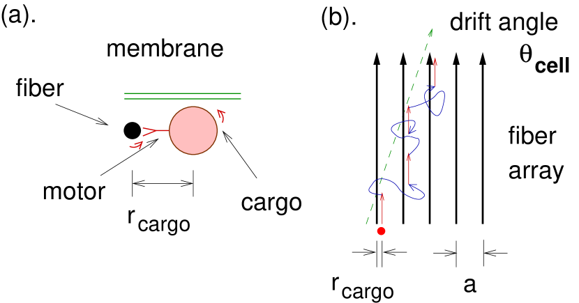
Active transport relies on motor molecules; since the fibers they move on are (microscopically) helical , their motion should (generically) be helical too [there’s the “ in (1)]. In fact, myosin V motors are known to spiral as they move on actin ali-helical . (However kinesin motors are tightly bound to the protofilaments that constitute a microtubule and those are, most often, quite straight.) Now, let’s imagine our motor spirals counter-clockwise (ccw); this will pull the cargo vesicle around the fiber till it gets jammed against membrane, and from then onwards the motor will just move lengthwise: in other words, it always travels “on the right side of the road” [see Fig.3(a,b)].
Each time the cargo detaches, diffuses, and reattaches during its progress, the most likely outcome is to reattach to the same fiber as it is closest. If it reattaches to a different fiber, it’s likelier to reattach to the fiber on its right side [Fig.3(b)] since that one is always closer than the one on the left. When we model this diffusion as confined to the plane (hence basically one dimensional), the probability of hopping one fiber to the right is , where is the fiber spacingberg ; it is still proportional to this ratio in more realistic models. If the cargo succeeds in hopping over, its path gets shifted by to the right: hence, the average non-random shift is .
The result is a mean transport current rotated rightwards by from , where
| (2) |
Here is the typical distance this motor goes before falling off (“processivity”), and is an effective distance of the cargo from the fiber axis. If we modeled the free diffusion interlude as a one-dimensional random walk transverse to the fiber array, the probability of reattaching to the fiber on your right is where is the separation; the likeliest outcome is to reattach to the same fiber, since it’s closer. Since m and nm, we find .
3.2 Collective level
This array is found only on (say) the ventral side of cells on (say) the embryo’s ventral side (the “” at organism level). Imagine the signal chemical gets released from (say) the anterior (head) end (this is the A/P or “” asymmetry at last). If the signaling chemical has a sideways bias relative to the array in each cell, it’s easy to see the macroscopic transport will have a bias of similar magnitude (see Fig.4).
4 Cell division
I now turn to an example that is not yet understood, but we know so much from experiments that we should be above to pin down the mechanism. This is (to use the categories of Sec. 1.3) the early-stage, cell-level mechanism of twisting cell division (“spiral cleavage” to the mollusc community), apparently due to an array of actin filaments. First I will review some experimental facts.
For molluscs, specifically snails, kuroda-snail-actin after the first two divisions an embryo consists of four cells in a square. These give rise to four (smaller) daughter cells by dividing along the axis normal to the square, but break the symmetry by twisting to the right till the new cells sit on top of the furrows between the first four cells. This determins the handedness of the grown snail (one evidence is that mutants which divide at this stage with the opposite twist, also have macroscoically reversed handedness.)
In C. elegans embryos, too, the embryo with four cells (planar but less symmetric) is the stage from which the ultimate handedness is determined. wood-review ; wood-review-2005 ; wood-orig ; poole-hobert ; hobert-brain-review . This was proven by manipulations wherein the cells get physically switched, leading to a mirror-reversal in the grown animal wood-orig . Furthermore, the motions of the cells in these divisions can be described by a general twisting tendency like the mollusc case wood-review-2005 .
4.1 Organism-level story
Putting this all together, we have a mechanism for the organism level. We hypothesize a torque which always drives the same twist of two daughter cells about their axis of division. If the cells were already latently polarized along parallel axes for the subsequent division, those axes get tilted by the torque as observed. Then right after the symmetry-breaking division at the four-cell stage, some chemical signal is passed between the cells, depending on which cell neighbors. Since the neighbor relation has become asymmetric, this tells the cells which is L and which is R; that “‘bit” of information is preserved in subsequent divisions for the descendents of these cells, and is expressed functionally at a much later stage.
4.2 Microscopic level story?
Experimentally it was shown that the twist depends on actin but not on microtubules, kuroda-snail-actin somewhat surprisingly since microtubules have the more prominent role in cell division (forming the “spindle” between the two new cell nuclei.) The role of actin in cell division is to form the “contractile ring”, an array of roughly parallel filaments that contract to pinch off the two cells from each other contractile-ring ; contractile-PRL .
Meanwhile, in frog eggs (under the influence of a certain drug), a twist of just this sort is observed and was shown to depend on an actin array. danilchik-xenopus-twist . Bundles of parallel actin fibers shear past each other, always in a clockwise sense. It appears myosin is responsible, rather than actin polymerization, as shown by turning off the latter with a poison. (In the case of Drosophila, a late-style mechanism which might or might not be related to this one, myosin I D is responsible speder-drosophila-orig ; hozumi-drosophila-orig ; speder-noselli-review0 .)
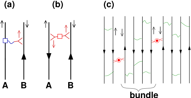
I do not have a satisfactory microscopic model for this case. The experiment shows the actin array’s shear motion is driven by myosin, and in turn the myosin moving on one actin fiber must be linked (directly or indirectly) to another fiber, in order to make any shear (see Fig. 5). So, the actin array somehow becomes organized — either (i) driven by the myosin motors, or (ii) during the array’s formation — such that whenever a motor is attached (in any fashion) to actin fiber and walking along actin fiber (as shown in Fig. 5), the polarity of fiber is towards the left as seen from the – link. 666 A special case of this is where the linkage connects motors walking on both actin fibers [e.g. Fig. 5(b)], as in muscle.
This arrangement might be produced (i) conceivably, by the spiraling of processive motors, as in the hypothetical mechanism of Sec. 3. But (ii) a more plausible mechanism would depend on the linker proteins that bind actin fibers into bundles (Fig. 5(c) A “basic” linker would join fibers with the same polarity. But a “special” linker, joining fibers with the opposite polarity, would be a dimer of membrane anchored proteins oriented to hold the motors oriented as shown, so they drive relative motion only if both fibers have polarity to the left as seen from the link.
5 Plants: rotating mt array?
Finally, I turn briefly to the case of plants. Like the previous story (cell division), this involves a dynamic, cortical fiber array — this time of microtubules (mt).
5.1 Organism scale mechanism
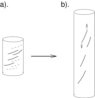
Plant cells are cylindrical and elongated in the growth direction. An mt array hashimoto-mt-review forms around their cell walls, roughly parallel and nearly transverse to the cylinder, but with a typical helical pitch (say a pitch angle away from transverse). In turn the microtubules orient a helical array of cellulose fibers around the cells, which stiffens the cell walls.
Now, the shoots (and roots) of many plant species have a consistent helical sense, particularly evident in climbing vines that twine around vertical supports. Experiments on arabidopsis confirmed the sense of the plant’s macroscopic twist corresponds to that of the microtubule array’s microscopic twist on the membrane: mutations that reverse the latter also reverse the former hashimoto .
My conjecture for how this happens comes from a paper about fungi gamow , where chitin plays the role of cellulose. (A related but not identical mechanism was proposed for helical twisting in chains of elongating bacteria without flagella wolgemuth .) Imagine a cellulose fiber of finite length anchored in the membrane (Fig. 6). The cell grows by elongation but the fibril can’t. Hence it feels opposing longitudinal forces at the ends, roughly proportional to , where is the fraction elongation per unit time. Since the fiber isn’t longitudinal, these forces exert a twist torque on the fiber (and vice versa) proportional to . Thus all the individual cells get a microscopic torque stress of the same sign, adding up to a macroscopic one on the whole shoot. Now elasticity theory tells us there can be an instability: if the centerline deviates from straight to helical, this can relieve the torque stress and decrease the energy (Such a “twist-to-writhe” conversion. is also responsible for supercoiling of, e.g., DNA.)
5.2 Cell level mechanism: mt arrays
Microtubule arrays get oriented in a collective process with the following rules: mt-rules
-
•
Rule (i) The end of the mt (mostly) grows, while the end depolymerizes (but not as fast).
-
•
Rule (ii) Furthermore, new mt nucleate on existing mt and grow at a specific branching angle .
-
•
Rule (iii) When a growing mt hits another, if the relative angle is less than it bends (this demands a linker to exert a force, since mt are rather stiff) and aligns (this process forms bundles).
-
•
Rule (iv) If the angle is larger, the growing mt suffers a “catastrophe” meaning it depolymerizes from the end (and disappears).
Note that either process (iii) or (iv) tend to drive the mt towards a steady state phase in which the alignment has long range order, as found in a simulation mt-simulation that omits branching (ii) and bending (iii)]. Also, the collective state is symmetry-broken: within the plane of the membrane, the mt array’s orientation is (so far) arbitrary.
It seems difficult to engineer such a mechanism to directly orient the mt with respect to the elongation axis. Instead, I speculate there is a spontaneous (slow) rotation of the array. The overall mean orientation angle follows the dynamics
| (3) |
Here is the spontaneous rotation rate (see below for possible mechanisms). The term proportional to expresses how elongation passively carries the mt to a higher angle. The consequence of (3) is the angle will evolve to a steady value satisfying
| (4) |
To explain the small (=nearly transverse) observed , we must posit the additional transverse bias , which tries to pull back towards 0 or . (Here is one speculative mechanism for : say there are membrane anchoring proteins that slightly bend the mt away from the membrane, so in effect the mt has a spontaneous curvature. If so, the mt has the least strain when it aligns along the direction of the membrane’s maximum curvature.)
Such dynamic rotation is suggested by morphologies of some plant cells that have multiple layers of cellulose fibers, each rotated relative to the one underneath preston82 . Indeed, domains undergoing such a rotation were seen directly by video imaging in growing plant cells mt-video (under the influence of a drug?). The authors do not infer a particular sense, but about 70% of the domains rotated clockwise (see Fig. 1 of Ref. mt-video ).
So the term in (3) is the key parameter determining L/R; what are the mechanisms for it? Recall that, by our basic symmetry arguments, the membrane must be involved in order to define a sense of rotation. The speculative ways this could happen may be classified according to the rules of microtubule growth mentioned in the preceding subsection. (a). Perhaps the microtubule-associated protein that nucleates a branch is also membrane associated [see Rule (ii), above]. (b) Perhaps the outcome of an mt-mt collision depends on which side the growing mt is impinging from [Rule (iv)], conceivably by the growing tip following a helical path of the filaments and getting pushed into or away from the membrane, but just as likely by another membrane/microtubule associated protein.
Imagine the mt-rotation mechanism (b) [the one based on collisions] in more detail. If new fibers have an orientation such that they hit old fibers from the right, then they grow longer. Thus, the new fibers’ orientation tends to be rotated counter-clockwise relative to the old fibers.
6 Discussion and conclusions
In conclusion, I reiterate: symmetry is key to recognizing which mechanisms can possibly be responsible for L/R asymmetry. Any such mechanism must explicitly connect three ingredients: two axes, and some chiral molecule that implements the “right hand rule”. Every story has a cell-level half and an organism half. At the cell level, a natural axis is the membrane normal, thus most of these mechanisms involved aligned “cortical” (adjacent to membrane) arrays of fibers.
I conjecture that the mechanism is always cytoskeletal, involving microtubules or actin fibers. As for exactly how chirality enters: the most elegant “physics” answer would be the fiber itself, via some screw mechanism whereby motion along a long helical fiber — walking by a motor, or microtubule collisions 777 The stories in this paper omit a third kind of screw mechanism, namely the change in pitch of a fiber (e.g. actin) under a change of its strains, or in its chemical environment. See e.g. upmanyu . gets converted into rotation around the axis. However, a “biology” answer, a molecule anchored in the membrane and binding the fibers too, may be more plausible. The membrane-anchoring mechanism furnishes a hint to biologists as to which proteins to focus on, in genetic or protein-expression studies aiming to discover the master L/R determining gene. On the other hand, the screw-motion hypothesis suggests that, if a processive motor is involved, the mutations which reverse (or affect) L/R determination were those that reversed (or affected) the motor’s screw motion on its fiber.
Twice we were led into self-organized cortical arrays of approximately parallel fibers confined to the plane adjacent to a membrane. In both cases, the conjectured cell-level mechanism did not depend on a globally defined axis, nor did it even need an ordering of the fibers’ polarization axes. Instead, it used the orientation axis to define a relative rotation, so the asymmetry was manifested in a rotation or shear rate.
The brain asymmetry, which supplies the very terminology (“handedness”) for this subject, is the most mysterious. Since nerve cells migrate far in the developing brain, one would hunt for an L/R asymmetry in cell locomotion. But that would occur at a later stage, in a more three-dimensional embryo, so it is less clear what the locally defined and could be. One possibility is that brain asymmetry is actually a very early mechanism, perhaps the same one discussed in Sec. 4, as suggested by some left-right anomalies seen in twins levin-birthdefects
It is left for future research to turn all these ideas into quantitative estimates, a necessary condition before any physics may be considered as completed.
References
- (1) N. A. Brown and L. Wolpert, Development 109, 1-9 (1990), “The development of handedness in left/right asymmetry”.
- (2) W. B. Wood, Annu. Rev. Cell. Dev. Biol. 13, 53-82 (1997), “Left-right asymmetry in animal development.”
- (3) M. Levin, Mech. Dev. 122, 3-25 (2005) “Left-right asymmetry in embryonic development: a comprehensive review”.
- (4) W. B. Wood, PLoS Biology 3, e292 (2005), “The left-right polarity puzzle: determining embryonic handedness.”
- (5) F. C. Frank, Biochim. Biophys. Acta 11, 459-464 (1953), “On spontaneous asymmetric synthesis.”
- (6) V. A. Avertisov, V. I. Goldanskii, and V. A. Kuz’min. Physics Today, July 1991, pp 33-41, “Handedness, origin of life, and evolution”.
- (7) A. García-Bellido, PNAS 93, 14229-14232 (Dec. 1996), “Symmetries throughout organic evolution”.
- (8) A. R. Palmer, Science 306, 828-833 (Oct. 2004). “Symmetry breaking and the evolution of development.”
- (9) J. Cooke, Biol. Rev. 79, 377-407 (2004), “Developmental mechanism and evolutionary origin of vertebrate left/right asymmetries”
- (10) N. Hirokawa, Y. Tanaka, Y. Okada, and S. Takeda Cell 125, 33-45 (Apr 7, 2006). “Nodal Flow and the Generation of Left-Right Asymmetry.”
- (11) C. McManus, Right hand, left hand: the origins of asymmetry in brains, bodies, atoms, and cultures (Harvard Univ. Press, Cambridge MA, 2002), and references therein.
- (12) T. Sun and C. A. Walsh, Nature Revs. Neuroscience 7, 655 (2006), “Molecular approaches to brain asymmetry and handedness.”
- (13) Y. B. Malashichev and R. J. Wassersug, Bioessays 26, 512-22 (2004), “Left and right in the amphibian world: which way to develop and where to turn?”.
- (14) M. E. Halpern, J. O. Liang, and J. T. Gamse, Trends Neurosci. 26, 308-313 (2003), “Leaning to the left: laterality in the zebrafish forebrain.”
- (15) Ye. B. Malashichev and A. W. Deckel, ed., Behavioral and morphological asymmetries in vertebrates (Landes Bioscience, Georgetown TX, 2006).
- (16) W. B. Wood, Nature 349, 536-8 (1991). “Evidence of handedness in C. Elegans embryos for early cell interactions determining cell fates.”
- (17) R. J. Poole and O. Hobert, Current Biology 16, 2279-2292 (2006), “Early embryonic patterning of neuronal left/right asymmetry in C. elegans.”
- (18) O. Hobert, R. J. Johnston Jr, and S. Chang, Nature Revs. Neuroscience 3, 629-640 (2008), “Left-right asymmetry in the nervous system: the Caenorhabditis elegans model”.
- (19) P. Spéder, G. Adam, and S. Noselli, Nature 440, 803-807 (Apr. 2006), “Type ID unconventional myosin controls left-right asymmetry in Drosophila”.
- (20) S. Hozumi, R. Maeda, K. Taniguchi, et al. Nature 440, 798-802 (Apr. 2006), “An unconventional myosin in Drosophila reverses the default handedness in visceral organs”.
- (21) P. Spéder and S. Noselli, Curr. Opinion in Cell Biol. 19, 82-87 (Feb. 2007), “Left-right asymmetry: class I myosins show the direction”.
- (22) R. Ueshima and T. Asami, Nature 425, 679 (2003), “Single-gene speciation by left-right reversal: A land-snail species of polyphyletic origin results from chirality constraints on mating”.
- (23) M. Hoso, T. Asami, and M. Hori, Biology Letters 3, 169-172 (2007), “Right-handed snakes: convergent evolution of asymmetry for functional specialization”.
- (24) Mechanics of Motor Proteins and the Cytoskeleton by Jonathon Howard (Sinauer Associates, Sunderland MA U.S.A., 2001).
- (25) Principles of Development, by L. Wolpert, J. Smith, T. Jessell, P. Lawrence, E. Robertson, and E. Meyerowitz. (3rd ed., Oxford University Press, Oxford, 2006).
- (26) J.H.E. Cartwright, O. Piro, and I. Tuval, PNAS 101, 7234-7239 (2004), “Fluid-dynamical basis of the embryonic development of left-right asymmetry in vertebrates”.
- (27) C. J. Brokaw, Cell Motil. Cytoskel. 60, 35-47 (2005), “Computer simulation of flagellar movement. IX Oscillation and symmetry breaking in a model for short flagella and nodal cilia”; correction at http://www.cco.caltech.edu/ brokawc/pubs.html.
- (28) M. Levin, Crit. Rev. Oral Biol. Med. 15, 197-206 (2004), “The embryonic origins of left-right asymmetry.”
- (29) S. Aw, D. S. Adams, D. Qiu, and M. Levin, Mech. Devel. 125, 353-372 (2008), “H,K-ATPase protein localization and Kir4.1 function reveal concordance of three axes during early determination of left-right asymmetry.”
- (30) M. Levin, G. A. Buznikov, and J. M. Lauder, Developmental Neuroscience 28, 171-185 (2006), “Of Minds and Embryos: Left-Right Asymmetry and the Serotenergic Controls of Pre-Neutral Morphogenesis.”
- (31) M. Y. Ali, Nature Struc. Biol. 9, 464 (2002), “Myosin V is a left-handed spiral motor on the right-handed actin helix”.
- (32) H. C. Berg, Random Walks in Biology, second edition (Princeton University Press, 1993); see Fig. 3.2
- (33) Y. Shibazaki, M. Shimizu, and R. Kuroda, Current Biology 14, 1462-1467 (2004), “Body handedness is directed by genetically determined cytoskeletal dynamics in the early embryo”
- (34) I. Mabuchi, J. Cell Science 107, 1853-1862 (1994), “Cleavage furrow: timing of emergence of contractile ring filaments and establishment of the contractile ring by filament bundling in sea urchin eggs.”
- (35) D. Biron, E. Alvarez-Lacalle, T. Tlusty, and E. Moses, Phys. Rev. Lett. 95, 098102 (2005), “Molecular model of the contractile ring”.
- (36) M. V. Danilchik, E. E. Brown, and K. Riegert, Development 133, 4517-4526 (2006), “Intrinsic chiral properties of the Xenopus egg cortex: An early indicator of left-right asymmetry?”
- (37) T. Hashimoto and T. Kato, Curr. Opinion in Plant Biol., 9, 5-11 (2006), “Cortical control of plant microtubules”.
- (38) I. Furutani, Y. Watanabe, R. Prieto, M. Masukawa, K. Suzuki, K. Naoi, S. Thitamedee, T. Shikanai,and T. Hashimoto, Development 127, 4443-53 (2000); “The SPIRAL genes are required for directional control of cell elongation in Arabidopsis thaliana. S. Thitamadee, K. Tuchihara, and T. Hashimoto, Nature 417, 193 (2002), “Microtubule basis for left-handed helical growth in Arabidopsis;”
- (39) M. P. Wold and R. I. Gamow, J. Theor. Biol. 159, 39-51 (1992), “Fiber-composite model for helical growth in the Phycomyces cell wall”.
- (40) C. W. Wolgemuth, Y. F. Inclan, J. Quan, et al. S. Mukherjee, G. Oster, and M. A. R. Koehl, Physical Biol. 2, 189-199 (2005), “How to make a spiral bacterium”.
- (41) D. W. Ehrhardt, Curr. Opinion Cell Biol. 20, 107-116 (2008), “Straighten up and fly right-microtubule dynamics and organization of non-centrosomal arrays in higher plants”.
- (42) V. A. Baulin, C. M. Marques, and F. Thalmann, Biophys. Chemistry 128, 231-244 (2007), “Collision induced spatial organization of microtubules”.
- (43) R. D. Preston, Planta 155, 356-363 (1982), “The case for multinet growth in growing walls of plant cells”.
- (44) J. Chan, G. Calder, S. Fox, and C. Lloyd, Nature Cell Biol. 9, 171 (2007), “Cortical microtubule arrays undergo rotary movements in Arabidopsis hypocotyl epidermal cells”.
- (45) M. Upmanyu, H. L. Wang, H. Y. Yang, and R. Mahajan, J. R. Soc. Interface 5, 303-310 (2008), “Strain-dependent twist-stretch elasticity in chiral filaments”.
- (46) M. Levin, Birth Defects Research (C) 78, 191-223 (2006), ”Is the early left-right axis like a plant, a kidney, or a neuron? The integration of physiological signals in embyronic asymmetry”.