Rapid Sequencing of Individual DNA Molecules in Graphene Nanogaps
Abstract
I propose a technique for reading the base sequence of a single DNA molecule using a graphene nanogap.
One of the greatest challenges of biotechnology is establishing the base sequence of individual molecules of DNA without the need for PCR amplification or other modification of the molecule. The Sanger method sanger_dna_1977 has proven extremely powerful and has resulted in the recent sequencing of the human genome in a monumental collaborative effort. lander_e.s._initial_2001 ; venter_sequence_2001
Sequencing human DNA occurs through shotgun sequencingventer_sequence_2001 ; lander_e.s._initial_2001 which is a strategy around the technique introduced more than 30 years ago by Sanger et al. sanger_dna_1977 It consists of breaking the sample into small random fragments and amplifying them, sequencing these fragments using the Sanger method, and merging these sequences by determining overlapping areas by their base sequence. There are many challenges to making current sequencing technology more cost effective and comprehensive. 1) The total process is time and resource intensive because the Sanger read length is short, requiring many small sequencing steps, many overlapping reads, and a lot of computational power to merge the sequences. 2) DNA amplification is required. Bacterial cloning with E. Coli sometimes contaminates read sequences with bacterial material. PCR sometimes creates artificially long repetitive segments due to polymerase stuttering bilsel_polymerase_1990 , or merges two unrelated sequences thereby creating a DNA segment that does not occur in the original sequence. In addition, it is a time and cost-intensive process and since it is at the heart of the sequencing process, it quickly increases the overall cost and time required for whole-genome sequencing. 3) The samples need to be tagged with fluorescent or radioactive labels to image the DNA fragments after gel electrophoresis. 4) It is not possible to sequence large homopolymeric segments, e.g. telomeres, of the genome due to the finite Sanger read length.
Using the requirement of the X-prize, to sequence 100 genomes in 10 days archon , as a benchmark for future sequencing technology with a single device that will sequence all of these genomes sequentially, without any pre- or post-processing, a s read time per base is required.
Numerous improvements are being developed, optimizing various aspects of the sequencing process. shendure_advanced_2004 ; fredlake_what_2006 Miniaturization with microfluidics is being developed to improve the readout speed, reduce the volume of material needed, and reduce the cost per base sequenced, while still relying on the proven Sanger method. emrich_microfabricated_1993 ; koutny_eight_2000 Also, reversible terminators are being developed which will allow for sequencing of homopolymeric sequences. metzker_termination_1994 ; welch_synthesis_1999 Finally, several single-molecule sequencing techniques are being developed. These represent a different strategy that deviate from the Sanger method. They require very little genome material and therefore no amplification. One such method demonstrated single-nucleotide microscopy of fluorescently labeled nucleotides that were inserted into individual DNA molecules. braslavsky_sequence_2003
Nanopore-based sequencing is a single-molecule sequencing technique that is especially promising. It is believed that a large read length and high throughput can be achieved simultaneously.zwolak_colloquium:_2008 The first translocation studies of individual DNA molecules were conducted with naturally occurring alpha-hemolysin (HL ) proteins that spontaneously embed themselves in a lipid bilayer and form a nanopore. This HL pore is studied using electrophysiology, in which a patch-clamp amplifier records the current through the protein pore while a DNA molecule translocates through it under the influence of an applied transmembrane electric field acting on the negatively-charged backbone. henry_blockade_1989 ; bayley_triggers_1994 ; bezrukov_counting_1994 ; bustamante_patch_1995 ; kasianowicz_characterization_1996 ; bezrukov_dynamics_1996 ; howorka_sequence-specific_2001 ; heng_electromechanics_2006 ; zhao_single-strand_2007 Both single-stranded DNA (ssDNA) and double-stranded DNA (dsDNA) have been studied. The minimum pore size that ssDNA can translocate through is 1.5 nmzhao_single-strand_2007 while it is 3 nm for dsDNA. heng_electromechanics_2006
Biological nanopores and the lipid bilayer membrane they are embedded in are only stable within a small range of temperature, pH, chemical environments, and applied electric fields, limiting practical applications. Solid-state nanopores do not suffer from this. Solid-state nanopores have been fabricated in Si3N4 membranes li_ion-beam_2001 , SiO2 membranes storm_fabrication_2003 , and polymer filmsmara_asymmetric_2004 . Translocation studies of dsDNA showed very high velocitiesstorm_fast_2005 , owing to the much reduced interaction of DNA with solid-state nanopores as compared to HL pores zhang_effective_2007 ; hu_theory_2008 .
Nanopore-based sequencing using a transverse conductance measurement of a DNA molecule while it translocates through the nanopore has been suggested as an alternative to the Sanger method. zwolak_electronic_2005 ; lagerqvist_fast_2006 The idea is that different bases have different local electronic densities of states with different spatial extent owing to their different chemical composition. If the bases are passing through a voltage-biased tunnel gap one by one, they will periodically alter the current based on whether the localized states in the bases are contributing to the tunnel current. Analyzing the current as a function of base is then expected to reveal the base sequence. However, making nano-electrodes that are aligned with the nanopore is very challenging.
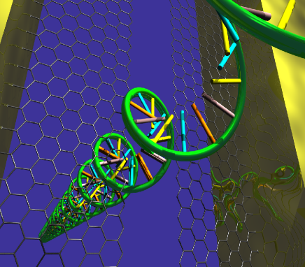
Here, I propose to use graphene nanogaps for DNA sequencing, using the graphene as the electrode as well as the membrane material. The experimental layout is drawn in figure 1. Graphene, a single-atom thick hexagonal carbon lattice that has recently been discovered novoselov_electric_2004 , can be synthesized in a variety of manners. berger_ultrathin_2004 ; gilje_chemical_2007 ; li_chemically_2008 It is an ideal material for making nanogaps for sequencing due to its single-atom thickness , its ability to survive large transmembrane pressures bunch_impermeable_2008 ; poot_nanomechanical_2008 ; lee_measurement_2008 , and its intrinsic conducting properties. The last property is especially advantageous because the membrane is the electrode, automatically solving the problem of having to fabricate nanoelectrodes that are carefully aligned with a nanogap. Contacts to the graphene sheet can be fabricated using standard electron-beam lithography, metal evaporation and lift off. The graphene sheet is covering a nm micropore in a Si/SiO2 wafer and the wafer is mounted in a PDMS fluid cell with integrated Ag/AgCl electrodes for ion current measurement as well as contacts to the Au electrodes for transverse conductance measurement.
Various methods can be used to obtain graphene nanogaps. They may be fabricated by nanolithography with a scanning tunneling microscope (STM), in a method similar to that used for cutting carbon nanotubes. venema_length_1997 ; rubio_a._mechanism_2000 Recently, STM nanolithography on the top graphene layer of grapite was demonstrated. tapaszto_tailoring_2008 The ideal nanogap width is nm, allowing for ssDNA to pass through it in an unfolded state zhao_single-strand_2007 as well as assuring the largest transverse current. The transverse conductance of DNA molecules can then be measured while they translocate through a nanogap in the graphene membrane, revealing the base sequence of the molecule.
The DNA translocation speed is typically much larger in solid-state nanopores than in biological nanopores, owing to their large difference in size and aspect ratio. storm_fast_2005 ; chauwin_strong_1998 ; ghosal_electrokinetic-flow-induced_2007 For pore sizes that are small compared to the ssDNA width, the bases stick to the side of the nanogap, lagging behind the backbone, while the molecule moves through the gap. sigalov_detection_2008 For large gap sizes, the bases’ orientation can vary significantly, but they can be aligned by the electric field due to the applied bias voltage across the gap. lagerqvist_fast_2006 ; lagerqvist_comment_2007
When large ( kbp) dsDNA translocates through solid-state nanopores with a diameter much wider than the molecule, the velocity depends as a power-law on the length
where is the Flory exponentsmith_dynamical_1996 and the required applied electric field strength is relatively low, V/m. storm_fast_2005 In contrast, ’long’ ( nt) ssDNA translocates through a much more narrow (1.8 nm) and nm deep HL nanopore with length-indepent velocity. The velocity depends quadratically on a much larger required driving voltage as
where mV, nm/us, and nm/usV2. meller_voltage-driven_2001 The electric field threshold for DNA translocation , depends on the pH and pore geometry and is due to a stretching transition of the molecule into the pore. heng_electromechanics_2006 ; zhang_effective_2007
The HL pore geometry is very close to that proposed here, since 1) the ideal graphene nanogap width of 1.5 nm is similar and 2) the narrowest region of the HL pore and the graphene nanogap are similar in thickness. This may result in similar DNA-graphene nanogap interaction strengths although a full model is required. gowtham_physisorption_2007 An advantage of graphene nanogaps is then that their local atomic configuration can be imaged directly with the STM after the gap has been fabricated allowing for a comprehensive comparison of measurements with theoretical calculations. Assuming an average field strength in the HL pore of , we can extrapolate that an applied voltage of 30 mV across the graphene membrane with effective thickness of 0.6 nm will yield an average translocation time of 3.6 us/nt. The voltage that is applied across the nanogap to read the DNA’s transverse conductance is expected to slightly alter the translocation velocity. hu_theory_2008
It has been suggested that the conduction mechanism that allows one to distinguish between the different bases depends on the spatial extend of the HOMO and LUMO levels (which are typically far away from the fermi level of the leads) and their overlap with the electrode wavefunction. zwolak_electronic_2005 ; lagerqvist_fast_2006 ; zwolak_colloquium:_2008 More recently, it was found that poly(GC) and poly(AT) can be distinguished electronically through measurement of localized states around . xu_electronic_2007 One can then estimate the current due to the bases by evaluating
where is the effective transmission of the electronic base states, and are the densities of states of the left and right electrodes, respectively. shapir_electronic_2008 For a realistic description of the tunnel current in the proposed experiment, both the distance dependence for this resonant-tunneling regimepayne_transfer_1986 , counter ions shapir_electronic_2008 , and the unique density of states of graphenenovoselov_electric_2004 ; berger_ultrathin_2004 ; gilje_chemical_2007 ; li_chemically_2008 need to be taken into account. Future studies of this system will also need to include the doping due to adsorbed water molecules on the graphene membrane and its reduction in the absense of an underlying SiO2 substrate. wehling_first-principles_2008
Although preliminary experiments indicate otherwisexu_electronic_2007 , it has been argued that transverse electronic transport cannot be used for DNA sequencing. zhang_first-principles_2006 As an alternative, the layout proposed here can also be used to directly detect voltage fluctuations due to the local and unique dipole moments of the bases. gracheva_electrical_2006 ; gracheva_simulation_2006 This capacitive detection approach is not preferred, however, due to its reliance on the relatively long-range capacitive interaction, possibly limiting the spatial resolution with which individual bases can be resolved.
Acknowledgments: I thank Michael Dickson, Karapet Karapetyan, Hanyu Lee, George Gomes, Konstantin Daskalov, Michael Zwolak, and Marc Bockrath for discussions.
References
- (1) F. Sanger, S. Nicklen, and A. R. Coulson, Proc. Nati. Acad. Sci. 74 (Dec. 1977).
- (2) E.S. Lander, Nature 409, 860 (2001).
- (3) J. C. Venter et al., Science 291, 1304 (2001).
- (4) P. A. Bilsel and S. T. Nichol, Journal of Virology 64, 4873 (Oct. 1990).
- (5) Archon x prize for genomics, http://genomics.xprize.org/.
- (6) J. Shendure, R. D. Mitra, C. Varma, and G. M. Church, Nature Reviews Genetics 5, 335 (2004).
- (7) C. P. Fredlake, D. G. Hert, E. R. Mardis, and A. E. Barron, Electrophoresis 27, 3689 (2006).
- (8) C. A. Emrich, H. Tian, I. L. Medintz, and R. A. Mathies, Science 261, 895 (1993).
- (9) L. Koutny et al., Anal Chem 72, 3388 (2000).
- (10) M. L. Metzker et al., Nucleic Acids Res 22, 4259 (1994).
- (11) M. B. Welch and K. Burgess, Nucleosides, Nucleotides and Nucleic Acids 18, 197 (1999).
- (12) I. Braslavsky, B. Hebert, E. Kartalov, and S. Quake, Proc. Natl. Acad. Sci. U.S.A. 100, 3960 (2003).
- (13) Michael Zwolak and Massimiliano DiVentra, Reviews of Modern Physics 80, 141 (2008).
- (14) J.-P. Henry, J.-F. Chich, D. Goldschmidt, and M. Thieffry, Journal of Membrane Biology 112, 139 (1989).
- (15) H. Bayley, Journal of Cellular Biochemistry 56, 177 (1994).
- (16) S.M. Bezrukov, I. Vodyanoy, and V. A. Parsegian, Nature 370, 279 (1994).
- (17) J. O. Bustamante, H. Oberleithner, J. A. Hanover, and A. Liepins, Journal of Membrane Biology 146, 253 (1995).
- (18) J. Kasianowicz, E. Brandin, D. Branton, and D. Deamer, Proc. Nati. Acad. Sci. 93, 13770 (1996).
- (19) S.M. Bezrukov, I. Vodyanoy, R.A. Brutyan, and J.J. Kasianowicz, Macromolecules 29, 8517 (1996).
- (20) J. B. Heng et al., Biophys. J. 90, 1098 (2006).
- (21) X. Zhao, C.M. Payne, P.T. Cummings, and J.W. Lee, Nanotechnology 18, 424018 (2007).
- (22) S. Howorka, S. Cheley, and H. Bayley, Nature Biotechnology 19, 636 (2001).
- (23) J. Li et al., Nature 412, 166 (2001).
- (24) A. J. Storm, J. H. Chen, X. S. Ling, H. W. Zandbergen, and C. Dekker, Nat Mater 2, 537 (2003).
- (25) A. Mara, Z. Siwy, C. Trautmann, J. Wan, and F. Kamme, Nano Letters 4, 497 (2004).
- (26) A.J. Storm et al., Nano Letters 5, 1193 (2005).
- (27) J. Zhang and B. I. Shklovskii, Physical Review E 75, 021906 (2007).
- (28) T. Hu and B. I. Shklovskii, Physical Review E 78, 032901 (2008).
- (29) M. Zwolak and M. DiVentra, Nano Letters 5, 421 (2005).
- (30) J. Lagerqvist, M. Zwolak, and M. DiVentra, Nano Letters 6, 779 (2006).
- (31) K. S. Novoselov et al., Science 306, 666 (2004).
- (32) C. Berger et al., J. Phys. Chem. B 108, 19912 (2004).
- (33) S. Gilje, S. Han, M. Wang, K.L. Wang, and R.B. Kaner, Nano Letters 7, 3394 (2007).
- (34) X. Li, X. Wang, L. Z. S. Lee, and H. Dai, Science 319, 1229 (2008).
- (35) J. S. Bunch et al., Nano Letters 8, 2458 (2008).
- (36) M. Poot and H. S. J. van der Zant, Applied Physics Letters 92, 063111 (2008).
- (37) C. Lee, X. Wei, J.W. Kysar, and J. Hone, Science 321, 385 (2008).
- (38) L.C. Venema et al., Applied Physics Letters 71, 2629 (1997).
- (39) A. Rubio, S. Apell, L.C. Venema, and C. Dekker, The European Physical Journal B 17, 301 (2000).
- (40) L. Tapaszto, G. Dobrik, P. Lambin, and L.P. Biro, Nat Nano 3, 397 (2008).
- (41) J.-F. Chauwin, G. Oster, and B.S. Glick, Biophys. J. 74, 1732 (1998).
- (42) S. Ghosal, Physical Review E 76, 061916 (2007).
- (43) G. Sigalov, J. Comer, G. Timp, and A. Aksimentiev, Nano Lett 8, 56 (2008).
- (44) J. Lagerqvist, M. Zwolak, and M. Di Ventra, Physical Review E 76, 013901 (2007).
- (45) D. E. Smith, T. T. Perkins, and S. Chu, Macromolecules 29, 1372 (1996).
- (46) A. Meller, L. Nivon, and D. Branton, Physical Review Letters 86, 3435 (2001).
- (47) S. Gowtham, R.H. Scheicher, R. Ahuja, R. Pandey, and S.P. Karna, Physical Review B 76, 033401 (2007).
- (48) M. Xu, R.G. Endres, and Y. Arakawa, Small 3, 1539 (2007).
- (49) E. Shapir et al., Nat Mater 7, 68 (2008).
- (50) M. C. Payne, Journal of Physics C 19, 1145 (1986).
- (51) T.O. Wehling, M.I. Katsnelson, and A.I. Lichtenstein, arxiv.org/0809.2894 (2008).
- (52) X.-G. Zhang, P.S. Krstic, R. Zikic, J.C. Wells, and M. Fuentes-Cabrera, Biophys. J. 91, L04 (2006).
- (53) M.E. Gracheva, A. Aksimentiev, and J.-P. Leburton, Nanotechnology 17, 3160 (2006).
- (54) M. E. Gracheva et al., Nanotechnology 17, 622 (2006).