Pyrophosphate Groups in Liquid Crystalline Phases of the DNA
Abstract
We study electrostatic interaction between molecules of the DNA in which a number of phosphate groups of the sugar-phosphate backbone are exchanged for the pyrophosphate ones. We employ a model in which the DNA is considered as a one-dimensional lattice of dipoles and charges corresponding to base pairs and (pyro)phosphate groups, respectively. The interaction between molecules of the DNA is described by a pair potential of electrostatic forces between the two sets of dipoles and charges belonging to respective lattices describing the molecules. Minima of potential indicate orientational ordering of the molecules and thus liquid crystalline phases of the DNA. We use numerical methods for finding the set of minima in conjunction with symmetries verified by potential . The symmetries form a noncommutative group of 8-th order, . Using the group we suggest a classification of liquid crystalline phases of the DNA, which allows of several cholesteric phases, that is polymorphism. Pyrophosphate forms of the DNA could clarify the part played by charges in its liquid crystalline phases, and make for experimental research, important for nano-technological and bio-medical applications.
pacs:
61.30Cz, 64.70MdI Introduction
According to the familiar legend the discovery of the cholesteric phase of the DNA was due to a happy chance that occurred to C.Robinson, who was sharp enough to see it, robinson . The story resembles that of Fleming’s discovering the penicilin. Since then cholesteric phases of the DNA have been the subject of numerous experimental and theoretical investigations owing to their variety and regularity, livolant1 . It has been established that the formation of the phases depends on chemical properties of an ambient solution and ions, the ingenious experimental technique has been worked out, livolant1 , livolant2 , and liquid crystalline phases of the DNA have become instrumental in studying the DNA itself. Another important development began surfacing in chemical physics of nucleic acids early in the 90-th. The group led by Z.A.Shabarova at the Lomonosov Moscow University, s1 — s5 succeeded in synthesizing the DNA in which some inter-nucleotide phosphate groups are exchanged for the pyro-phosphate ones, and thus considerably extended the field of research, providing new insights into the chemistry of nucleic acids, as well as new possible bio-medical applications. In this paper we aim at making it clear that pyrophosphate forms of the DNA could be helpful in studying liquid crystalline phases of the DNA.
Theoretical work on the physics of liquid crystalline phases dates from the seminal paper by Onsager, onsager , which relies on the picture of hard rods representing molecules in solvent. Applications of the model require the use of phenomenological constants and theoretical assumptions, difficult to verify in specific situations. Cholesteric phases need even more careful investigating owing to their chirality. In a series of papers Kornyshev, Leikin, and their collaboratorslk_model , lk1 - lk3 , kim , osipov , put forward the theory of cholesteric liquid crystalline phases of the DNA that relies on the helical distribution of charges of the DNA. Within the framework of this theory one can employ various approaches and approximations and investigate specific conformations. Generally, a molecule of the DNA is considered as a charged rod or cylinder, the charge being distributed continuously over the surface of the rod, complying with the helical symmetry, theoretical tools employed being of analytical character. In the present paper we use a discrete approximation for the charge’s distribution and rely a computer simulation for finding molecular conformations. It should be noted that the distribution of charge in the DNA molecule is essentially discrete being caused by (1) the dipole moments of the base pairs, (2) the charges of the phosphate groups, (3) counterions which are not uniformly distributed round the DNA molecule. The electrostatic interaction between two DNA molecules is due to this essentially non-uniform distribution of charges. Our approach, still remaining within the framework of papers lk_model , lk1 , provides new details of the phenomenon. It is worth noting that we aim only at a qualitative description, which could be useful for explaining experimental data.
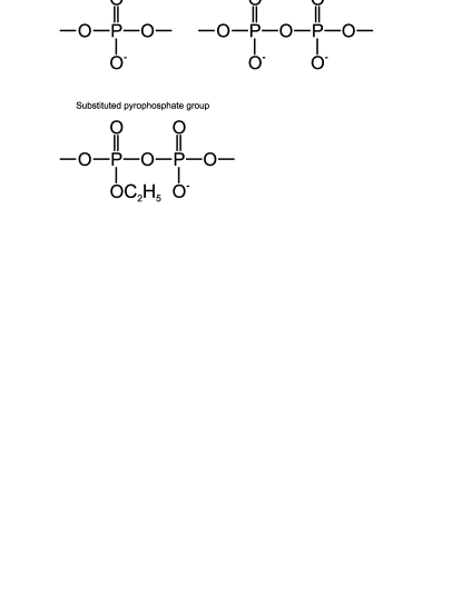
Since the current theory considers electrostatic interaction as a cause for the formation of liquid crystalline phases the DNA , it is interesting to investigate opportunities that can be provided by the use of the DNA containing a number of pyrophosphate groups, PP-forms, instead of the usual phosphate ones, P-forms, see Figure 1, so as to have a means of changing the charge conformation of the molecule. It is important that synthetic forms of the DNA can contain PP-groups in the duplex of the DNA instead of the usual phosphate ones in such a way that the structures of the pyro-modified and usual phosphate molecules, remain rather close,s5 , the inter-nucleotide distance, the stacking, and the Watson-Crick interaction suffering no change.
Synthetic forms of the DNA are instrumental in the study of fundamental problems in molecular biology, biochemistry, medicine, ferments’ activity in nucleotide exchange, protein-nucleic acids complexes, structural functioning of biopolymers, and regulation of the genetic expression. The modification of inter-nucleotide groups is of particular importance owing to its preserving the ability of molecules of the DNA to penetrate cell membranes and regulate gene expression, while retaining the basic function of the DNA, that is to interact with the complimentary sequences of nucleotides. It is possible to synthesize the DNA so as to have the exchanged pyrophosphate groups located at prescribed sites of the sugar-phosphate spine, each pyrophosphate group bringing forth an additional negative charge.
Minima of the potential U for pair-interaction of molecules of the DNA should correspond to orientational ordering of the molecules in solvent and therefore liquid, or solid, crystalline phases. Special means are required to find the minima of . At this point the symmetry of U provides valuable information. As was found in the previous paper gkv , is invariant under the action of a group of discrete transformations, and therefore its minima form a set having the same symmetry. The circumstance reduces the amount of numerical work, which is quite large. But we feel that the symmetry of the pair-interaction is by far of more general importance for understanding the physics of liquid crystalline phases of the DNA than one can infer from its numerical applications.
II Preliminaries.
We shall recall certain basic facts of the DNA. A molecule of the DNA can attain several hundred in length. If we neglect details that have a size of one thousand Å, or more, we can visualize it as a soft shapeless line and conclude that on this scale it behaves like an ordinary polymer. In contrast, looking at its smaller segments, of one hundred Åor less, we observe that it tends to be straight. Thus, borrowing a comparison from everyday life, we may say that a molecule of the DNA looks like a piece of steel wire whose long segments are flexible and the short ones are stiff. The elastic properties of the DNA are intimately related to its being a double helix. The latter imposes severe constraints on deformations which can be effected without destroying the molecule and to a large extent determines its mechanical properties. In fact, the two strands comprising the molecule of DNA have just small bending rigidities, just as usual polymers. But the formation of the two-stranded structure drastically changes the DNA by making it both stiff and capable of forming sophisticated spatial shapes.
As was mentioned above, the double helix of DNA consists of long chains, or strands, which have the backbones composed of sugar and phosphate residues, and special chemicals, bases, keeping the two strands together (the structure is illustrated in Figure 2). The fundamental building blocks of the strands are nucleotides, joined to each other in polynucleotide chains. The nucleotide consists of a phosphate joined to a sugar (2’-deoxyribose), to which a base is attached. The sugar and base alone are called a nucleoside. The chains, or strands, of the DNA wind round each other in a spiral forming a double helix, the bases being arranged in pairs: adenine - thymine (AT), guanine - cytosine (GC), so that the sequence of bases in one strand determines the complimentary sequence of bases in the other and constitutes the genetic code stored by the molecule of DNA. There are several forms of the DNA, denoted by A,B, and Z. The most common one in nature, is the so-called B-form. One turn of the helix of the B-form, corresponds approximately to base-pairs, and the distance between adjacent pairs of bases is approximately Å. In real life there are considerable deviations from the canonical B-form of the DNA. Therefore, there is a need for a special nomenclature for describing its conformations (see calladine for the details),and generally a considerable set of parameters is required. It is worth noting that the deviations from the canonical form are by no means small, and may have a size of tens of degrees.
Synthetic analogues of nucleic acids (NA) containing modified internucleotide groups are useful for solving various problems of molecular biology, biotechnology, and medicine. Shabarova et al, s1 , s2 ,developed a novel type of modified DNA duplexes containing pyrophosphate ( PP) and substituted pyrophosphate ( SPP) internucleotide groups at the definite position of the sugar-phosphate backbone s1 - s5 (see Figure 1). The PP-group bears one additional negative charge in comparison with a natural internucleotide group; SPP group contains no additional charges. The introduction of PP-groups into DNA leads to an increase of total negative charge of a molecule of the DNA. The study of oligonucleotide duplex containing a PP and SPP groups has revealed that stacking and Watson-Crick interactions are not significantly affected. By flipping out of the disubstituted phosphate these groups fit into the helix structure without elongation of the internucleotide distance. The analysis of helical parameters of base-pairs , internucleotide distances, and overall global structure, reveals a close similarity of the initial and modified duplexes.
The location of PP-groups of the DNA are to verify certain conditions:
-
1.
their total number does not exceed 10 % of the total number of phosphates;
-
2.
they are not allowed to be located opposite each other;
-
3.
they are not allowed to occupy the ends of a molecule;
-
4.
two adjacent pyrophosphate groups are to be separated by at least 10 phosphate ones.

III Model.
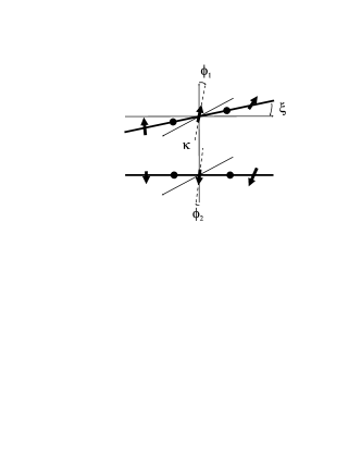
Theoretical study of liquid crystalline phases of the DNA generally uses models that are necessarily based on very crude simplifications. The first point at issue is the right choice of a potential of interaction. In this paper we model the molecular of the DNA on a 1-dimensional lattice of charges and dipoles with an elementary cell of size Å. It mimics the spatial conformation of charges of phosphate groups and dipoles of base-pairs. We consider short segments of the DNA, approximately Å, that is of the size of persistence length, so that to a good approximation they are segments of straight lines, and assume that both molecules have the same number, 151, of base-pairs that can be visualized as points on a straight line parallel to the axis of the molecule, one base-pair being located at the center of a corresponding molecule (see Figure 3). The centers of the straight lines belong to a straight line perpendicular to plane x-y which is parallel to either of them. We shall denote by the angle between the straight lines describing the molecules. Both molecules are of the same helicity, which is determined by the rotation of the frame of the dipole moments. Thus, we model a molecule of the DNA on a one-dimensional lattice having at its sites either vectors of dipoles of the base pairs or scalars of the phosphate charges. It is important that the values of the dipoles and charges are renormalized owing to screening effects caused by counterions and ions adsorbed at the molecule. Therefore, we consider effective phosphate charges and dipoles of base pairs. The case of total neutralization of phosphate charges was considered in paper kik .
The dipoles are suggested to have the helix symmetry with rotation / bp, corresponding to the structure of the ideal double helix of the DNA. Of course, it is necessary to take into account the structure of DNA being not uniform and the relative positions of the base pairs varying slightly from base pair to base pair. Hence, the dipole moments of the base pairs do not form a precise lattice structure. Even more so they should depend on the local DNA sequence. Therefore, our assumption of the regular dipole positions is a crude approximation.
The distance, , between the centers of the lattices, which is fixed, is an important parameter of the model. In what follows we use the distance between adjacent base-pairs, that is Å, as a unit of length, take a unit of charge for which the dipole moment of equals , and perform calculations in the dimensionless units generated by these quantities.
The energy of electrostatic interaction of two molecules can be cast in the sum
| (1) |
in which is the dielectric permeability of solvent and is the self energy of the pair, which does not influence its conformation, the first term describes the interaction between dipoles of the first molecule and those of the second; the second term - dipoles of the first and phosphate charges of the second; the third - charges of the first and dipoles of the second; the fourth - charges of the first and the second. The interactions are given by the equations
| (2) | |||||
| (3) | |||||
| (4) | |||||
| (5) |
in which is the inverse Debye length , and
We shall take the screening functions in Schwinger’s form
The important point about the electro-statical interaction between molecules of the DNA is a wise choice of the screening factor. The common practice is to employ the Debye-Hückel theory, or its modifications that might accommodate the dipole charges, the so-called Schwinger screening, podg . The full treatment of this problem requires a separate investigation. In this paper we confine ourselves to the Debye-Hückel and the Schwinger theories, podg .
It is worth noting that the pair potential is invariant: if we change the sign of angle between the axes of the two molecules, at the same time as the sign of helicity, the potential remains the same. There are symmetry rules for the helixes of the same kind. One may convince oneself that the following transformations
| (6) | |||||
| (7) | |||||
| (8) |
leave the potential invariant. The angles are defined within limits
values corresponding to the same configurations of the molecules. The transformations given by equations (6-8) verify the equations
where is a transformation that leaves all invariant. Using the above equations one can easily convince oneself that generate a non-commutative group of 8-th order, . Its maximal subgroup is a normal subgroup of 4-th order, commutative, and generated by the transformations
| (9) |
Elements in its turn generate subgroups and of , respectively. It is worth noting that are of second order, both. They are conjugate subgroups of S, that is for an element of we have , or we may state , in the notations of group theory, which can be cast in the form of the diagram
| (10) |
The element
| (11) |
generates subgroup of . It is important that is a normal subgroup of , that is for any element of . Thus, we have the diagram of subgroups inside the symmetry group
| (12) |
in which the arrows signify the embedding of subgroups.
The group of symmetries, , plays the key role in finding minima of the potential . The following general arguments, based on the theory of groups, are quite useful in this respect. Consider a point of space of the angles . Suppose that is a minimum of . Then points
called the orbit of the point under the action of the group , are also minima of . The number of points of the orbit can vary. In fact, let us consider all transformations of that leave invariant, that is . It is alleged to be known that the transformations form a subgroup of , called stationary subgroup . The stationary subgroups, and , for points and of an orbit, are conjugate, that is for an element of . The number of different points equals to the ratio of the orders of and , that is to 2 or 4, depending on the choice of point . To be specific, consider a point having a stationary subgroup that coincides with the subgroup . The latter is a normal subgroup of of index 2, that is the factor set consists of two elements. Thus, the orbit of under the action of consists of only two points that correspond to the same value of and have the same stationary subgroup , because the latter is a normal subgroup of . The situation is quite different if we take a point having stationary subgroup , which is different from . The subgroups do not coincide in , even though they are conjugate. The orbit of under the action of indicated above consists of four points that we may sort out as follows: two points having the stationary subgroup and two points having . This is due to the fact that for one thing the subgroup is commutative and therefore its elements generate points of the orbit but with the same stationary subgroup, that is , and for another there is an element that gives points of the orbit having the stationary subgroup . In contrast, a point having the stationary subgroup has the orbit consisting of four points which have the same stationary subgroup , because the latter is a normal subgroup of .
IV Numerical simulation
It is to be noted that numerical evaluation of the minima runs across a poor convergence of standard algorithms for minimization, owing to flat surfaces of constant value for the function of three variables, . To some extent, one may get round the difficulty by observing that for points remaining fixed with respect to a subgroup of , the minimization problem is reduced to that for a smaller number of variables. This is due to the necessary conditions for extremum being verified automatically for degrees of freedom perpendicular to the set of invariant points, so that one needs only to study the conditions for longitudinal variables, that is to solve a smaller system of equations.
To see the point let us consider a function of variables even in , so that . The set of invariant points is plane, and we may look for minima of the function , thus we need to solve only two equations
The number of variables necessary for calculations can be reduced even further in case of larger groups of symmetries. It is easy to convince oneself that the sets of fixed points , that is invariant under the action of a subgroup of , read as follows
| (13) | |||||
| (14) | |||||
| (15) |
in which the are invariant under the action of subgroups , respectively. The above symmetries are illustrated in Figure 4.

The analysis of symmetries of given above, cf. p. III, enables us to sort out the minima according to the effective value of the phosphate charge . The dependence of the values of minima on the effective charge is illustrated in Figure 5.


It should be noted that minima of the pair-interaction depend on the distance between molecules , and the effective phosphate charge . The latter is the control parameter we employ in numerical simulation. It is also useful for the description of possible experimental results. In this paper we are considering to within Å. Effective charge of phosphate groups determines the neutralization; it varies to within , in dimensionless units, corresponding to the total neutralization. Charges that correspond to the charge inversion, have not been considered. The Debye length, , has been varied to within Å, depending on the ion strength of solution.
The value of effective charge is determined by its electrostatic surrounding. It depends not only on ion charges in solvent, but also on those adsorbed by a molecule of the DNA.It seems that the conventional Debye — Hückel theory does not work in this situation, podg . At any rate, it does not accommodate the adsorbed charges.According to livolant2 the effective charge is small.
The numerical data and the symmetry analysis given above suggest that there should be the following three types of the minima.
-
1.
Type I characterized by molecules having a cross-like conformation, ” snowflakes”, that is being close to . It exists for large enough. Its symmetry subgroup depends on and may take values . Therefore, we may claim that there exist four sub-types of minima I: , each of them consisting of two subtypes which are given by specific conformations of the angle variables.
-
2.
Type II for which takes values to within ; the symmetry subgroups are and , either type consists of two sub-types.
-
3.
Type III for which is to within , that is larger than for II. The symmetry subgroup is , and there are four constituent types of the same symmetry.
As can be inferred from the considerations given above, the study of the pair-interactions between molecules of the DNA requires a means to vary the charges of a molecule and their positions in it. By now the only method available to that end is to vary the ion composition of solvent, the molecules itself being intact. The use of the DNA containing PP-groups, should provide new opportunities for the research, for it could change in a prescribed way the conformation of charges of the molecule. Thus one may compare the formation of liquid crystalline phases for the same solutions but for a different charge conformation of the DNA. Our numerical simulation suggests that the effect could be tangible enough to be looked in experiment. The dependence of the minima on effective charge taking into account PP-groups is indicated in Figure 6. The behavior of is illustrated in Figure 7 by means of iso-energy surfaces. Another point in favor of working with the pyro-phosphate forms of the DNA is that one can vary the effective charges of a molecule. In the case of the usual phosphate DNA the phosphate charges are all equal, and therefore one may suggest that the effective charges, which enter in our simulation, are also equal. Using the pyro-phosphate forms we may expect to achieve even the regime for which all the phosphate charges are neutralized whereas the pyro-phosphate ones still remain, even though being small. Such an experiment would be helpful in determining the nature of intra-molecular pair interaction that leads, according to the accepted physical picture, lk_model , to the formation of cholesteric phases of the DNA.
The main point about our numerical simulation is the choice of values for effective charges and dipoles. In case there are PP-groups we may consider the effective charges in a way described above. The situation is more subtle as far as dipoles are concerned. We assume their numerical values being of the first order in the units indicated above. This is as much as to say that the charges of base pairs that constitute dipoles, are screened much less than the phosphate ones. If we proceed otherwise and take small values of the dipoles, there are no minima small but nonzero value of , that is no cholesteric phases. In fact, they are, livolant1 . Thus, it is reasonable to assume that the charges of base pairs are screened in a manner different from that for the phosphate ones. One may suggest to the effect that the renormalization of charges is due mainly to adsorption of ions from the solvent, and not to the screening clouds of ions in the solvent. If so, it is likely that the charges of P-groups, due to ions of , are in a different positions than those of the base pairs, and the renormalization of charges of P-groups and base pairs follow different paths. For this argument we are indebted to Yu.M. Yevdokimov.
The important point is that PP-groups make for the formation of cholesteric angles different from those of P-groups, and thus provide a means of the identification of new phases. Summing up:
-
•
the phase of ”snowflakes” sustains the presence of PP-groups;
-
•
the PP-groups may result in splitting energy levels of minima, so that minima corresponding to the same values of become different, when PP-groups are present;
-
•
minima of are separated by low potential barriers; iso-energy surfaces of constant values of , being like galleries between halls that illustrate the minima;
-
•
minima corresponding to cholesteric phases are very narrow, whereas those of snowflakes are broad and sloping; energy barriers separating minima corresponding to snowflakes and cholesteric phases, respectively, are of the order ;
-
•
values of angles are subject to constraints inside galleries joining two minima,

V Conclusions: New opportunities for studying the liquid crystalline phases of the DNA.
The pyro-phosphate DNA may serve a valuable probe into the physics of cholesteric phases of the DNA. providing a unique opportunity for changing the effective charge of a molecules of the DNA. The use of PP-forms may result in appreciable experimental effects, which in its turn could throw light on the nature of intramolecular interaction in solution of the DNA. The charge screening still poses a number of questions. The usual Debye - Hückel theory does not seem to be an adequate solution, podg , especially as the screening of electrical dipole moments is concerned. A theory that could give a reasonable agreement with experiment, should be that of finite number of particles, whereas the Debye - Hückel theory relies on the use of macroscopical considerations. The study of the cholesteric phases of the DNA with pyrophosphate inter-nucleotide insertions could provide a means to find characteristics that indicate a way to understanding the phenomenon. An important issue is the different screening of the phosphate charges and the dipoles of the base pairs. It is should be noted that the screening is caused by a non-uniform adsorption of counterions at a molecule of the DNA, so that the phosphate charges and the base pair dipoles are not screened in the same manner.
Our calculations rely on a model that is based on general and qualitative assumptions of the helical charge distribution of the DNA. We feel that it accommodates a picture of the DNA, without going into finer details, and agrees with the approach of paper lk_model in which the continuous approximation plays the directive part. The choice of the pair-potential for the intramolecular interaction is important. The shape of the pair potential chosen in this paper enabled us to accommodate the two different sets of charges — the Coulomb and the dipole ones, and also take into account a finer detail of the pyrophosphate charges, which could turn out to be a valuable instrument for further investigating the cholesteric phases. The symmetry constraints have played an important part in finding the minima of the pair-potential. It seems that their meaning could be greater than a simple arithmetic device for simplifying calculations, and could indicate certain symmetry law peculiar to the cholesteric phases of the DNA. It is reasonable to expect the polymorphism liquid crystalline phases of the DNA. New artificially synthesized phases of the DNA could be a fruitful instrument to that end.
We are thankful to F.Livolant for the useful correspondence. This work was done within the framework of the Interdisciplinary Programme of the Lomonosov Moscow State University, and partially supported by RFBR Grant # NS—660.2008.1.
References
- (1) C.Robinson, Tetrahedron, 13, 219 (1961).
- (2) F.Livolant, J.de Phys. (Paris), 47, 1605 (1986).
- (3) F.Livolant, Biophys. J., 65, 56 (1993).
- (4) Onsager L., Ann.Acad.Sci. NY, bf 51, 621 (1999).
- (5) A.A. Purmal, V.L. Druca, Z.A. Shabanova, Bio-Org.Chem. 10, 394 (1984) (in Russian).
- (6) S.A. Kuznetsova, C. Clusel, E. Ugarte, I. Elias, M. Vasseur, M. Blumenfeld, and Z.A. Shabanova, Nucl.Acids Res. 24, 4783 (1996).
- (7) G.Ya. Sheflyan, E.A. Kubareva, S.A. Kuznetsova, A.S. Karyagina, I.I. Nikol’skaya, E.S. Gromova, and Z.A. Shabanova, FEBS Lett. 390, 307 (1996).
- (8) A.A. Purmal, Z.A. Shabanova, R.I. Gumport, Nucl.Acids Res. 20, 3713 (1992).
- (9) M.V. Rogacheva, A.V. Bochenkova, S.A. Kuznetsova, M.K. Saparbaev, and A.V. Nemukhin, J.Phys.Chem. 111, 432 (2007).
- (10) A.A.Kornyshev and S.Leikin, J.Chem.Phys. 107, 3656 (1997).
- (11) A.A.Kornyshev and S.Leikin, Phys.Rev.Lett., 84, 2537 (2000).
- (12) A.A.Kornyshev, D.J.Lee, S.Leikin, A.Wynveen, and S.B.Zimmerman, Phys.Rev.Lett., 95, 148102-1, (2005).
- (13) A.A.Kornyshev and S.Leikin, Proc.Natl.Acad.Sci.U.S.A. 95, 13579 (1998).
- (14) Y.H.Kim, J. de Phys.(Paris) 43, 559 (1982).
- (15) B.Samorĭ, M.Osipov, I.Domini, and A.Bartolini, Int.J.Macromol., 15, 353 (1993).
- (16) V.L.Golo, E.I.Kats, and I.P.Kikotx, Pisma ZhETF 84, 334 (2006).
- (17) V.L. Golo, E.I.Kats and, Yu.S.Volkov, Pis’ma ZhETF 86, 311 (2007),
- (18) M.A.El Hassan and C.R.Calladine 1995 J.Mol.Biol. 251 648
- (19) Punkkinen1 O., Naji A. , Podgornik R., . Vattulainen1 I., and Hansen P.-L., EPL, 82, 48001, (2008).