Excited-state spectroscopy of single NV defect in diamond using optically detected magnetic resonance
Abstract
Using pulsed optically detected magnetic resonance techniques, we directly probe electron-spin resonance transitions in the excited-state of single nitrogen-vacancy (NV) color centers in diamond. Unambiguous assignment of excited state fine structure is made, based on changes of NV defect photoluminescence lifetime. This study provides significant insight into the structure of the emitting excited state, which is invaluable for the development of diamond-based quantum information processing.
Over the last decade, the negatively charged nitrogen-vacancy (NV) color center in diamond has attracted a lot of interest because it can be optically addressed as single quantum system [1] and exhibits several important properties for quantum information science applications. First, its perfect photostability at room temperature enables to realize a practical NV-based single photon source [2, 3] for quantum cryptography applications [4, 5]. Second, NV color centers have a paramagnetic ground state which spin can be optically polarized, read-out and exhibits long coherence time even at room temperature [6, 7]. Coherent manipulation of electron and nuclear spins of single NV color centers has been used to realize solid-state quantum physics experiments, ranging from coherent coupling of a single NV color center to other single spins in the diamond crystalline matrix [7, 8, 9], to the implementation of a quantum register [10] and conditional two-qubit CNOT gates [11], and very recently the generation of Bell and GHZ states with long coherence times [12].
Despite these results, which make the NV color center a competitive candidate for solid-state quantum information processing, the excited-state structure of the defect is not yet fully understood [13, 14]. This knowledge is however crucial for single-spin high-speed coherent optical manipulation through -based transitions [15, 16, 17] as well as for future implementation of quantum information protocols like quantum repeaters [18, 19] that can be used as a building block for a quantum network [20].
Resonant optical excitation of single NV color centers at low temperature [21] and cw electron-spin resonance experiments [22] have recently provided new insights into the structure of the excited-state, showing that its fine structure is strongly affected by local strain in the diamond matrix [21, 22]. Recent ensemble experiments have also studied the behaviour of an infrared emission line that gives a better understanding of the metastable state responsible for spin polarization [23].
Here we develop a new approach to probe the excited-state fine structure of single NV color centers. Using pulsed optically detected magnetic resonance techniques, we directly probe electron-spin resonance transitions of the excited-state. Unambiguous assignment of excited state fine structure is made, based on changes of NV defect photoluminescence lifetime.
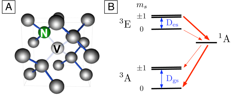
The NV defect center in diamond consists of a substitutional nitrogen atom (N) associated with a vacancy (V) in an adjacent lattice site of the diamond crystalline matrix (Fig. 1-(A)). For the negatively charged NV color center, which is addressed in this study, the ground state is a spin triplet state , originating from six unpaired electron spins. Owing to symmetry of the NV center, ground state spin sublevels are degenerate and the zero-field splitting from is GHz (Fig. 1-(B)). The excited state is also a spin triplet, associated with a broadband photoluminescence emission with zero phonon line at eV. The order of other energy levels is still under debate but it is now well established that at least one metastable state is lying between the ground and excited triplet state [13, 23]. This metastable state plays a crucial role in spin dynamics of the NV color center. Indeed, whereas the optical transitions E are spin conserving, non-radiative inter-system crossing transitions to the metastable state A are strongly spin selective as the shelving rate from the sublevel is much smaller than those from . Conversely, the metastable state decays preferentially towards the ground state sublevel, leading to a strong spin polarization into after a few optical excitation-emission cycles [24].
Another consequence of this spin-selective process is that the photoluminescence intensity is higher when the state is populated, allowing optical detection of spin-rotation of a single NV center at room temperature by optically detected magnetic resonance (ODMR) [25, 26]. Indeed, if a single NV center, initially prepared in the state through optical pumping, is driven to the spin sublevels by applying a resonant microwave frequency, a decrease in photoluminescence signal is observed. This technique is now routinely used for single-spin readout in solid-state quantum optics experiments using single spins in diamond as quantum bits. Until now only the ground state spin sublevels have been detected using ODMR. However, as optical transitions are spin conserving, spin rotations in the excited states should be also detected, allowing to probe the structure of spin sublevels.
We first investigate single NV color centers artificially created in a ultra-pure type IIa diamond sample (Element6), by implanting MeV isotopically pure atoms and by annealing the sample for two hours in vacuum at [27]. NV centers are then optically addressed at room temperature using a standard confocal microscope coupled to a Hanbury-Brown and Twiss setup used to measure the photoluminescence second order corrrelation function and verify that an individual NV center is adressed.
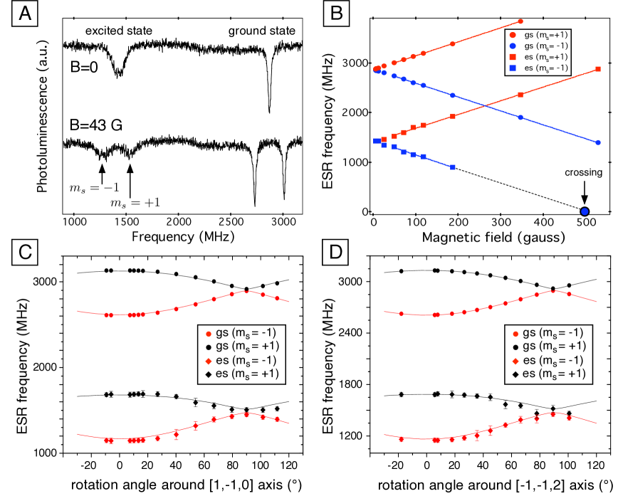
Electron spin resonance (ESR) spectroscopy of single NV centers is realized by applying microwaves, using a copper microwire (m diameter) close to the NV center, and by monitoring the photoluminescence intensity. When the microwave frequency is resonant with the transition between and sublevels, spin rotation is evidenced as a dip of the photoluminescence signal as explained above. Figure 2-(A) (upper trace) describes a typical ODMR signal obtained by sweeping the microwave frequency without any applied magnetic field. The well-known ground state transition between and sublevels is detected at GHz, and an additional broad line around GHz is observed. By applying a magnetic field to the sample, the degeneracy of is lifted by the Zeeman effect, leading to the appearance of two lines in corresponding resonances of the ODMR spectrum (see figure 2-(A) bottom trace). As an hypothesis, we attribute the broad resonance to spin sublevels of an excited state of the NV color center. In the following, we will demonstrate that this excited state actually corresponds to the emitting excited state .
We first study in more details the ESR frequency positions as a function of the magnitude of a magnetic field () applied along the NV symmetry axis which corresponds to a [111] crystal axis. The results of this experiment are depicted on figure 2-(B). Neglecting electron-nuclear spin coupling, the excited-state spin Hamiltonian of the NV defect can be written as:
| (1) |
where is the excited-state zero-field splitting, , is the excited-state strain-induced splitting coefficient, the excited state g-factor and the Bohr magneton. By considering magnetic field magnitudes such that the strain-induced fine structure splitting is negligible compared to Zeeman splitting (), the resonant frequencies associated to eigenstates are given by
| (2) |
For the same NV center considered in this study, it was not possible to observe any strain-induced splitting in the excited state, the ODMR dip being very broad with a FWHM on the order of MHz (see figure 2-(A)). As a result, it is reasonable to consider that the strain-induced splitting coefficient is such that MHz [28]. Following this consideration the experimental results depicted on figure 2-(B) can be fitted using equation (2) for magnetic field magnitudes bigger than G, corresponding to a Zeeman splitting on the order of MHz (). The results of the fit lead to MHz and an isotropic g-factor which is similar to the ground state g-factor. This isotropy indicates that the orbital angular momentum does not play a significant role in the excited state. Finally, the positions of ESR frequencies were measured by rotating a magnetic field of magnitude G around a [1,-1,0] crystal axis (figure 2-(C)) and around a [-1,-1,2] crystal axis (figure 2-(D)). The experimental results provide a strong evidence that the ground and excited states exhibit the same orientations.
It is interesting to note that a level anti-crossing between the and sublevels of the excited state is expected for a magnetic field amplitude on the order of G (see figure 2-(B)). This explains why a decrease of the NV color center photoluminescence has been observed in ensemble experiments at such magnetic field [23, 29]. Indeed, when a level anti-crossing occurs in the excited state, the electron spin polarization of the center is significantly reduced. This effect is identical to the well-known photoluminescence dip occuring at G, when the and sublevels of the ground state are crossing.
The cw experiments reported above prove that the ESR signal corresponds to an excited state, but do not provide ultimate proof that this state is responsible for fluorescence emission. For example, other dark state involved in spin polarization pathway can influence the spin polarization of NV defect [23]. We now demonstrate that the excited state observed in ODMR spectra actually corresponds to the emitting excited state . Following a method introduced in Ref. [31], the experiment is based on observing a modification of photoluminescence decay by manipulating the excited state spin sublevels with resonant microwave pulses.
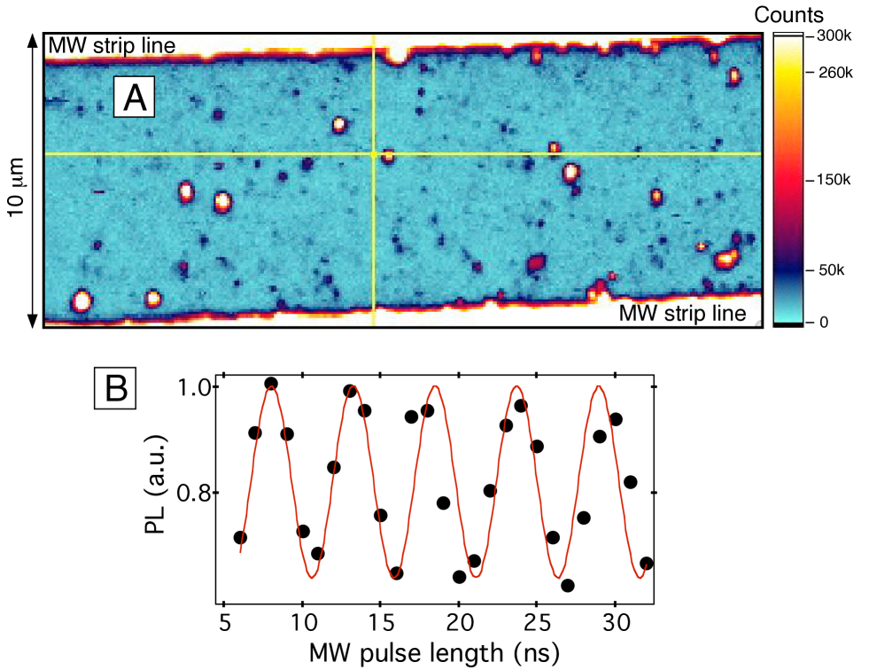
For that purpose, a large microwave driving field is required in order to reach a significant change in the population of the excited-state spin sublevels within the radiative lifetime. In order to more easily meet that requirement, we used single NV color centers in diamond nanocrystal, for which the photoluminescence decay is known to be much longer than in bulk diamond sample [32]. In addition, the nanodiamonds were spin-coated on a microscope cover glass on which gold strip-line microwave wires had been deposited using shadow mask photolithography and metal electrodepositing in order to reach high ESR Rabi frequencies. Typical dimensions of the wires were m width and m thick. Figure 3-(A) shows a photoluminescence map of the sample, the yellow cursors indicating the individual NV center studied in the following. For this NV center, we measured a Rabi nutation between ground state spin levels at a frequency of MHz, corresponding to a pulse of ns (figure 3-(B)).
As depicted on figure 4-(A), the ODMR spectrum shows a huge energy splitting between ESR lines, even at small magnetic field magnitude ( G). Such splitting, which can not be explained by the Zeeman effect, results from strain-induced splitting which is known to be much stronger in diamond nanocrystals than in bulk samples [30]. However, note that strain has almost no effect on the values ( and ) whereas it does cause the values ( and in the Hamiltonian described by equation (1)) to become non-zero. In the following, we use resonant microwaves with the ground () and excited state () transitions, at MHz and MHz respectively (see figure 4-(A)).
As a first step, the excited state lifetime associated with each spin sublevels was measured. The NV center was first polarized into the ground state sublevel using an optical pulse of duration s at the wavelength nm. After a time delay of s, which ensures that the NV center has relaxed to the ground state, an optical pulse (40 ps, nm) much shorter than the radiative lifetime was used to excite the NV center. As the optical transition is spin-conserving, such a sequence allows to build up the photoluminescence decay of the excited-state sublevel using a standard start-stop technique for lifetime measurements, the start being a part of the pulsed excitation and the stop being the single-photon counter signal. For measuring the decay of sublevel, an additional microwave pulse resonant with ground state spin transition () was introduced before the ps optical pulse.
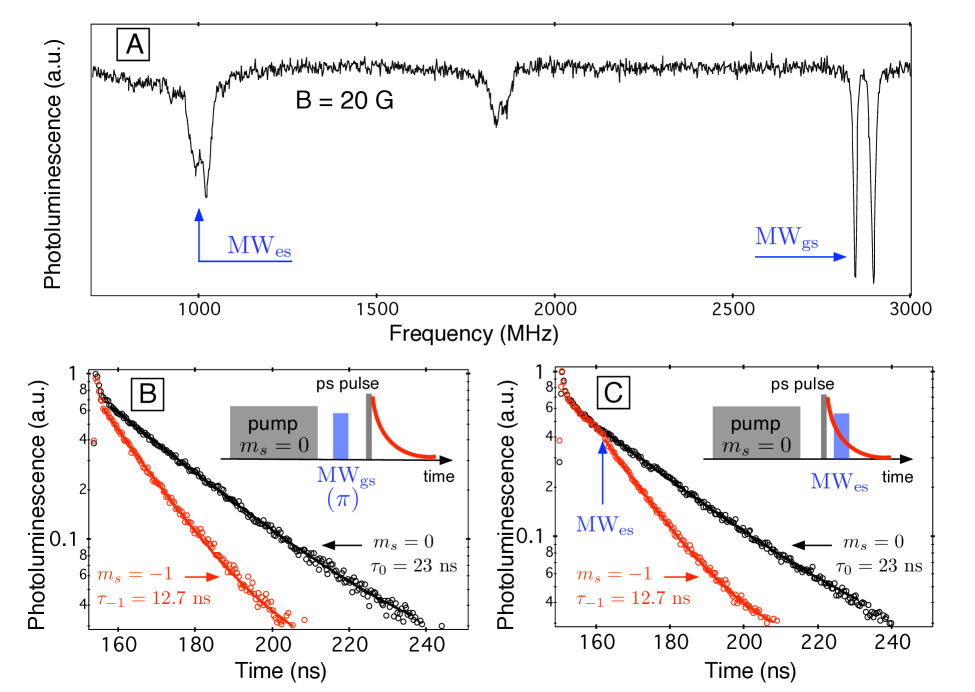
The results of such measurements are depicted on figure 4-(B). Fluorescence decays follow single exponential decay associated to a lifetime ns for sublevel and ns for . As expected, these values are much bigger than the ones measured in bulk samples [31]. As the photoluminescence is always smaller from sublevels compared to , the measured lifetime difference results from spin-selective non-radiative inter-system crossing transition to the metastable state, and not from a modification of the transition strength. It is interesting to notice that the measured lifetime ratio, , is in good agreement with recent theoretical predictions [13] and previously reported measurements in bulk samples [31].
In order to check if the resonance lines observed in ODMR spectra actually correspond to spin transitions in the emitting excited state, the same experiment is performed by applying a microwave pulse resonant with the excited state spin transition (), just after the picosecond optical pulse. The results, depicted in figure 4-(C), indicate a drastic change of the photoluminescence decay when the microwave pulse is applied. Before the microwave excitation, the photoluminescence decay follows the single exponential decay associated with the excited-state sublevel ( ns). The microwave excitation then suddenly rotates the spin in the excited state leading to the exponential decay associated with the excited-state ( ns). These results unambigously evidence that the new ESR lines observed in ODMR spectra are related to fine structure of the emitting excited state .
It was not possible to detect Rabi nutations on the excited state by varying the resonant microwave pulse duration. This appears as a difficult issue because of many factors. Among them are the large width of the excited-state ESR resonance, a strong hyperfine coupling owing to high spin density of the excited-state wavefunction at the nitrogen nucleus [33] and the short radiative lifetime of NV color centers.
Finally, we would like to shortly discuss our observations in the context of previously reported models of the excited-state structure. Resonant optical excitation of single NV color centers at low temperature has recently indicated that the excited-state is actually an orbital doublet, split into two orbital singlet branches by local strain [21]. Then, we would expect to observe four excited-state resonances when a magnetic field is applied to the NV center, while we only ever observed two lines (figures 2-(A) and 4-(A)). We tentatively identify the observed excited-state features to the upper branch because this branch shows high difference in inter-system crossing rate to the metastable state [13, 31], as observed in experiments (see figure 4-(B) and (C)). However, previous model of the excited-state structure has predicted non vanishing in the upper branch [21]. The observations in bulk dimond reported in this paper () bring then into question the completeness of currently available models for NV center excited-state structure.
Summarizing, we have performed the excited-state spectroscopy of single NV color centers in diamond using cw and pulsed ESR techniques. This work provides significant insight into the structure of the emitting excited state, which might be useful for diamond-based quantum information processing using -based transitions for high-speed coherent optical manipulation of single spins [17] as well as for entanglement protocols used in quantum repeaters [18, 34].
References
References
- [1] Gruber A, Drbenstedt A, Tietz C, Fleury L, Wrachtrup J and von Borczyskowski C 1997 Scanning confocal optical microscopy and magnetic resonance on single defect centres Science 276 2012 2014
- [2] Kurtsiefer C, Mayer S, Zarda P and Weinfurter H 2000 A robust all-solid-state source for single photons Phys. Rev. Lett. 85 290-293
- [3] Brouri R, Beveratos A, Poizat J-P and Grangier P, 2000, Single photon emission from colored centers in diamond, Opt. Lett. 25 1294
- [4] Beveratos A, Brouri R, Gacoin T, Villing A, Poizat J-P and Grangier P 2002 Single photon quantum cryptography Phys. Rev. Lett. 89 187901
- [5] Alléaume R, Treussart F, Messin G, Dumeige Y, Roch J-F, Beveratos A, Brouri-Tualle R, Poizat J-P and Grangier P 2004 Experimental open-air quantum key distribution with a single-photon source New J. Phys. 6 92
- [6] Jelezko F, Gaebel T, Popa I, Gruber A, and Wrachtrup J 2004 Observation of Coherent Oscillations in a Single Electron Spin Phys. Rev. Lett. 92 076401
- [7] Gaebel T et al 2006 Room-temperature coherent coupling of single spins in diamond Nat. Phys. 2 408 413
- [8] Hanson R, Mendoza F M, Epstein R J and Awschalom D D 2006 Polarization and readout of coupled single spins in diamond Phys. Rev. Lett. 97 087601
- [9] Childress L, Dutt M V G, Taylor J M, Zibrov A S, Jelezko F, Wrachtrup J, Hemmer P R and Lukin M D 2006 Coherent dynamics of coupled electron and nuclear spin qubits in diamond Science 314 281 285
- [10] Dutt M V G, Childress L, Jiang L, Togan E, Maze J, Jelezko F, Zibrov A S, Hemmer P R and Lukin M D 2007 Quantum register based on individual electronic and nuclear spin qubits in diamond Science 316 1312 1316
- [11] Jelezko F, Gaebel T, Popa I, Domhan M, Gruber A, and Wrachtrup J 2004 Observation of Coherent Oscillation of a Single Nuclear Spin and Realization of a Two-Qubit Conditional Quantum Gate Phys. Rev. Lett. 93 130501
- [12] Neumann P, Mizuochi N, Rempp F, Hemmer P, Watanabe H, Yamasaki S, Jacques V, Gaebel T, Jelezko F and Wrachtrup J 2008 Multipartite Entanglement Among Single Spins in Diamond Science 320 1326-1329
- [13] Manson N B, Harrison J P and Sellars M J 2006 Nitrogen-vacancy centre in diamond: model of the electronic structure and associated dynamics Phys. Rev. B 74 104303
- [14] Manson N B and McMurtrie R L 2007 Issues concerning the nitrogen-vacancy center in diamond J. Lumin. 127 98-103
- [15] Hemmer P R, Turukhin A V, Shahriar M S and Musser J A 2001 Raman-excited spin coherences in nitrogen-vacancy color centres in diamond Opt. Lett. 26 361 363
- [16] Santori C et al 2006 Coherent population trapping in diamond N-V centres at zero magnetic field Opt. Express 14 7986 7993
- [17] Santori C et al 2006 Coherent population trapping of single spins in diamond under optical excitation Phys. Rev. Lett. 97 247401
- [18] Childress L, Taylor J M, Sorensen A S, and Lukin M D 2006 Fault-Tolerant Quantum Communication Based on Solid-State Photon Emitters Phys. Rev. Lett. 96 070504
- [19] Childress L, Taylor J M, Sorensen A S, and Lukin M D 2006 Fault-tolerant quantum repeaters with minimal physical resources and implementations based on single-photon emitters Phys. Rev. A 72 052330
- [20] Cirac J I, Zoller P, Kimble H J, and Mabuchi H 1997 Quantum state transfer and entanglement distribution among distant nodes in a quantum network Phys. Rev. Lett. 78 3221
- [21] Tamarat P et al 2008 Spin-flip and spin-conserving optical transitions of the nitrogen-vacancy centre in diamond New J. Phys. 10 045004
- [22] Fuchs G D, Dobrovitski V V, Hanson R, Batra A, Weis C D, Schenkel T, Awschalom D D 2008 Excited-state spectroscopy using single-spin manipulation in diamond, Phys. Rev. Lett. 101 117601
- [23] Rogers L J, Armstrong S, Sellars M J and Manson N B 2008 New infrared emission of the NV centre in diamond: Zeeman and uniaxial stress studies, New J. Phys. 10 103024
- [24] Jelezko F, Popa I, Gruber A, Tietz C, Wrachtrup J, Nizovtsev A and Kilin S 2002 Single spin states in a defect centre resolved by optical spectroscopy Appl. Phys. Lett. 81 2160 2162
- [25] Wrachtrup J, von Borczyskowski C, Bernard J, Orrit M and Brown R 1993 Optical detection of magnetic resonance in a single molecule Nature 363, 244
- [26] Khler J, Disselhorst J A J M, Donckers M C J M, Groenen E J J, Schmidt J and Moerner W E 1993 Magnetic resonance of a single molecular spin Nature 363, 242-244
- [27] Rabeau J R et al 2006 Implantation of labelled single nitrogen vacancy centers in diamond using 15N Appl. Phys. Lett 88 023113
- [28] Note that surprisingly, for most of the single NV centers () studied in our implanted sample it was not possible to observe a strain-induced splitting in the excited-state.
- [29] Epstein R J, Mendoza F M, Kato Y K and Awschalom D D 2005 Anisotropic interactions of a single spin and dark-spin spectroscopy in diamond Nat. Phys. 1 94-98
- [30] Jelezko F, Tietz C, Gruber A, Popa I, Nizovtsev A, Kilin S and Wrachtrup J 2001 Spectroscopy of Single N-V Centers in Diamond Single Mol. 2 255-260
- [31] Batalov A, Zierl C, Gaebel T, Neumann P, Chan I Y, Balasubramanian G, Hemmer P R, Jelezko F and Wrachtrup J 2008 Temporal Coherence of Photons Emitted by Single Nitrogen-Vacancy Defect Centers in Diamond Using Optical Rabi-Oscillations Phys. Rev. Lett. 100 077401
- [32] Beveratos A, Brouri R, Gacoin T, Poizat J-P and Grangier P 2001 Nonclassical radiation from diamond nanocrystals Phys. Rev A 64 061802R
- [33] Gali A, Fyta M, and Kaxiras E 2008 Ab initio supercell calculations on nitrogen-vacancy center in diamond: Electronic structure and hyperfine tensors Phys. Rev. B 77 155206
- [34] Moehring D L, Maunz P, Olmschenk S, Younge K C, Matsukevich D N, Duan L-M and Monroe C 2007 Entanglement of single-atom quantum bits at a distance Nature 449 68-71