Current address: ]London Center for Nanotechnology, University College London, London-WC1H0AH, UK.
Electron Spin Relaxation and 39K Pulsed ENDOR Studies on Cr5+ doped K3NbO8 at 9.7 and 240 GHz.
Abstract
Cr5+ doped K3NbO8, considered to be useful as a electron spin qubit, has been investigated by pulsed X-band ( GHz) and 240 GHz electron paramagnetic resonance and electron nuclear double resonance (ENDOR). Comparison of the low temperature electronic spin-lattice relaxation rate 1/ at 9.7 and 240 GHz shows that it is 250 times faster at 240 GHz than at X-band. On the other hand, the spin-spin relaxation rate 1/ appears largely frequency independent and is very likely related to the superhyperfine (SHF) coupling of the Cr5+ electron with the surrounding potassium and niobium nuclei. This coupling was investigated by HYSCORE at 9.7 GHz and pulsed Mims ENDOR at 240 GHz. The high frequency and field enabled us to unambiguously measure the hyperfine and quadrupole couplings of the 39K in spite of its small magnetic moment. We find that the largest 39K SHF coupling is positive, with 0.522 MHz and 0.20 MHz as its isotropic and dipolar parts respectively. 93Nb ENDOR was dominantly due to its quadrapolar interaction, with a coupling of about 0.8 MHz, and a SHF coupling of about 0.08 MHz. The significance of these data to spin qubit studies is pointed out.
pacs:
76.30.-v, 61.72.Hh, 76.70.Dx,7 6.60.Es, 31.30.Gs, 63.20.kdI Introduction
The Cr5+ ion diluted in the diamagnetic host K3NbO8 (henceforth noted as Cr:K3NbO8) has been suggested as a magnetic field calibration standard for high field electron paramagnetic resonance (EPR) experiments Cage99 . This system exhibits a small linewidth ( 0.15 - 0.2 mT depending on the orientation) and the g-value just below the free-electron g-value, ge. The (50,52,54Cr) isotopes give rise to a single line that serves as a g-marker, while the 53Cr (9.5% ) yields four hyperfine lines that can be used to calibrate the linearity of the field Cage99 .
Recently Cr:K3NbO8 has also been suggested Nellutla07 as a new transition metal-ion based single electron-spin qubit system for quantum information processing. Quantum computation requires, among others, a long spin-lattice relaxation time as well as a long spin-spin or spin-memory relaxation time . The latter time provides a measure of the decoherence processes in the material and is used to calculate the figure of merit of a qubit, which is defined as the number of operations that can be performed before the phase information is lost. On the other hand, a ’reset’ of the qubit to a well defined starting state takes a time proportional to . Hence, shorter time means enhanced computing speed. It is therefore desirable to understand the and relaxation processes via their temperature and frequency dependence.
The present work reports on our detailed measurements of the electronic and relaxation in Cr:K3NbO8 from ambient temperatures down to 4 K at X-band ( GHz, tesla (T)) and at 240 GHz ( T). We find that the low temperature relaxation time is strongly frequency dependent: it decreases by a factor of 250 on going from 9.7 GHz to 240 GHz. On the other hand, was found to be frequency independent in the investigated temperature range.
In order to understand the process(es) controlling the we first employed hyperfine sublevel correlation (HYSCORE) spectroscopy Hoefer86 at X-band, and found that the nuclei responsible for are the neighboring 39K nuclei. But the magnitudes of 39K Zeeman, superhyperfine (SHF) and quadrupole interactions are such that the analysis at X-band becomes very complicated for any definitive analysis. On the other hand, the utilization of the high frequency (240 GHz) pulsed EPR/ENDOR spectrometer yielded ENDOR peaks that could easily be assigned to the neighboring 39K as well as to the 93 Nb nuclei, showing the advantage of the higher frequency/field EPR/ENDOR spectroscopy. The data clearly suggests that the process, and thus also the linewidths of the continuous wave (cw) EPR peaks, are related to the unresolved SHF coupling to the 39K and 93Nb nuclei. We note that there are few low frequency electron spin echo envelope modulation Barkhuijsen84 and cw-ENDOR DuVarney79 ; Dalal89 ; Dmitry08 studies where the 39K hyperfine couplings were resolved.
This article is organized as follows. Section II provides the experimental details on crystal structure, EPR and the ENDOR spectrometers. The results and analysis are presented in section III, and the salient points are summarized in section IV.
II Experimental details
The Cr:K3NbO8 sample was synthesized according to the published procedure Cage99 . A typical synthesis involves addition of CrO3 to a solution of KOH and Nb2O5 at -15∘C to form a 50% ice slurry followed by drop-wise addition of cold 30% H2O2. Light yellow crystals of Cr:K3NbO8 were obtained after keeping the final reaction mixture at 5∘C for 2-3 days. The Cr5+ concentration in the studied Cr:K3NbO8 samples was determined to be mol% by comparing its EPR signal intensity with a reference sample (BDPA in polystyrene) of known number of spins.
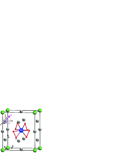
The crystal structure of Cr5+ doped K3NbO8 is shown in Fig. 1. This system has body-centered tetragonal crystal lattice ( space group) with cell parameters Å, Å and . The Cr5+ ion occupies the Nb5+ position and for convenience is shown at the body-center position in Fig. 1. Each Cr5+/Nb5+ ion is surrounded by four peroxy (O22-) groups (shown as red balls in Fig. 1) in a dodecahedral arrangement.
The 9.7 GHz cw- and pulsed-EPR measurements were performed on a commercial Bruker Elexsys 580 instrument with a Flexline dielectric resonator using a single crystal of approximate size mm3. The data at 240 GHz were collected on a home-built superheterodyne high field cw-EPR instrument Vantol05 which is recently extended to pulsed operation Morley08 . The single crystals used were typically mm3 in size. A semi-confocal Fabry-Perot resonator was used with a 12.5 mm spherical gold-coated mirror and a semi-transparent flat mirror, consisting of a gold mesh deposited on a 160 thick quartz cover slip. The maximum available power at this frequency is of the order of 20 mW and the typical pulse length is 200 ns.
The ENDOR coil for the 240 GHz spectrometer consisted of a single copper wire parallel to the mesh, which was led through a quartz capillary (0.3 mm or 0.25 mm OD). The single crystal was attached to the outside of the quartz capillary. A typical pulse sequence for the Mims ENDOR experiment consisted of a stimulated echo sequence of three pulses of about 240 ns length separated by 600 ns () and 200 s () respectively. A 150 s radio frequency (RF) pulse was applied between second and third millimeterwave pulses. About 10 W of power was applied to the untuned ENDOR ’coil’. The pulsed ENDOR measurements were performed at around 5-6 K. Typically 20 shots were averaged per RF frequency and the total acquisition time of the ENDOR spectra was of the order of 20-30 minutes.
III Results and Discussion

Figure 2 shows a typical 240 GHz electron-spin-echo (ESE) detected EPR spectrum obtained with a two pulse Hahn echo sequence ( - - - echo) and measuring the integrated echo intensity. The ESE-EPR spectra of Cr:K3NbO8 show intense central peak from the isotopes (4.34% 50Cr, 83.8% 52Cr, 2.36% 54Cr) at for the field and for . The -values indicate that the unpaired electron resides in a 3 orbital Cage99 ; Nellutla07 ; Dalal81 . The four peaks flanking the central peak are the expected four nuclear transitions from the of 53Cr nucleus. The hyperfine coupling is mT for and mT for . All relaxation and ENDOR measurements described below were performed on the central peak.
III.1 Spin-lattice relaxation
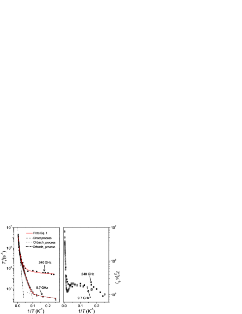
The spin-lattice relaxation time of Cr:K3NbO8 was at 9.7 and 240 GHz measured by an inversion-recovery method employing the sequences - - - FID and - t - - - - - echo, respectively. The left panel in Fig. 3 shows the temperature dependence of 1/ for at 9.7 and 240 GHz. At 9.7 GHz the increases from about 500 ns at room temperature to about 1 s at 4 K. Above 25 K at 240 GHz is same as the 9.7 GHz but it reaches to ms at 4 K. We attempted to fit the 9.7 and 240 GHz data to different models Abragam70 that included various combinations of: (a) direct process with , (b) Raman process with , where m is the spectral dimensionality that depends on the spin system and (c) Orbach process with . The best-fit to the data was found to be with the model given in Eq.(1).
| (1) |
As can be seen from the solid lines in Fig. 3, the temperature dependence of the spin-lattice relaxation rate 1/ at 9.7 and 240 GHz was successfully modeled with Eq.(1) and the best-fit parameters are listed in Table 1.
| Parameter | Best-fit value | |
|---|---|---|
| 9.7 GHz | 240 GHz | |
| C1 | 0.070(8) s-1 | 250(8) s-1 |
| C2 | 6(1)*104 s-1 | 6(1)*104 s-1 |
| 75(3) cm-1 | 75(3) cm-1 | |
| C3 | 3.35(73)*106 s-1 | 3.35(73)*106 s-1 |
| 255(20) cm-1 | 255(20) cm-1 | |
It is noteworthy that the spin-lattice relaxation rate 1/ at low temperatures (4 - 10 K) is considerably faster at 240 GHz as compared to that at 9.7 GHz which indicates that the direct process is important at these temperatures. However, the ratio of the direct process contribution to 1/ at 240 and 9.7 GHz is not as large as the expected dependence for a Kramer’s system Abragam70 but it is closer to dependence in non-Kramer’s spin systems Abragam70 . For example, at 4 K the observed ratio of the direct process contribution between 240 and 9.7 GHz is about 1/4 of the calculated value whereas it is about 4*10-4 times the value. This difference in the frequency dependence has been observed before Eaton01 ; Aminov97 and needs further theoretical analysis which is beyond the scope of this undertaking.
As can be seen from Fig. 3, the spin-lattice relaxation rate 1/ at 9.7 and 240 GHz frequencies for 10 K is well described by two Orbach relaxation pathways involving thermally accessible energy levels at cm-1 and cm-1. Based on the earlier infrared and Raman studies Haeuseler03 , the 75 cm-1 mode can be ascribed to the translational and librational modes, found to be below 200 cm-1 in the peroxychromate ion. Similarly, the 255 cm-1 mode compares with the bending vibrational modes reported in the 200 - 400 cm-1 region in the peroxychromate ion.
III.2 Spin-spin relaxation
The standard 2-pulse Hahn echo sequence was used to obtain the spin-spin relaxation time at both 9.7 and 240 GHz. The right panel of Fig. 3 shows the 1/ rate as a function of temperature for . At 9.7 GHz the increases from ns at room temperature to s at 50 K, then decreases to s at 20 K and finally increases to s at 4 K. In contrast to , the time does not show a strong field dependence. However, the temperature dependence of at both frequencies shows a minimum at about 20 K. We tentatively ascribe this minimum to some sort of motional narrowing caused by the increase in the spin fluctuations of K and Nb hyperfine fields as the temperature is raised above about 20 K. This type of temperature dependence has previously been reported for dilute solutions of free radicals Brown74 ; Brown76 ; Stillman80 and other Cr5+ complexes Nakagawa92 . Above 50 K, the spin-spin relaxation time is mainly governed by the spin-lattice relaxation processes.
III.3 Superhyperfine (SHF) interactions
As mentioned earlier the hyperfine interaction of the unpaired electron with the 53Cr in Cr:K3NbO8 was explored previously Dalal81 , and therefore in this report we will only address the SHF coupling with the potassium ( 39K) and niobium ( 93Nb) nuclei. We believe that the electronic spin-spin relaxation processes in Cr:K3NbO8 are most likely influenced by these interactions and in order to characterize them we used HYSCORE spectroscopy at 9.7 GHz and pulsed Mims ENDOR at 240 GHz.

The HYSCORE spectra of Cr:K3NbO8 at 40 K are shown in Fig. 4 for the orientation along the -axis (bottom panel) and an orientation in the -plane (top panel). The spin echo modulations due to the interactions with neighboring potassium nuclei are very large, which resulted in a good quality HYSCORE spectrum. However, the spectra were found to be too complex for analysis, possibly because at 9.7 GHz, the Zeeman, quadrupole and SHF interactions become comparable and lead to a complex interplay of these interactions. We therefore resorted to ENDOR measurements at 240 GHz ( T) where the Zeeman terms become dominant.
At 240 GHz, pulsed ENDOR signals of 39K were measured at various crystal orientations (see Fig. 5). A systematic study of orientation dependence of the ENDOR could not be performed due to the unavailability of a goniometer for this spectrometer at the present time. While the angle () of relative to the crystal -axis can be accurately determined from the resonance field of the EPR transition (via the g-tensor anisotropy), the axial g-tensor symmetry does not help with the precise determination of the angle of in the -plane. However, a closer look at the crystal structure (see Fig. 1) reveals that only the eight potassium nuclei labeled as K2 and K2′ have an appreciable overlap with the Cr5+ 3 orbital and therefore are expected to be responsible for the 39K ENDOR signals. These eight equivalent nuclei are related by symmetry (), and at some orientations eight sets of signals can indeed be distinguished (e.g. spectrum in Fig. 5). The potassium ions labeled as K1 and K3 are in the nodal planes of the Cr unpaired electron. They should thus exhibit negligible isotropic SHF couplings.
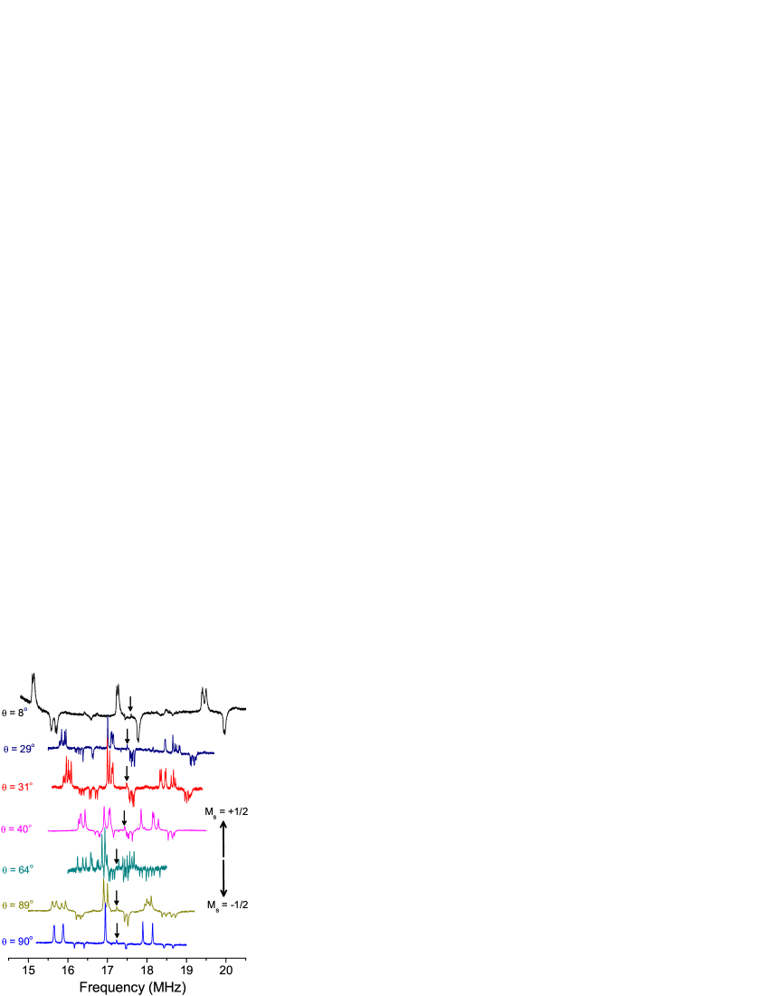
It should be noted that both negative (smaller stimulated echo) and positive (larger stimulated echo) signals are observed, and that the signal intensities are quite large. When on ENDOR resonance, the observed echo height varied from 40% to 200% of the echo height observed without RF power or out of resonance. The ENDOR intensity is largest when the shot-repetition time is of the order of . This implies that the 39K nuclei are strongly polarized by the pulse sequence, leading to an increase in echo intensity when a nuclear transition in the electron manifold is addressed, while the echo intensity becomes smaller when a transition in the state is excited, as explained in Ref. Bennebroek97 . These anomalous ENDOR intensities occur when the microwave quantum () is of the same order or larger than the thermal energy and allows for the determination of the sign of the hyperfine coupling in both pulsed Bennebroek97 ; Epel01 and cw-ENDOR Vantol99 . The observed sets of 39K ENDOR signals are sufficient for a unique assignment of the SHF and quadrupole tensor values and their relative orientations. The data are analyzed by the diagonalization of the usual effective spin Hamiltonian given in Eq.(2) with and .
| (2) |
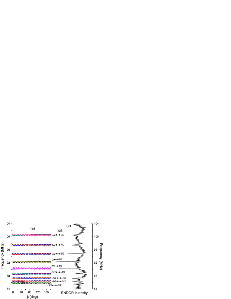
Here, is the electron Zeeman tensor, and are, respectively, SHF and quadrupole tensors of the surrounding nuclei. The principal values and their directions of the tensor (namely A1, A2, A3) and tensor (namely Q1, Q2, Q3) for K2′ nucleus obtained from the analysis are given in Table 2. The tensors for the other chemically equivalent K2 nuclei can be obtained via the afore-mentioned crystal symmetry operations. The isotropic SHF interaction is +0.522 MHz, and can be interpreted as a spin density of 0.23% in the orbital when compared to the hyperfine coupling in the ground state of atomic potassium Li94 . While we cannot make a completely unambiguous assignment of the specific potassium nucleus associated with each of the eight sets of SHF and quadrupole tensors, it is natural to assume that the anisotropic contribution to the SHF interaction should reflect the dipolar interaction between the potassium nuclear spin and the chromium electron spin. We therefore assign the SHF tensor given in Table 2 to the potassium for which the direction is close to the Cr-K direction (labeled as K2′ in the Fig. 1).
| Value(MHz) | |||
|---|---|---|---|
| A1 | +0.407(5) | 147 | 90 |
| A2 | +0.425(5) | 90 | 0 |
| A3 | +0.733(5) | 57 | 90 |
| Q1 | -0.423(4) | 87.6 | 65.6 |
| Q2 | -0.329(4) | 90.6 | -24 |
| Q3 | 0.752(4) | 2.5 | -100 |
The quadrupole tensor of K2′ is not entirely axial and has the direction of its largest principal value close to the -axis. The slight deviation from the -axis indicates that the local surrounding of the Cr5+ ion is slightly distorted as compared to that for Nb5+ ion in the undoped lattice.
At some crystal orientations 93Nb ENDOR is also observed. ENDOR spectra for field about 40∘ from the crystal -axis is shown in Fig. 6(b). We speculate that at most orientations the SHF interaction is too small to give an appreciable ENDOR intensity, but it can be detected when the field is along the Nb-Cr dipolar direction because of the largest dipolar interaction in this direction. Since Cr and Nb ions are fairly well separated (Cr-Nb: Å), the dipolar contribution to the Cr-Nb SHF interaction can be estimated using a point dipolar approximation as b[3-1] with b MHz, where is the angle between Cr-Nb dipolar axis and . While the limited data prevents a detailed analysis, the 93Nb ENDOR spectrum in Fig. 6 can be simulated with a quadrupole tensor Q[3-1] with Q = 0.82 MHz under the assumption that the quadrupole tensor is nearly axial with the principal direction along the crystal -axis.
IV Summary
We have measured the electron spin relaxation times in Cr:K3NbO8 using pulsed EPR at 9.7 and 240 GHz. The SHF couplings of the electron spin with the neighboring potassium nuclei have also been determined using pulsed Mims ENDOR at 240 GHz. The ENDOR results show that the SHF interaction of the Cr unpaired electron with neighboring potassium (labeled as K2 and K2′ in Fig. 1) and the niobium nuclei is responsible for the electron spin-spin relaxation process. An unpaired spin density of about 0.2% on the K2 and K2′ nuclei has been estimated from the isotropic 39K SHF coupling constant. While the time in Cr:K3NbO8 is largely frequency independent, its temperature dependence shows a minimum around 20 K that has been tentatively ascribed to the change in the rate of surrounding nuclear spin flip flops. At both 9.7 and 240 GHz the spin-lattice relaxation time is dominated by a direct and two Orbach processes. The modes involved in the Orbach processes are assigned to bending vibrations, translational lattice vibrations and/or libration modes of the CrO ion. Furthermore, the direct process contribution to at 240 GHz is found to be about more than two orders of magnitude larger than the contribution at 9.7 GHz. Surprisingly, the contribution ratio is closer to an dependence in a non-Kramer’s system than it is to the dependence expected for a Kramer’s system. Further theoretical analysis is necessary to understand this divergence. Finally, the frequency dependence of relaxation times in Cr:K3NbO8 shows that it is possible to tune with the frequency/field while keeping relatively the same, a feature useful in transition metal-ion based spin qubits for quantum information applications.
Acknowledgements.
We thank Prof. Ronald J. Clark of Chemistry and Biochemistry department of the Florida State University for crystal structure determination of KNbO8. SN and JvT acknowledge the State of Florida and NSF Cooperative Agreement grant DMR0654118 and NSF grant DMR0520481 for financial support. NSD acknowledges NSF for funding through grant DMR0506946.References
- (1) B. Cage, A. Weekley, L. -C. Brunel and N. S. Dalal, Anal. Chem. 71, 1951 (1999).
- (2) S. Nellutla, K.-Y. Choi, M. Pati, J. van Tol, I. Chiorescu and N. S. Dalal, Phys. Rev. Lett. 99, 137601 (2007).
- (3) P. Hoefer, A. Grupp, G. Nebenfuehr and M. Mehring, Chem. Phys. Lett. 132, 279 (1986).
- (4) H. Barkhwjsen, R. de Beer, A. F. Deutz, D. van Ormondt and G. Völkel, Solid State Comm. 49, 679 (1984).
- (5) R. C. DuVarney and J. M. Spaeth, Solid State Comm. 32, 1237 (1979).
- (6) N. S. Dalal and P. K. Kahol, Solid State Comm. 70, 623 (1989).
- (7) D. Zverev, H. Vrielinck, F. Callens, P. Matthys, S. Van Doorslaer and N. M. Khaidukov, Phys. Chem. Chem. Phys. 10, 1789 (2008).
- (8) J. van Tol, L. C. Brunel and R. J. Wylde, Rev. Sci. Inst. 76, 074101 (2005).
- (9) G. M. Morley, L.C. Brunel and J. van Tol, Rev. Sci. Inst. (to be published). Article is avaliable at http://arxiv.org/abs/0803.3054.
- (10) N. S. Dalal, J. M. Millar, M. S. Jagadeesh and M. S. Seehra, J. Chem. Phys. 74, 1916 (1981).
- (11) A. Schweiger and G. Jeschke, Principles of Pulse Electron Paramagnetic Resonance (Oxford University Press, Oxford, 2001).
- (12) A. Abragam and B. Bleaney, Electron Paramagnetic Resonance of Transition ions (Clarendon Press, Oxford, 1970).
- (13) S. S. Eaton, J. Harbridge, G. A. Rinard, G. R. Eaton and R. T. Weber, Appl. Magn. Reson. 20, 151 (2001).
- (14) L. K. Aminov, I. N. Kurkin, S. P. Kurzin, D. A. Lukoyanov, I. Kh. Salikhov and R. M. Rakhmatullin, JETP 84, 183 (1997).
- (15) H. Haeuseler and G. Haxhillazi, J. Raman Spectrosc. 34, 339 (2003).
- (16) I. M. Brown, J. Chem. Phys. 60, 4930 (1974).
- (17) I. M. Brown, J. Chem. Phys. 65, 630 (1976).
- (18) A. E. Stillman, L. J. Schwartz and J. H. Freed, J. Chem. Phys. 73, 3502 (1980).
- (19) K. Nakagawa, M. B. Candelaria, W. W. C. Chik, S. S. Eaton and G. R. Eaton, J. Magn. Reson. 98,81 (1992) and the references therein.
- (20) M. T. Bennebroek and J. Schmidt, J. Magn. Reson. 128, 199 (1997).
- (21) B. Epel, A. Pöppl, P. Manikandan, S. Vega and D. Goldfarb, J. Magn. Reson. 148, 388 (2001).
- (22) J. van Tol, L. C. Brunel and P. Wyder, Bull. Am. Phys. Soc. 44, 1130 (1999).
- (23) S. Li and J. F. Clauser, Phys. Rev. A. 49, 2702 (1994).