Low energy defibrillation in human cardiac tissue: a simulation study
Final version: November 19, 2008)
Abstract
We aim to assess the effectiveness of feedback controlled resonant drift pacing as a method for low energy defibrillation.
Antitachycardia pacing is the only low energy defibrillation approach to have gained clinical significance, but it is still suboptimal. Low energy defibrillation would avoid adverse side effects associated with high voltage shocks and allow the application of ICD therapy where it is not tolerated today.
We present results of computer simulations of a bidomain model of cardiac tissue with human atrial ionic kinetics. Re-entry was initiated and low energy shocks were applied with the same period as the re-entry, using feedback to maintain resonance. We demonstrate that such stimulation can move the core of re-entrant patterns, in the direction depending on location of electrodes and a time delay in the feedback. Termination of re-entry is achieved with shock strength one order of magnitude weaker than in conventional single-shock defibrillation.
We conclude that resonant drift pacing can terminate re-entry at a fraction of the shock strength currently used for defibrillation and can potentially work where antitachycardia pacing fails, due to the feedback mechanisms. Success depends on a number of details which these numerical simulations have uncovered.
Keywords Re-entry; Bidomain model; Resonant drift; ICD; Defibrillation; Antitachycardia pacing; Feedback.
Introduction
Several clinical trials established that the timely application of an electric shock, particularly with ICD, is the only reliable therapy to prevent sudden cardiac death (1). However, the strong shocks required are reported to have serious adverse effects, most prominently via electroporation, alterations of the action potential waveform and duration (2), depolarization of the resting potential (3), increased pacing thresholds (4), loss of excitability (5), and transient ectopic afterdepolarizations which may initiate postshock arrhythmias (6). Other studies reported mechanical aftereffects such as mechanical dysfunction (stunning), increases in contractility (7) and hemodynamically mediated symptoms (8). Psychological effects on patients play a non-negligible role (9), and despite long-term survival benefits, patients suffering from arrhythmias which are not immediately life-threatening do not tolerate ICD therapy (10). Biphasic (11), multiphasic (12), and truncated exponential (13) shock waveforms defibrillate at a lower threshold, but still too high for painless defibrillation.
Several approaches to minimize defibrillation energy by employing smarter protocols are under examination, but so far only antitachycardia pacing has gained clinical significance. Antitachycardia pacing is a series of weak shocks applied at a frequency higher than the intrinsic frequency of the arrhythmia. This therapy has conventionally been applied to slower, presumably hemodynamically tolerated, ventricular tachycardias. Fast ventricular tachycardias ( to beats/min) typically receive high amplitude shock therapy, even though antitachycardia pacing may work (14). Although the mechanisms responsible for antitachycardia pacing failure are not fully understood, an inherent weakness is evident: a fixed pacing frequency is likely to be suboptimal depending on the arrhythmia. No arrhythmia-specific input is used to form or adjust the antitachycardia pacing sequence.
In this study we assume that arrhythmias are sustained by re-entry and consider a method which employs feedback-driven pacing to control and eliminate the re-entry cores, by moving them until they hit inexcitable obstacles or each other, and annihilate. The method relies on a phenomenon of “resonant drift” (15, 16): the drift of re-entrant waves when periodic, low energy, shocks are applied in resonance with the period of the re-entry. A feedback algorithm (17) is used to maintain the resonance. Resonant drift and its feedback control, herein referred to as resonant drift pacing, have only been studied experimentally in the Belousov-Zhabotinsky reaction medium and in simulations of simplified models of cardiac tissue (e.g. (18, 19, 20)).
The goal of the present simulation study is to investigate the effectiveness of using resonant drift pacing for low-voltage defibrillation. Previous studies of resonant drift were in models which were very different from modern models of cardiac tissue in many important respects, and their relevance for low-voltage defibrillation is debatable. Here we use an anisotropic bidomain model of cardiac tissue with microscopic heterogeneities and realistic cellular kinetics of human atria. Shocks are applied by injecting current into, and withdrawing from, the extracellular space. Such description was used in simulations before, but only to study high-voltage single-shock defibrillation (21)
Our results show that in this model setting, resonant drift pacing can be used to move the core of re-entrant activation patterns. Termination can be achieved with high probability, and within the time deemed acceptable by clinicians for antitachycardia pacing to work, at a fraction of the conventional single shock defibrillation strength, by moving the cores until they hit an anatomical boundary, or annihilate with each other. We show that in a realistically anisotropic model, direction of movement depends on electrode location and time delays of the shock application, which is in agreement with previous studies e.g. (15, 19). Knowing the electrode location and the anatomy of the heart, the best delay could be estimated to move the core in the direction of a suitable anatomical structure or boundary (i.e. an inexcitable piece of tissue) which is likely to terminate the re-entrant wave.
Methods
Governing equations
The bidomain model of cardiac tissue is most widely used to study defibrillation-related phenomena (22). The system can be written as
| (1) | |||||
| (2) |
where and are the intracellular and extracellular potential distributions, is the average cell’s surface to volume ratio, is the membrane capacitance per unit area, is the transmembrane potential, and are the intracellular and extracellular conductivity tensors respectively, is the extracellular current, and is the ionic current density through the membrane.
In absence of extracellular current , if variations of are negligible compared to , or if the anisotropy ratios in both domains are the same, then (1) and (2) can be replaced by a simpler monodomain equation,
| (3) |
where is the monodomain conductivity tensor.
We used a bidomain description of cardiac tissue, with the exception of monodomain description later than after shocks when determining single-shock defibrillation thresholds. A recent comparison of the monodomain and bidomain models suggests that they yield very similar results as long as no strong electric fields are applied (23). To allow comparison with (21), we have used the Courtemanche et al. human atrial model (24) for the ionic currents, with the alterations described below. This model is well established and very detailed, taking into account all major ionic transport mechanisms and intracellular calcium handling. Note that resonant drift in monodomain models was investigated both with atrial (25) and ventricular (19) cellular kinetics and results were similar, modulo the difference between steadily rotating and meandering spirals. So we expect that our present result should be interesting for ventricular fibrillation too, subject to a proper account of other important differences between ventricles and atria.
Numerical methods and parameters
All simulations were performed by the Cardiac Arrythmia Research Package (CARP) (26, 27, 28). We used a numerical setup similar to Plank et al. (21). We used a thin sheet of cardiac tissue with the fibres along the axis, no-flux boundary conditions and no surrounding bath.
As in (21), the intracellular conductivities in bidomain calculations were fluctuating, with conductivities at different points being uncorrelated random numbers within of the average,
| (4) |
where , the level of fluctations was fixed to , and are independent equidistributed random numbers. The justification for introducing such fluctuations is that although cardiac tissue at a macroscopic scale is frequently approximated as a homogeneous bisyncytium, this is not valid at a microscopic scale, and this makes an essential difference when an external electric field is applied. As the simulation study (21) showed, the presence of fluctuations was fundamental for the mechanism of defibrillation, although the exact value of was less important. Apart from the which was the maximal considered in (21), we also tried in a few test simulations and found the difference not principal.
| Object/quantity | Notation/value |
|---|---|
| Bidomain model | |
| Monodomain model | |
| Intra- and extra-cellular potentials | and |
| Transmembrane potential | |
| Transmembrane ionic current density | , as in (24), with alterations |
| External extracellular current density | , by stimulating electrodes |
| Average cell’s surface to volume ratio | |
| Membrane capacitance per unit area | |
| Extracellular conductivity, tensor | |
| …, along the fibers | |
| …, across the fibers | |
| Intracellular conductivity, tensor | |
| …, fluctuations intensity | |
| …, uncorrelated equidistributed noise | |
| …, average, along the fibers | |
| …, average, across the fibers | |
| Monodomain tissue conductivity, tensor | |
| …, along the fibres | |
| …, across the fibres | |
| Space discretization step | |
| Time discretization steps, most of the time | |
| …, during and after single shocks |
Values of the numerous parameters used in this study are presented in Table 1.
Visualization
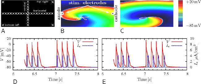
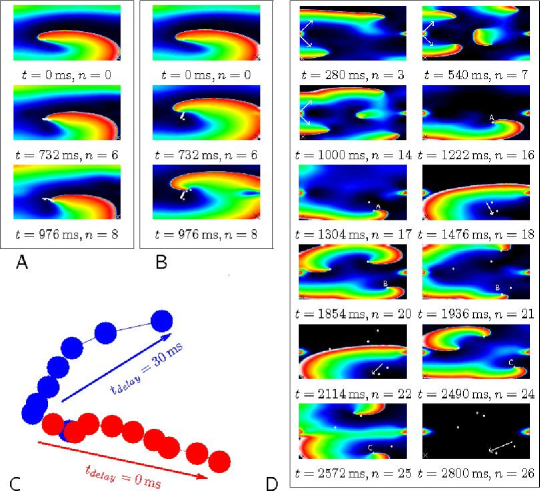
We used CARP’s transmembrane voltage output to visualize the results of simulations, using the colour-coding shown in Fig. 1C. The same information was used to detect the wave tips. The wavefront at time was defined as the line . Then the wave tips for that time were defined as the intersections of the front at time with the front at time . The time delay of was chosen purely empirically.
To visualize drift, we used a “stroboscopic” method: we showed positions of the tip synchronized with the signals detected by the registration electrodes.
For meandering spirals this required further refinement, since the stroboscopic selection of tips produced a congested picture, even without any stimulation: so, a five-petal meandering pattern (as shown in Fig. 1B) produced five clusters of tips. With stimulation, the tip picture becomes even more complicated and unreadable. So we showed only every -th stroboscopic tip position. Since the meandering patterns were affected by stimulation, we found that the optimal value for was different from 5; we used .
A possible alternative to the stroboscopic method is sliding averaging of the trajectories. However, it requires careful choice of the averaging window, which may be different in different situations, so we found it less convenient.
Generation of re-entry patterns
We used two alterations of the Courtemanche human atrial model (24) of the ionic currents, :
-
(i)
To prevent the transmembrane voltages from rising to non-physiological values during the defibrillation shocks, an electroporation current was included, see (21) and references therein. In addition, a formulation for a acetylcholine-dependent potassium current, , was added (29, 21). This ionic model was used to generate a meandering re-entry, see Fig. 1B.
-
(ii)
In addition to electroporation and , we used a block of the slow inward L-type current coupled with a nine-fold increase in the slow delayed outward current and the rapid delayed outward current as suggested by Xie et al. (30). This ionic model was used to generate a stationary rotating re-entry, see Fig. 1C.
To initiate re-entry, we used an S1-S2 protocol.
Single shock defibrillation benchmark
Monophasic current shocks were injected into, and withdrawn from, the extracellular space via volumes , “stimulating electrodes”, centered along the left and right edges of the slab (Fig. 1B). The shocks were of rectangular waveform, duration and varied amplitude .
Shocks were applied at twelve different timings , separated by intervals, after the same initial conditions. This covered an entire single rotor cycle, . A single shock was deemed successful if no re-entry was detectable at the moment after its end. We define the single shock success threshold as the shock amplitude which gives a % success rate across the twelve timings.
Resonant drift pacing
Repetitive low-amplitude shocks of the same waveform and via the same stimulating electrodes as above, were applied at the time moments determined by signals received via registration electrodes.
Six different locations for the registration electrodes were used: “point” electrodes in the top left, top right, bottom left and bottom right corners and “line” electrodes (18) (of cross-section ) through the whole medium either horizontally, along the fibres or vertically, across the fibres, see Fig. 1A.
The signal from a registration electrode was defined as the average potential at all the nodes covered by it. The signal triggered a shock application when it exceeded . A shock was applied with a delay after it had been triggered. In most cases we set , see Fig. 1(D). To demonstrate its effect on the direction of the drift, we set , about a quarter of the re-entry period, see Fig. 1(E).
In all simulations, the registration electrodes were de-activated for a “blanking time” after a shock application.
For the meandering re-entry, resonant drift pacing was considered successful if the re-entry was terminated sooner than the self-termination time of the re-entry, . For the stationary rotating re-entry, this time was extended to .
Results
Single shock defibrillation results
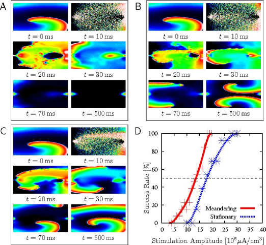
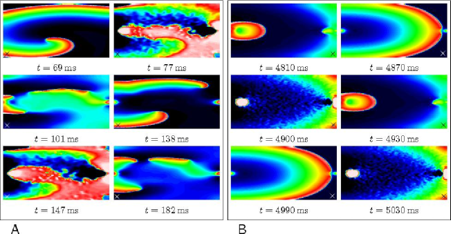
We have varied the timings, and strength , of the single shocks to assess the variability of the outcomes. Consistent with previous observations (21), there were three typical single-shock defibrillation outcomes for both the meandering and stationary-rotating patterns:
We have found the single shock defibrillation threshold to be for the meandering patterns and for the sationary-rotating patterns (Fig. 2D).
Resonant drift pacing results
We tested resonant drift pacing on the meandering, and stationary rotating, re-entrant patterns with shock amplitudes lower than the corresponding single shock defibrillation success thresholds.
Meandering re-entry
Point registration electrodes.
Depending on the shock strength, the following outcomes were observed.
-
•
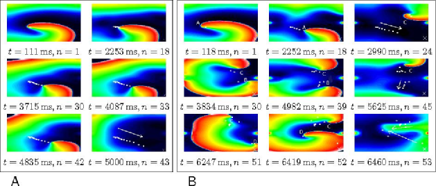
The original re-entry drifted, roughly towards the registration electrode, until termination on an inexcitable boundary, see Fig. 3A.
-
•
Secondary re-entrant patterns were generated near stimulating electrodes; both the original and newly generated re-entries drifted. An example is shown in Fig. 3B. The original re-entry “A” was terminated by the first 24 shocks. However, a secondary re-entrant pattern “C” was generated by a shock in the process. After “A” terminated, it was “C” that triggered the subsequent shocks. A third re-entrant pattern, “B” was generated by a further shock. Due to the proximity of “B” to the registration electrode, it took over the control of the shock applications. Re-entry “B” drifted to the boundary and terminated after a further 15 shocks. Then “C” was again the solitary re-entry and took over the control. A fourth re-entry, “D” was generated by a shock and, due to its proximity to the registration electrode, took over the control. After 3 shocks, “D” collided with “C” and they annihilated each other without reaching an inexcitable boundary.
Note that mutual annihilation of re-entrant patterns in the bulk of the tissue could be a plausible explanation why Pak et al. (31) saw no evidence of annihilation at the ventricular boundaries when testing a protocol similar to the resonant drift pacing.
Line registration electrodes
-
•
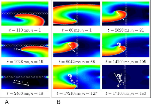
Horizontal electrode: With the registration electrode along the fibres, the re-entry drifted at a small angle to it (Fig. 4A).
-
•
Vertical electrode: With the registration electrode across the fibres, the drift was usually more complicated. An example is shown in Fig. 4B. The re-entry initially drifts upwards along the electrode. After 21 shocks, the drift turns downwards, until it reaches an inexcitable boundary after a further 109 shocks and terminates. The trajectory of the drift crosses itself, which is not allowed in the asymptotic theory of drift of rigidly rotating spiral waves (17) and was not observed in our simulations of a stationary rotating re-entry, so we consider it a new feature due to meander.
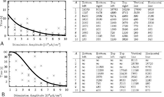
The time taken for termination of all re-entrant activity using different locations of the registration electrode is shown in Fig. 5A. It shows that resonant drift pacing can successfully terminate meandering re-entrant patterns using shocks 14 times weaker than the single shock defibrillation threshold. Termination was achieved in all simulations in this series.
Among the registration electrodes, the vertical line was the most successful, as it produced the largest proportion of successful terminations in the fastest time except at the smallest amplitudes. Using a point-electrode in the top right location was the least successful.
We conclude that the termination time depends on the mutual position of the registration electrode, anode, cathode, initial position of the re-entry, tissue size and fibre orientation.
For amplitude , re-entry termination time for the meandering pattern, in most cases, was longer than its self-termination time . Thus, we did not consider lower values for the stimulation amplitudes.
Stationary rotating re-entry
Fig. 5B shows the times taken for annihilation of all re-entrant waves for the different locations of the registration electrode. Resonant drift pacing can terminate stationary-rotating patterns using amplitudes 18 times lower than the single shock threshold. The combination of shock strength with the location of the registration electrode affects the probability of success and the time taken for termination.
Termination of stationary rotating re-entry was not always observed within the limit. Sometimes the algorithm gets caught in an infinite loop (Fig. 6), when the original re-entrant pattern has been terminated, but shocks produce new wavefronts which trigger further stimulation producing further wavefronts and so on.
Direction of drift
The theory of the resonant drift (15) predicts that the drift direction depends on the stimulation phase. This dependence leads to the relationship between the drift direction and the delay in the case of a feedback controlling forcing (17, 32). A delay by a certain fraction of a spiral’s period causes a change in the drift direction by the same fraction of . We have verified that it works in this realistically anisotropic bidomain model as well.
Using a time delay to induce success
We have mentioned in section Stationary rotating re-entry that resonant drift pacing may fail through an infinite loop of wavefront eliminations and creations. If newly created wavefronts are unbroken as in Fig. 6B then the re-entrant activity will vanish if the stimulation is stopped at any time, i.e. the success is achieved.
A genuine failure happens when newly created waves are broken and form new re-entries, as in Fig. 6A. The chances of getting in such a loop depend on the drift trajectory, which depends on the location of the registration and stimulation electrodes and the feedback delay. Hence such loops should be avoidable by an appropriate choice of the electrode locations and/or of . In a real-life situation, such choice would require plenty of information and may be difficult to make. Not all theoretical solutions (e.g. optimal positions of the electrodes) may be possible to implement in practice. Further, the optimal parameters may vary from one arrhythmia episode to another so difficult to predict a priori.
Of all the mentioned parameters, is the easiest to change, and it may even be adjusted in real time, during pacing. Hence we propose that a change of the feedback delay may be used to discontinue an infinite loop even after it has already started.
Discussion
Summary of results: Resonant drift pacing works in a realistic model of electric field action on cardiac tissue
This simulation study presents results which can be used as experimentally testable hypotheses. It has been based on the understanding of the mechanism by which external electric current affects excitation and propagation via non-uniformity of the electric field and the heterogeneity of the tissue conductivities. This mechanism implies both a strong action near electrodes due to the electric field inhomogeneity, and a weaker, but far reaching action in the bulk of the tissue due to omnipresent tissue heterogeneities. This model has been used before to simulate single-shock defibrillation (21). Here we have demonstrated that the far-field action of the electric current can cause, under repetitive stimulation, a drift and elimination of the re-entrant sources. In our simulations, resonant drift pacing can eliminate re-entry, including multiple re-entries, with high probability and within acceptable time, at amplitudes much lower than single shock defibrillation. If we allow 10 s for low-voltage re-entry termination, as is the case for antitachycardia pacing, the required shock strength with resonant drift pacing is 12 to 15 times smaller than with a single shock.
Our simulations have shown that a major possible obstacle to elimination of re-entrant sources is the possibility that the electric shocks create new re-entrant waves while eliminating existing ones, which may lead to infinite loops of annihilation and creation. The creation of the new sources occurs near the shock electrodes where the electric field is highly inhomogeneous, and the 12 to 15-fold decrease in defibrillation threshold is observed notwithstanding this effect. The highly inhomogeneous electric field is created by point electrodes, so bigger electrodes, which create more homogeneous fields, should perform even better.
Our simulations have also demonstrated yet another way to overcome infinite annihilation-creation loops. This is due to the dependence of the direction of resonant drift on the time delay in the feedback loop. This dependence has been predicted and observed in isotropic monodomain models with simplified description of the electric field action (15, 17, 32, 25, 19). Here we demonstrated that this dependence is still observed in the present more realistic model and, moreover, it can be used to discontinue an infinite annihilation-creation loop, via a change in the feedback delay once such a loop has been detected.
Comparison with previous research
Anti-tachycardia pacing
In clinical practice, antitachycardia pacing is as efficient and as safe as single shock defibrillation, even for fast ventricular tachycardias with up to 250 beats/min (14). The exact mechanisms underlying success or failure of antitachycardia pacing are widely unexplored. The traditional understanding implies stimulation which is faster than the anatomical re-entry and which engages all the larger area of tissue until it reaches an “isthmus” of the re-entry and blocks it. It is conceivable, however, that in some cases antitachycardia pacing may be unwittingly applied to a functional rather than anatomical re-entry and then its mechanism could be the resonant drift. In such cases, feedback-controlled resonant drift pacing will do a better job than pacing with prescribed frequency used in antitachycardia pacing. Although the probability of antitachycardia pacing success is high, when it fails a single strong shock has to be applied.
Unpinning
It has been speculated that anatomically or functionally anchored ventricular tachycardias are less likely to be terminated by antitachycardia pacing. For these cases, it has been suggested that a weak shock, with an amplitude in the range used in this study, can be applied to unpin the re-entry from the obstacle by a virtual electrode polarization mechanism (33, 34). Such a stimulus must be correctly timed, which can be achieved by synchronizing it to the signal from a registration electrode, with a correctly chosen delay with respect to that signal, which in practice may require scanning through possible delays. The resonant drift pacing also requires synchronization with a registered signal, and we have seen that change in the delay during the course of pacing may also be beneficial. This makes the unpinning protocol operationally very similar to our resonant drift pacing protocol, and in any particular case it may not be possible to say with certainty which mechanism has worked.
Experiments with feedback-controlled pacing
Pak et al. (31) eliminated ventricular fibrillation in rabbit by multisite pacing synchronized with optical signal from a fixed reference site. Their stimulation protocol was similar to resonant drift pacing considered here, except they used more pacing electrodes and aimed to deliver shocks only when pacing site was in an excitable gap, under the implicit assumption that shock delivered to a site in an absolute refractory state could not possibly affect the re-entry, i.e. a priori ignoring far-field effects. Despite this artificial self-limitation, they were successful at defibrillating with shock strengths an order of magnitude lower than single shock defibrillation and with better success rate than overdrive and high-frequency pacing. It was argued in (31) that earlier mathematical modeling of resonant drift only demonstrated termination of a single re-entrant wave (see, however, (32)) and their experiments did not show evidence of extinction of re-entries on ventricular borders. Our present simulations indicate that
-
•
far-field effects can make it worthwhile issuing shocks regardless of the tissue state at the pacing site,
-
•
the resonant drift pacing can work for multiple re-entrant waves (providing that the difficulties of new wavefronts being initiated by the shocks are overcome), and
-
•
annihilation at the boundaries is not a necessary feature of resonant drift elimination, as re-entry sources can annihilate with each other.
Thus, we find the results by Pak et al., contrary to their own interpretation, as strong experimental evidence of the resonant drift pacing being an efficient method of low voltage defibrillation.
Clinical implications
Resonant drift pacing may present an alternative or a supplement for existing (single shock defibrillation, synchronized cardioversion, antitachycardia pacing, overdrive pacing, and high-frequency pacing) and proposed (unpinning) therapies. The theoretical mechanism underlying this protocol is for functionally determined re-entries, i.e. probably for higher frequency tachycardias and fibrillations. Due to similarity between resonant drift and unpinning pacing protocols, the two protocols may be possible to combine into one, which would work via unpinning for pinned (monomorphic tachycardias), and via resonant drift for unpinned re-entrant waves (polymorphic tachycardias and fibrillations).
There is evidence that some atrial arrhythmias are not re-entrant, but are due to rapid ectopic focal activity (35). The proposed method might work on such arrhythmias via a completely different mechanism, say overdrive suppression, but this can only be clear after further studies.
As the study presented here is purely theoretical, the key question is whether the low-energy approach to defibrillation considered here could work in real heart. Clearly this question can only be answered by experimental studies, and there is clear scope to advance the field by interaction between modeling studies such as this one and experimental studies.
Study limitations
Apart from inevitable limitations inherent in mathematical modeling compared to experimental study, which are due to limited current knowledge, we have made a number of simplifications. Our model tissue was essentially two- rather than three-dimensional. Its geometry was not realistic (rectangular shape). The model tissue lacked any macroscopic inhomogeneities, such as trasmural, center-periphery or base-apex gradient of excitability properties, or variations of the conductivity tensor. The position and geometry of the electrodes was arbitrary. Values of some of the parameters lack reliable experimental foundation, including some of the most important ones, such as , the intracellular conductivity fluctuations amplitude. The spatially uncorrelated structure of conductivity fluctuations is an idealization. These limitations can be lifted as more experimental data become available and via further, more detailed, simulation studies.
Resonant drift pacing may potentially work for ventricular tachycardia and fibrillation too. However, the ventricles have thick walls and overall more complicated anatomy, which may pose extra difficulties for defibrillation. So any extrapolation of our present results to ventricles should be thought of with caution, bearing in mind possible complications. There are modeling studies suggesting that at least some of the new difficulties arising in resonant pacing of three-dimensional re-entries can be successfully overcome (see (36) and references therein), however those were in overly simplified models of exciable media, and more realistic computational studies are still desirable.
Conclusion
Our results show that in a bidomain model of cardiac tissue, with microscopic conductivity fluctuations, re-entrant waves can be annihilated using feedback-controlled repetitive stimulations by inducing resonant drift and directing the re-entry towards an inexcitable boundary. If associated difficulties are overcome, termination using this approach is achieved, with high probability and within acceptable time, at a fraction of the conventional shock strength. The direction of the drift can be managed by choosing the location of the electrodes and the time delay of the shock application.
Difficulties occur due to new wavefronts being initiated from the shock electrodes. However, numerical simulations allow a detailed insight into this problem and can be used as a tool to suggest solutions.
Our findings present experimentally testable hypotheses for what we expect to observe in real cardiac tissue. There is scope to advance the field of low-energy defibrillation by interaction between modeling studies such as this and experimental studies.
Acknowledgements
This study was supported in part by EPSRC grant EP/D500338/1, Royal Society grant 2005/R4, Liverpool University Research Development Fund grant 4431 and Austrian Science Fund FWF grant F3210-N18.
References
- Lee et al. (2003) Lee, D. S., L. D. Green, P. P. Liu, et al., 2003. Effectiveness of implantable defibrillators for preventing arrhythmic events and death: a meta-analysis. J. Am. Coll. Cardiol. 41:1573–1582.
- Malkin (2002) Malkin, R. A., 2002. Large sample test of defibrillation waveform sensitivity. J. Cardiovasc. Electrophysiol. 13:361–370.
- DeBruin and Krassowska (1999) DeBruin, K. A., and W. Krassowska, 1999. Modeling electroporation in a single cell. I. Effects of field strength and rest potential. Biophys J 77:1213–1224.
- Sambelashvili et al. (2004) Sambelashvili, A. T., V. P. Nikolski, and I. R. Efimov, 2004. Virtual electrode theory explains pacing threshold increase caused by cardiac tissue damage. Am. J. Physiol. Hear. Circ. Physiol. 286:H2183–H2194.
- Sharma et al. (2005) Sharma, V., R. C. Susil, and L. Tung, 2005. Paradoxical loss of excitation with high intensity pulses during electric field stimulation of single cardiac cells. Biophys J 88(4):3038–3049.
- Zivin et al. (1999) Zivin, A., J. Souza, F. Pelosi, et al., 1999. Relationship between shock energy and postdefibrillation ventricular arrhythmias in patients with implantable defibrillators. J Cardiovasc Electrophysiol 10:370–377.
- Kamm et al. (1997) Kamm, R. M., H. Garan, B. A. McGovern, et al., 1997. Transient right bundle branch block causing R wave attenuation postdefibrillation. Pacing Clin. Electrophysiol. 20:130–131.
- Tokano et al. (1998) Tokano, T., D. Bach, J. Chang, et al., 1998. Effect of ventricular shock strength on cardiac hemodynamics. J Cardiovasc Electrophysiol 9:791–797.
- Boriani et al. (2005) Boriani, G., M. Biffi, P. Silvestri, et al., 2005. Mechanisms of pain associated with internal defibrillation shocks: results of a randomized study of shock waveform. Heart Rhythm 2:708–713.
- Jung et al. (1997) Jung, J., A. Heisel, R. Fries, et al., 1997. Tolerability of Internal Low-Energy Shock Strengths Currently Needed for Endocardial Atrial Cardioversion — effect on atrial defibrillation threshold and pain perception. The American Journal of Cardiology 80:1489–1490.
- Walcott et al. (1995) Walcott, G. P., R. G. Walker, A. W. Cates, et al., 1995. Choosing the optimal monophasic and biphasic waveforms for ventricular defibrillation. J. Cardiovasc. Electrophysiol. 6:737–750.
- Zhang et al. (2006) Zhang, Y., B. Rhee, L. R. Davies, et al., 2006. Quadriphasic waveforms are superior to triphasic waveforms for transthoracic defibrillation in a cardiac arrest swine model with high impedance. Resuscitation 68:251–258.
- Sullivan et al. (2007) Sullivan, J. L., S. B. Melnick, F. W. Chapman, et al., 2007. Porcine defibrillation thresholds with chopped biphasic truncated exponential waveforms. Resuscitation 74:325–331.
- Wathen et al. (2004) Wathen, M. S., P. J. DeGroot, M. O. Sweeney, et al., 2004. Prospective randomized multicenter trial of empirical antitachycardia pacing versus shocks for spontaneous rapid ventricular tachycardia in patients with implantable cardioverter-defibrillators: Pacing Fast Ventricular Tachycardia Reduces Shock Therapies (PainFREE Rx II) trial results. Circulation 110:2591–2596.
- Davydov et al. (1988) Davydov, V. A., V. S. Zykov, A. S. Mikhailov, et al., 1988. Drift and resonance of spiral waves in active media. Radiofizika 31:574–582.
- Agladze et al. (1987) Agladze, K. I., V. A. Davydov, and A. S. Mikhailov, 1987. An observation of resonance of spiral waves in distributed excitable medium. JETP Lett. 45:767–770.
- Biktashev and Holden (1994) Biktashev, V. N., and A. V. Holden, 1994. Design principles of a low voltage cardiac defibrillator based on the effect of feedback resonant drift. J.theor.Biol 169:101–112.
- Zykov and Engel (2007) Zykov, V. S., and H. Engel, 2007. Analysis and control of complex nonlinear processes in physics, chemistry and biology, volume 5 of World Scientific Lecture Notes in Complex Systems. World Scientific.
- Biktashev and Holden (1996) Biktashev, V. N., and A. V. Holden, 1996. Re-entrant activity and its control in a model of mammalian ventricular tissue. Proc. Roy. Soc. Lond. ser. B 263:1373–1382.
- Panfilov et al. (2000) Panfilov, A. V., S. C. Muller, V. S. Zykov, et al., 2000. Elimination of spiral waves in cardiac tissue by multiple electrical shocks. Phys. Rev. E 61:4644–4647.
- Plank et al. (2005) Plank, G., L. J. Leon, S. Kimber, et al., 2005. Defibrillation depends on conductivity fluctuations and the degree of disorganization in reentry patterns. J. Cardiovasc. Electrophysiology. 16:205–216.
- Trayanova (2006) Trayanova, N., 2006. Defibrillation of the heart: insights into mechanisms from modeling studies. Exp.Physiol 91:323–337.
- Potse et al. (2006) Potse, M., B. Dube, J. Richer, et al., 2006. A comparison of monodomain and bidomain reaction-diffusion models for action potential propagation in the human heart. IEEE Trans. Biomed. Eng. 53:2425–2435.
- Courtemanche et al. (1998) Courtemanche, M., R. J. Ramirez, and S. Nattel, 1998. Ionic mechanisms underlying human atrial action potential properties: insights from a mathematical model. Am. J. Physiol. 275:H301–H321.
- Biktashev and Holden (1995a) Biktashev, V. N., and A. V. Holden, 1995. Control of re-entrant activity in a model of mammalian atrial tissue. Proc. Roy. Soc. Lond. ser. B 260:211–217.
- (26) Vigmond, E. J., and G. Plank. Cardiac Arrhythmia Research Package (CARP). http://carp.meduni-graz.at.
- Vigmond et al. (2003) Vigmond, E. J., M. Hughes, G. Plank, et al., 2003. Computational tools for modeling electrical activity in cardiac tissue. J. Elec. Supp. 36:69–74.
- Plank et al. (2007) Plank, G., M. Liebmann, R. Weber dos Santos, et al., 2007. Algebraic multigrid preconditioner for the cardiac bidomain model. IEEE Trans. Biomed. Eng. 54:585–596.
- Kneller et al. (2002) Kneller, J., R. Zou, E. Vigmond, et al., 2002. Cholinergic atrial fibrillation in a computer model of a two-dimensional sheet of canine atrial cells with realistic ionic properties. Circ. Res. 90:E73–E87.
- Xie et al. (2002) Xie, F., Z. Qu, A. Garfinkel, et al., 2002. Electrical refractory period restitution and spiral wave reentry in simulated cardiac tissue. Am. J. Physiol. Circ. Physiol. 283:448–460.
- Pak et al. (2003) Pak, H., Y. Liu, H. Hayashi, et al., 2003. Synchronization of ventricular fibrillation with real-time feedback pacing: implication to low-energy defibrillation. Am. J. Physiol. Heart Circ. Physiol. 285:H2704–H2711.
- Biktashev and Holden (1995b) Biktashev, V. N., and A. V. Holden, 1995. Resonant drift of autowave vortices in two dimensions and the effects of boundaries and inhomogeneities. Chaos Solitons Fractals 5:575–622.
- Ripplinger et al. (2006) Ripplinger, C. M., V. I. Krinsky, V. P. Nikolski, et al., 2006. Mechanisms of unpinning and termination of ventricular tachycardia. Am J Physiol Heart Circ Physiol 291:H184–H192.
- Cysyk and Tung (2008) Cysyk, J., and L. Tung, 2008. Electric field perturbations of spiral waves attached to millimeter-size obstacles. Biophys J 94(4):1533–1541.
- Nattel (2002) Nattel, S., 2002. New ideas about atrial fibrillation 50 years on. Nature 415:219–226.
- Morgan et al. (2008) Morgan, S. W., I. V. Biktasheva, and V. N. Biktashev, 2008. Control of scroll-wave turbulence using resonant perturbations. Phys. Rev. E 78:046207.