Origin of Plateau and Species dependence of Laser-Induced High-Energy Photoelectron Spectra
Abstract
We analyzed the energy and momentum distributions of laser-induced high-energy photoelectrons of alkali and rare gas atoms. For the plateau electrons with energies above , ( is the ponderomotive energy), in the tunneling ionization regime, we showed that they originate from the backscattering of laser-induced returning electrons. Using the differential elastic scattering cross sections between the target ion with free electrons, we explain experimental observations of whether the plateau electron spectra is flat or steeply descending, and their dependence on species and laser intensity. This quantitative rescattering theory can be used to obtain energy and momentum distributions of plateau electrons without the need of solving the time-dependent Schrödinger equation, but with similar accuracy.
pacs:
32.80.Rm, 32.80.Fb, 34.50.RkWhen atoms or molecules are placed in an intense laser pulse, an electron can be released through either a multiphoton or a tunneling mechanism. The distinction is based on the Keldysh parameter , where is the ionization energy and is the ponderomotive energy with is the peak value of the vector potential. In the multiphoton regime (), the electron spectra exhibit characteristic above-threshold ionization (ATI) peaks separated by photon energy, with yields decreasing monotonically with increasing electron energy Agostini . At higher intensities, in the tunneling region (), the spectra are notably different. First, the ionization yield drops steeply from the threshold, but from about 3 or onward, the yield flattens out significantly until at about where it drops precipitously again. The flattened spectral region from 4- is called the plateau. Similar plateau and cutoff are also well-known in the high-order harmonic generation (HHG). Despite this canonical description, the HHG and electron spectra in the plateau region are not always flat. For electron spectra, earlier experiments show pronounced enhancement in the plateau region in potassium, but not in sodium Gaarde . Similarly, using nearly identical lasers, clear plateau shows up in Xe target, but not in Kr and Ar Paulus_PRL94 . Experimentally the energy spectra of plateau electrons have been observed to depend on laser intensities Grasbon . The origin of plateau electrons and their dependence on target species and laser intensity is explained in this Letter.
Considerable understanding of laser-induced ATI spectra from atoms has been achieved since 1990’s yang93 ; Paulus_JPB94 ; Walker . While the observed spectra in general can be reproduced from solving the time-dependent Schrödinger equation (TDSE) within the single active electron approximation, interpretation of the plateau electrons is based on the rescattering model. In this model electrons that are released earlier by tunneling ionization can be driven back by the laser field to recollide with the target ion. The plateau electrons are understood to originate from the elastic back scattering of the returning electrons by the target ion. However, existing rescattering model does not predict how they depend on target species and laser intensity. In this Letter we provide such a quantitative rescattering theory (QRS) where the energy and momentum spectra of the plateau electrons can be directly calculated using the elastic scattering cross sections between the target ion and free electrons, in combination with laser-induced returning electron wave packet. We also show that the returning electron wave packets depend largely on the laser parameters only. Thus the behavior of the plateau electron spectra is determined entirely by the energy and species dependence of the elastic scattering cross sections between the returning electrons and the target ion.
To understand electron energy spectra, we first examine how electron-ion collisions contribute to the photoelectron momentum distributions. In Fig. 1 we show a typical two-dimensional (2D) electron momentum spectra for an atomic target calculated by solving the TDSE chen06 ; toru07 . (We use linearly polarized electric field for the laser pulse along the axis with the carrier-envelope phase set to zero chen06 ). The horizontal axis is the direction of laser’s polarization and the vertical axis is any direction perpendicular to it (the electron spectra has cylindrical symmetry). Due to the short laser pulse, the right-hand side () and the left-hand side () spectra are not symmetric. In Fig. 1, four semi-circles with photoelectron energies of 2, 4, 6 and , respectively, are shown. On the right-hand side, three other semi-circles are given, each with its center shifted along the axis. Each of this circle can be expressed vectorially by . Measured from its own center, the circle maps out the momentum space of the elastically scattered electron with incident momentum . The center is shifted since collision occurs in the presence of the laser field. If the electron-ion collision occurs at time when the vector potential is , the electron will reach the detector gaining an additional momentum . (Atomic units are used in the above expressions such that the momentum gain in the laser field is directly given by the vector potential .) In the figure, we have chosen the “incident” electrons to be from the right, i.e., with negative . The direction of and the elastic scattering angles are measured from this “incident” direction. This choice would result in high-energy or plateau electrons to emerge with positive after the electron undergoes large-angle backscatterings (). Note that electrons scattered into the forward directions will emerge with low energies. Clearly, similar circles can be drawn for electrons “incident” from the left.
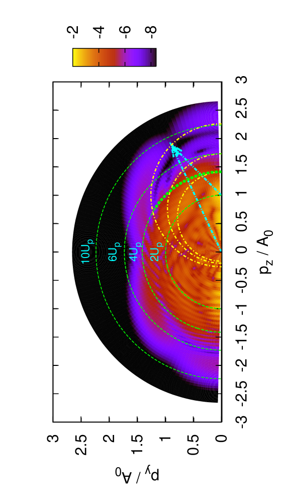
According to the classical model, electrons that return to the ion core at the time when the vector potential is at the peak, , it will have maximum kinetic energy of , i.e., with . If the electron is backscattered by , the emerging photoelectron would have a momentum of , or energy of . In a recent paper toru08 , we examined the photoelectron distributions after the electrons have been elastically scattered into the backward directions. The electron yield along this ring (called BRR, or back rescattering ridge) has been shown to be given by
| (1) |
This equation is based on the concept of rescattering theory. It states that photoelectron yields along the BRR are proportional to the elastic differential cross section (DCS), , multiplied by a returning electron wave packet, represented by , with the target ion. The validity of Eq. (1) was established in toru08 based on accurate TDSE results. Its validity has also been confirmed experimentally in two recent reports Japan_08 ; USA_08 .
To examine the plateau electrons, elastic scattering by electrons with returning energy less than must be included. These are electrons that return to the parent ion when is not at the peak. We set the returning electron’s momentum and using the rescattering model, Eq. (1). The validity of this model can be checked by comparing the extracted from the calculated photoelectron momentum spectra , with those directly calculated from electron-ion collisions, as carried out in Morishita et al. toru08 . Alternatively, we can first use Eq. (1) at an arbitrary angle, say, for photoelectron with momentum vector 10∘ from the polarization axis, to derive the wave packet . We then obtain the rescattering “generated” photoelectron spectra by using Eq. (1). This way we have a quantitative rescattering theory which can be used to obtain the momentum and energy spectra for plateau electrons. We remark later that can also be obtained by other approximations.

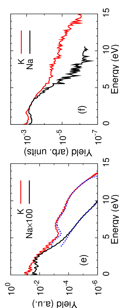
In Figs. 2(a,b) electron momentum spectra () for sodium and potassium atoms exposed to a 5-cycle laser pulse with wavelength of 3200 nm and peak intensity of 1 TW/cm2 are presented. On the left-hand half of the 2D plot, the results calculated from TDSE are shown, and on the right-hand half the same distributions obtained from the present QRS model are presented. If the two calculations agree well, then there should exist a good reflection symmetry in the figure. This is clearly the case for each target, confirming the validity of the QRS model at the level of electron momentum distributions for the plateau electrons. In Figs. 2(c,d) the DCS for e-Na+ and e-K+ collisions are shown for the range of momenta that contribute to the 4- photoelectrons. As the collision energy increases, the DCS decreases. However, for the same , the DCS for e-K+ is about 100 times larger. Furthermore, the angular dependence of the DCS in Fig. 2(c,d) are clearly reflected in the momentum distributions in Figs. 2(a,b). In other words, the elastic scattering cross sections between free electrons and target ion can be “read” out directly from the ATI electron spectra in the momentum space. This is possible because of the other important feature of QRS: the returning electron wave packet depends mostly on the lasers only. This fact has been noted in the high energy region before chen07 .
From the momentum spectra the ATI energy spectra can be calculated. Since the right-hand side and left-hand side 2D momentum spectra are not symmetric for short pulses, we calculate the energy spectra from each side separately and the sum of them reproduces the whole energy spectra in which the left-hand side makes more contribution to the lower energy part. In Fig. 2(e), the results from TDSE and from QRS are compared. We limit the QRS model to electron energy above only. Clearly the QRS model reproduces the TDSE results well. These calculations can also be compared to the experimental data reported in Gaarde et al. Gaarde , as shown in Fig. 2(f). There is a close similarity in the relative yields between our calculations and experiment, even though our calculations used a 5-cycle (20 fs FWHM) pulse while the experiment used somewhat different intensities for a pulse with duration of 1.9 ps. The QRS results clearly establish that the experimental plateau electron spectra for Na and K are due to their elastic scattering cross sections in the momentum range of 0.3 to 0.5. Although it has long been understood (or speculated) that the behavior of plateau electrons are related to elastic scattering cross sections Walker ; Gaarde , no direct quantitative connection between the two has ever been established until now.
As a side note, we comment that the lower end of the plateau electrons has been set at in this Letter. According to the classical theory, direct ionization by tunneling (without rescattering) will give maximum electron energy of . Between 2 and , the direct ionization part could still interfere with the rescattering part chen07 , thus we chose as the lower end of the QRS model.
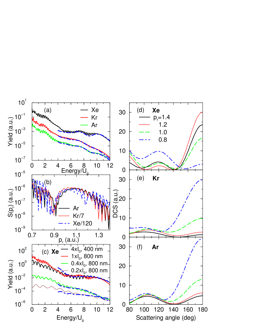
We have also studied the plateau electrons for Ar, Kr and Xe atoms. Experimental data for these systems have been reported using lasers of different wavelengths and pulse durations since the 1990’s Paulus_PRL94 ; Grasbon . In fact, the plateau in the high-energy ATI spectra was first observed in Xe. In Fig. 3(a) we show the calculated ATI spectra vs electron energy in units of at the same laser intensity of W/cm2 for a five-cycle, 800 nm pulse. In the case of Xe, the plateau is clearly seen – it remains almost constant from to , while for Kr and Ar, each drops about two to three orders of magnitude in the same energy range. Since the momentum distributions of the returning electron wave packets are essentially identical (up to a normalization due to the different tunneling ionization rates) for the same laser pulse, as shown in Fig. 3(b), the differences in the electron energy spectra are attributed to differences in the DCS at large angles among the three targets. In Figs. 3(d-f) their DCS are shown for the relevant range of . Note that they cover the same range of magnitude, but at large angles, say 160∘-180∘, the DCS for Xe behaves “anomalously” for it increases with increasing energies instead of otherwise, as in Ar and Kr. Since higher returning energies contribute more to the plateau electron yields (see Fig. 1), this “anomalous” energy dependence explains the flat plateau in Xe. Likewise, the “normal” energy dependence of DCS in Ar and Kr can be used to explain the steep drop of their electron spectra at high energies, see Fig. 3(a). We comment that the QRS results shown in Fig. 3(a) are again in good agreement with those obtained from TDSE.
According to the QRS model, for different lasers with identical , their returning electron wave packets will have the same range of kinetic energy, thus similar plateau electron momentum and energy distributions. In Fig. 3(c), we show the electron spectra for Xe using 800 nm and 400 nm lasers, respectively, but with the latter having four times the peak intensity. The spectra in the plateau region indeed agree quite well quantitatively after they are normalized to each other at around . Both results are obtained from solving the TDSE.
We also comment that the QRS model is based on the rescattering theory, thus it would fail at lower intensities in the multiphoton ionization regime. In Fig. 3(c) we show the comparison of results from TDSE and from QRS at two lower laser intensities, with Keldysh parameters of and 2.24, respectively. We see clear evidence of the failure of the QRS model at . This sets a rough upper limit for the validity of QRS at near about 2.0.
The above examples illustrate that the behavior of laser-induced plateau electrons with energies from to are determined by electron-ion elastic scattering cross sections for electrons with energies between and (or momentum between 0.79-). Flat plateau is expected when the DCS at large angles (close to 180∘) increases with increasing kinetic energy and when they are highly peaked at large angles, as in the case of Xe [Fig. 3(d)]. Such conditions occur often in the DCS in low energy electron-ion collisions. Where does it occur depends on electron energies and target species. Based on the DCS shown in Fig. 3, the plateau will become more pronounced for Ar and Kr, but less so for Xe, as the laser intensity is decreased. This is consistent with earlier experimental results Grasbon .
In summary we studied laser-induced high-energy plateau electrons. Together with our previous results where the species dependence of HHG was traced to their photo-recombination cross sections toru08 ; AT_JPB ; AT_prl , we now have established a quantitative rescattering (QRS) theory for laser-induced ATI electron and HHG spectra in the plateau region. Based on the QRS model which is valid in the plateau region, there is no need to solve TDSE directly. For HHG one only needs to calculate photo-recombination cross sections and for plateau electrons one needs only to calculate elastic electron-ion scattering cross sections. The returning electron wave packets, as shown previously chen07 ; AT_prl , can be extracted from a companion target or from the second-order strong-field approximation. Conversely, the QRS theory allows one to extract electron and photon scattering information from laser-induced electron spectra or HHG, respectively. As proposed elsewhere toru_NJP , these cross sections can be further used to deduce the structure of the target, thus opening up the opportunity of using infrared laser pulses for determining the structural change of a dynamic system with temporal resolution of sub-femtoseconds to a few femtoseconds.
I Acknowledgment
This work was supported in part by Chemical Sciences, Geosciences and Biosciences Division, Office of Basic Energy Sciences, Office of Science, US Department of Energy. TM is also supported by a Grant-in-Aid for Scientific Research (C) from MEXT, Japan, and by a JSPS Bilateral joint program between US and Japan.
References
- (1) P. Agostini et al., Phys. Rev. Lett. 42, 1127 (1979).
- (2) M. B. Gaarde et al., Phys. Rev. Lett. 84, 2822 (2000).
- (3) G. G. Paulus et al., Phys. Rev. Lett. 72, 2851 (1994).
- (4) F. Grasbon et al., Phys. Rev. Lett. 91, 173003 (2003).
- (5) B. Yang et al., Phys. Rev. Lett. 71, 3770 (1993).
- (6) G. G. Paulus et al., J. Phys. B 27, L703 (1994).
- (7) B. Walker et al., Phys. Rev. Lett. 77, 5031 (1996).
- (8) Z. Chen et al., Phys. Rev. A 74, 053405 (2006).
- (9) T. Morishita et al., Phys. Rev. A 75, 023407 (2007).
- (10) T. Morishita et al., Phys. Rev. Lett. 100, 013903 (2008).
- (11) M. Okunishi et al., Phys. Rev. Lett. 100, 0143001 (2008).
- (12) D. Ray et al., Phys. Rev. Lett. 100, 0143002 (2008).
- (13) Z. Chen et al., Phys. Rev. A 76, 043402 (2007).
- (14) A. T. Le et al., J. Phys. B 41, 081002 (2008).
- (15) A. T. Le, T. Morishita, C. D. Lin, Phys. Rev. Lett. (submitted); arXiv:0712.3577v1.
- (16) T. Morishita et al., New J. Phys. 10, 025011 (2008).