Direct Observation of Dark Excitons in Individual Carbon Nanotubes:
Role of Local Environments
Abstract
We report the direct observation of the spin-singlet dark excitonic state in individual single-walled carbon nanotubes through low-temperature micro-photoluminescence spectroscopy in magnetic fields. A magnetic field up to 5 T, applied along the nanotube axis, brightened the dark state, leading to the emergence of a new emission peak. The peak rapidly grew in intensity with increasing field at the expense of the originally-dominant bright exciton peak and finally became dominant at fields 3 T. This behavior, universally observed for more than 50 nanotubes of different chiralities, can be quantitatively explained through a model incorporating the Aharonov-Bohm effect and intervalley Coulomb mixing, unambiguously proving the existence of dark excitons. The directly measured dark-bright splitting values were 1-4 meV for tube diameters 1.0-1.3 nm. Scatter in the splitting value emphasizes the role of the local environment surrounding a nanotube in determining the excitonic fine structure of single-walled carbon nanotubes.
pacs:
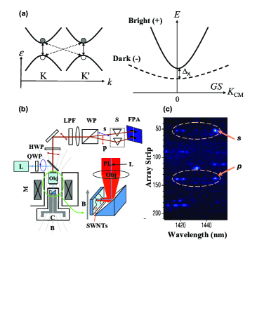
78.67.Ch,71.35.Ji,78.55.-mOptical properties of single-walled carbon nanotubes (SWNTs) are affected by their unique band structure together with strong Coulomb interactions characteristic of one-dimensional systems, as shown by recent theoretical Ando (1997); Spataru et al. (2004); Chang et al. (2004); Perebeinos et al. (2004) and experimental Wang et al. (2005); Maultzsch et al. (2005); Mortimer and Nicholas (2007); Shaver et al. (2007); Shaver and Kono (2007); Shaver et al. (2008) studies. In semiconducting SWNTs, there are two equivalent valleys (K and K′) in momentum space having opposite helicities about the tube axis. Short-range Coulomb interactions cause inter-valley mixing as well as singlet-triplet splitting, giving rise to exciton fine structure consisting of four singlet and twelve triplet, partially-degenerate states. Among the four singlet states, only one is predicted to be optically active (‘bright’) Zhao and Mazumdar (2004); Spataru et al. (2005); Perebeinos et al. (2005); Ando (2006), lying above the lowest energy optically-inactive (‘dark’) state, as shown in Fig. 1(a). However, there is no consensus as to the value of singlet dark-bright splitting, theoretical predictions ranging from a few to hundreds of meV Zhao and Mazumdar (2004); Spataru et al. (2005); Perebeinos et al. (2005); Ando (2006); Tretiak (2007); Jiang et al. (2007). A detailed study of the excitonic fine structure of SWNTs is thus essential for resolving these discrepancies and furthering our understanding of radiative and non-radiative energy relaxation processes in SWNTs.
A tube-threading magnetic flux lifts the valley degeneracy and produces two bright excitons due to the Aharonov-Bohm effect Ajiki and Ando (1993); Zaric et al. (2004, 2006). Previous temperature ()-dependent magneto-optical studies on SWNT ensembles performed at high magnetic fields Mortimer and Nicholas (2007); Shaver et al. (2007); Shaver and Kono (2007); Shaver et al. (2008) have revealed magnetic field ()-induced increase of the photoluminescence (PL) intensity. However, a direct observation of the dark state was obscured by the broad linewidths of ensemble samples, and the existence of dark excitons was only inferred from the - and -dependences of PL intensity. The most direct way to “observe” the dark excitons in SWNTs is to perform spectroscopy on single nanotubes at low such that the linewidths are smaller than the dark-bright splitting and then apply a magnetic field to brighten the dark excitons. Such methods have been used to prove the existence of dark excitons in semiconductor quantum dots BayeretAl00PRB . Spectroscopy on a single nanotube can also give us insight into the role of the local environment surrounding the nanotube, which can cause significant changes in emission energies Htoon et al. (2004) and radiative lifetimes Hagen et al. (2005).
Here, we present direct evidence for the existence of the lowest-lying dark exciton state using low- micro-PL spectroscopy of single SWNTs in up to 5 T. The dark exciton state acquired a finite oscillator strength in a parallel , appearing at a lower energy than the bright exciton peak. Due to the very narrow linewidths (1 meV) at low , we could directly measure the dark-bright splitting without the use of any spectral curve fitting. Brightening was found to be completely absent when the tube axis was perpendicular to , indicating the crucial role the Aharonov-Bohm phase plays. A simple model Shaver et al. (2007); Shaver and Kono (2007); Shaver et al. (2008) based on Ando’s theory of magnetic brightening Ando (2006) explains our results remarkably well. The measured values for a number of tubes show scatter, even for tubes with the same chirality. This clearly suggests the importance of the local environment, particularly local dielectric screening, in determining the excitonic fine structure of SWNTs. The values of are found to be 1-4 meV for most nanotubes studied.
We performed magneto-PL spectroscopy of individual micelle-encapsulated HiPco SWNTs deposited on quartz substrates. Figure 1(b) schematically depicts the setup. The sample was cooled in a liquid-helium-flow cryostat in the room temperature bore of a 5 T superconducting magnet. PL was excited using a tunable Ti:Sapphire laser, collected using a near-infrared objective lens (0.5NA), and detected using a liquid-nitrogen-cooled InGaAs 2D detector array. To determine the orientation angle of each SWNT with respect to , we relied on their linearly-polarized emission. A quarter-wave plate was used to circularly polarize the excitation beam to excite all tubes, which were randomly oriented. PL from SWNTs, linearly polarized along their axes, was sent through a half-wave plate set in such a way that it did not introduce any retardation for the light polarized parallel to . A Wollaston prism spatially separated the PL into its respective and components, which were focused onto different vertical locations along the spectrometer slit. This enabled us to simultaneously measure the and components [as shown in Fig. 1(c)] and determine the tube orientation . The substrate was mounted either parallel or perpendicular to . In the parallel case, due to the random orientation of nanotubes () on the substrate, we could find some nanotubes that were parallel to . However, in the perpendicular case, none of the nanotubes were parallel to .
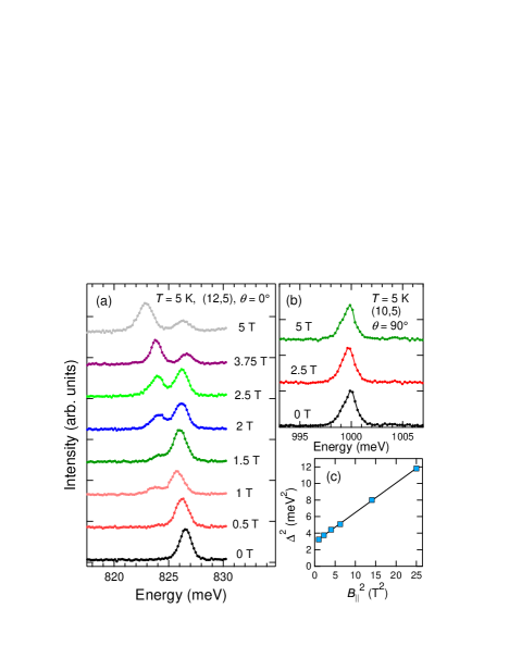
Figure 2(a) shows typical -dependent single-tube PL spectra taken at = 5 K. At = 0, a single, sharp peak exists. As is increased, a second peak appears at a lower energy than the first peak. With further increase of , this second peak gradually grows in intensity and finally dominates the spectrum. The splitting between the two peaks also increases with . This -induced appearance of a lower energy peak was universally observed in all of the 50+ nanotubes of different chiralities which were not completely perpendicular to . In Fig. 2(b), typical PL spectra for SWNTs oriented completely perpendicular to are shown. Clearly, there is no appearance of any lower energy peak even at the highest fields. We emphasize that we did not observe two PL peaks for nanotubes that were completely perpendicular to the field. Even in the parallel configuration, only the tubes which had showed any observable magnetic brightening of the dark state. Assuming a completely random distribution of , this corresponds to 72% of all the tubes. Indeed, out of the 75 nanotubes studied in the parallel configuration, 55 tubes (73%) showed magnetic brightening as is expected from a random distribution of . This also implies that all tubes in our sample emit from the higher energy state at zero field.
We attribute the appearance of the second peak to the -induced brightening of the lowest-energy singlet dark exciton state. Its absence in perpendicular to the tube axis convincingly suggests that brightening is induced by the Aharonov-Bohm phase. In the presence of , the valley degeneracy is lifted due to time-reversal symmetry breaking Zaric et al. (2006). As a result, the dark exciton state acquires a finite oscillator strength. At the highest fields, emission is dominated by the dark state, owing to the increase in splitting, which depopulates the bright state, as well as the increase in the oscillator strength of the dark state at the expense of the bright state oscillator strength.
To quantitatively explain our observations, we use a two-level model described in Refs. Shaver et al., 2007; Shaver and Kono, 2007; Shaver et al., 2008. We assume that at zero field the dark state is completely dark while the bright state has a relative oscillator strength of 1. The total dark-bright splitting in the presence of tube-threading magnetic flux is given by
| (1) |
where is the zero-field splitting, , is a coupling constant, = , is the tube diameter, , and is the disorder-induced zero-field splitting. In the following, we do not include , whose existence would imply zero-field brightening of the dark state. Indeed, we only see a single emission peak from the bright state when = 0 for any tube, suggesting that the effect of disorder in brightening the dark state is negligible in our sample. We use Eq. (1) to fit the splitting value versus , obtained directly from the PL spectra at different fields. The offset and slope of the linear fit between and give us and , respectively, as shown in Fig. 2(c).
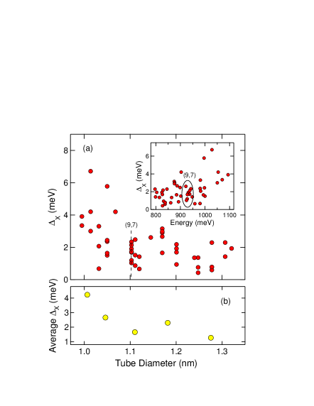
Figure 3(a) shows for 45 different tubes versus tube diameter and emission wavelength (inset). The value of is less than 4 meV for the majority of the tubes. These values (1-4 meV) agree well with those of Refs. Mortimer and Nicholas, 2007 and Shaver et al., 2007 but should be contrasted to the large (10-100 meV) values predicted by some theoretical studies Zhao and Mazumdar (2004); Spataru et al. (2005); Tretiak (2007); Jiang et al. (2007). A possible cause of this discrepancy could be the difference in the value of the dielectric constant () used in different calculations. An accurate prediction must not only take into account the dynamically-screened potential arising from the many-body interactions within the nanotube but also the local variations in due to the medium surrounding it (e.g., surfactant, water, quartz substrate). The dielectric confinement arising from the difference in between the nanotube and the surrounding medium causes image charges that can modify both the exciton binding energy and Shabaev and Efros (2004); Tran Thoai et al. (1990).
Another striking observation one makes from Fig. 3(a) is that the value of shows scatter even for tubes of the same chirality. This scatter must arise from the fluctuations in the local environment of the tubes. Micro-PL spectroscopy is a powerful probe for such fluctuations and enables us to study the role played by the local environment in determining the excitonic fine structure. Since is determined by the exchange interaction, it can vary from tube to tube if the local varies Ando (2006). Thus, such local variations of can lead to “inhomogeneous broadening” of . For example, according to a recent theoretical estimate Per , a change of = 3 to = 2 leads to a change in from 6 meV to 2 meV for an (11,7) nanotube. To extract any trend that is present in the dependence of , we averaged over five ranges. This average , plotted in Fig. 3(b), decreases with , which is reasonable because the short-range Coulomb interaction is predicted to decrease with Spataru et al. (2005); Perebeinos et al. (2005); Ando (2006). However, an exact functional form of the dependence is difficult to deduce from Fig. 3(b) due to the insufficient number of data points. From Eq. (1) and Fig. 3 we obtain values of of 0.4-1.0 meV/T-nm2 for most nanotubes. These values agree well with the theoretical predictions Ajiki and Ando (1993) as well as previous measurements on ensemble SWNT samples in high magnetic fields Zaric et al. (2004, 2006); Shaver et al. (2007); Shaver and Kono (2007); Shaver et al. (2008).
Finally, we analyze how the relative intensities of the two peaks change as a function of . Even though the absolute intensity of each peak was subject to blinking, the relative intensities were not affected by any intensity fluctuations. Within our two-level model Shaver et al. (2007); Shaver and Kono (2007); Shaver et al. (2008), the intensity ratio of the two peaks is given by
| (2) |
where is the Boltzmann constant and are the oscillator strengths of the dark () and bright () levels in the presence of , which vary as
| (3) |
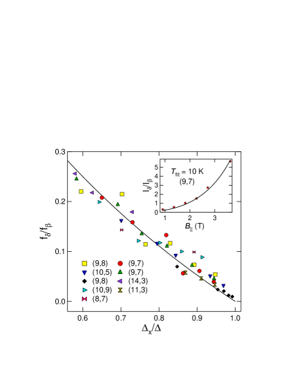
Equation (2) allows us to extract the oscillator strength ratio from the experimentally-measured intensity ratio () and splitting () for each . This quantity, when plotted versus , should exhibit the same functional form given by Eq. (3) for all nanotubes. Figure 4 shows the oscillator strength ratio as a function of for 9 different nanotubes of varying chiralities. We find that the data points for all nanotubes indeed follow the universal curve given by the right-hand side of Eq. (3), in remarkable agreement with the theory.
In conclusion, we have unambiguously demonstrated the existence of the theoretically-predicted spin-singlet dark exciton state below the bright state. The dark-bright splitting was directly measured and found to be 1-4 meV for tubes with diameters of 1.0-1.3 nm. Scatter in splitting value suggests the importance of the local environment in determining the excitonic fine structure.
Acknowledgements.
We thank the US Army Research Office (No. 49735-PH) and the Robert A. Welch Foundation (No. C-1509) for support and V. Perebeinos for helpful discussions. Magneto-PL studies reported here were conducted at the Center for Integrated Nanotechnologies, jointly operated for US Department of Energy by Los Alamos and Sandia National Laboratories.References
- Ando (1997) T. Ando, J. Phys. Soc. Jpn. 66, 1066 (1997).
- Spataru et al. (2004) C. D. Spataru, S. Ismail-Beigi, L. X. Benedict, and S. G. Louie, Phys. Rev. Lett. 92, 077402 (2004).
- Chang et al. (2004) E. Chang, G. Bussi, A. Ruini, and E. Molinari, Phys. Rev. Lett. 92, 196401 (2004).
- Perebeinos et al. (2004) V. Perebeinos, J. Tersoff, and P. Avouris, Phys. Rev. Lett. 92, 257402 (2004).
- Wang et al. (2005) F. Wang, G. Dukovic, L. E. Brus, and T. F. Heinz, Science 308, 838 (2005).
- Maultzsch et al. (2005) J. Maultzsch, R. Pomraenke, S. Reich, E. Chang, D. Prezzi, A. Ruini, E. Molinari, M. S. Strano, C. Thomsen, and C. Lienau, Phys. Rev. B 72, 241402(R) (2005).
- Mortimer and Nicholas (2007) I. B. Mortimer and R. J. Nicholas, Phys. Rev. Lett. 98, 027404 (2007).
- Shaver et al. (2007) J. Shaver, J. Kono, O. Portugall, V. Krstic, G. L. J. A. Rikken, Y. Miyauchi, S. Maruyama, and V. Perebeinos, Nano Lett. 7, 1851 (2007).
- Shaver and Kono (2007) J. Shaver and J. Kono, Laser & Photon. Rev. 1, 260 (2007).
- Shaver et al. (2008) J. Shaver, S. A. Crooker, J. A. Fagan, E. K. Hobbie, N. Ubrig, O. Portugall, V. Perebeinos, P. Avouris, and J. Kono, arXiv:0802.3203 (2008).
- Zhao and Mazumdar (2004) H. Zhao and S. Mazumdar, Phys. Rev. Lett. 93, 157402 (2004).
- Spataru et al. (2005) C. D. Spataru, S. Ismail-Beigi, R. B. Capaz, and S. G. Louie, Phys. Rev. Lett. 95, 247402 (2005).
- Perebeinos et al. (2005) V. Perebeinos, J. Tersoff, and P. Avouris, Nano Lett. 5, 2495 (2005).
- Ando (2006) T. Ando, J. Phys. Soc. Jpn. 75, 024707 (2006).
- Tretiak (2007) S. Tretiak, Nano Lett. 7, 2201 (2007).
- Jiang et al. (2007) J. Jiang, R. Saito, G. G. Samsonidze, A. Jorio, S. G. Chou, G. Dresselhaus, and M. S. Dresselhaus, Phys. Rev. B 75, 035407 (2007).
- Ajiki and Ando (1993) H. Ajiki and T. Ando, J. Phys. Soc. Jpn. 62, 1255 (1993).
- Zaric et al. (2004) S. Zaric, G. N. Ostojic, J. Kono, J. Shaver, V. C. Moore, M. S. Strano, R. H. Hauge, R. E. Smalley, and X. Wei, Science 304, 1129 (2004).
- Zaric et al. (2006) S. Zaric, G. N. Ostojic, J. Shaver, J. Kono, O. Portugall, P. H. Frings, G. L. J. A. Rikken, M. Furis, S. A. Crooker, X. Wei, et al., Phys. Rev. Lett. 96, 016406 (2006).
- (20) M. Bayer, O. Stern, A. Kuther, and A. Forchel, Phys. Rev. B 61, 7273 (2000).
- Htoon et al. (2004) H. Htoon, M. J. O’Connell, P. J. Cox, S. K. Doorn, and V. I. Klimov, Phys. Rev. Lett. 93, 027401 (2004).
- Hagen et al. (2005) A. Hagen, M. Steiner, M. B. Raschke, C. Lienau, T. Hertel, H. Qian, A. J. Meixner, and A. Hartschuh, Phys. Rev. Lett. 95, 197401 (2005).
- Shabaev and Efros (2004) A. Shabaev and A. L. Efros, Nano Lett. 4, 1821 (2004).
- Tran Thoai et al. (1990) D. B. Tran Thoai, R. Zimmermann, M. Grundmann, and D. Bimberg, Phys. Rev. B 42, 5906 (1990).
- (25) V. Perebeinos, private communications.