Optical Identification of a DNA-Wrapped Carbon Nanotube: Signs of Helically Broken Symmetry
pacs:
78.67.-n,78.67.Ch,87.14.gkHigh intrinsic mobilityrogers and small, biologically-compatible size make single-walled carbon nanotubes (SWNTs) in demand for the next generation of electronic devices. Further, the wide range of available bandgaps due to changes in diameter and symmetry give SWNTs greater versatility than traditional semiconductors.charlier Single-stranded DNA has been employed to make these desirable properties accessible for large scale fabrication of devices. Because single-stranded DNA can helically wrap a SWNT, forming a stable hybrid structure zhengSci ; zhengNat , DNA/polymer wrapping has been used to disperse bundles of intrinsically hydrophobic SWNTs into individual tubes in aqueous solution zhengSci ; numata . The ability to isolate individual tubes, make them soluble, and separate them according to symmetry would enable fabrication of SWNT optoelectronic devices that benefit from the unique electronic properties of specific nanotube structures avouris . Envisioning optoelectronic applications of nanotubes, we investigate whether the optical properties of DNA-wrapped SWNT materials are different than those of pristine SWNTspuller . Our previous work found that bandstructures of DNA-SWNTs were indeed affected by the charged wrap. That is, the direct optical bandgap, , decreases, but changes are fairly small snyder . This is consistent with the available experimental data in standard experimental geometry in which incident light is polarized along the SWNT axis chou . Here we consider optical absorption of light with perpendicular (or circular) polarization with respect to the tube axis, which has been measured experimentally for SWNTs dispersed using a surfactant maruyama ; kikkawa ; lefebvre . In this geometry we find qualitative changes in the absorption spectra of SWNTs upon hybridization with DNA, including strong optical circular dichroism in non-chiral SWNTs. These optical effects are predicted to serve as qualitative tools to directly identify the DNA wrapping.
In general, a helical wrap may break the symmetry of a bare SWNT. The potential of the ionized backbone of the DNA is too strong to justify a perturbative approach. In this Communication we numerically solve the joint Schrödinger-Poisson equations beyond the perturbation approximation to determine the modulation of optical properties resulting from hybridization.
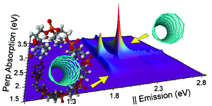
Our simulations predict new peaks in the cross-polarized absorption spectra of DNA-SWNTs with frequencies close to transitions, prohibited for pristine nanotubes in such polarization. In Figure 1 we plot simulated emission along the tube axis vs. simulated absorption of light polarized across the tube axis for the (7,0) SWNT with and without a DNA wrap. The figure shows a dramatic difference in luminescence-absorption maps in the region of the first van Hove singularity, , which is explained in the remainder of this Communication.
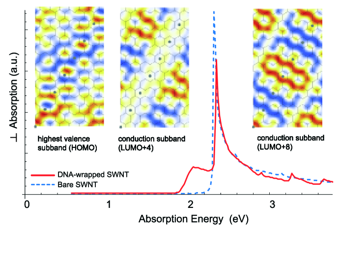
As we observed, absorption (and luminescence) of light with parallel polarization should not change significantly upon DNA hybridization snyder . In contrast, Figure 2 shows that for a semiconducting zigzag (7,0) DNA-SWNT hybrid with the wrap geometry shown in the inset, the optical absorption coefficients for light polarized across the SWNT axis (solid red curve) drastically differ from the bare tube absorption in the same polarization (dashed blue curve). The first absorption peak in cross-polarization for the bare SWNT corresponds to and transitions. This peak is also present for the DNA-SWNT hybrid, although it is shifted to higher frequency. In addition, a peak at lower frequency near that of the bare transitions appears as a consequence of the lifting of selection rules.
For cross-polarized transitions, the selection rule for angular momentum is for the bare tube. This is dictated by the odd symmetry of the momentum operator and the even symmetry of the product of wavefunctions with the same angular momentum for the electron and the hole.
In contrast to the bare tube, the subbands of the wrapped SWNT do not have definite angular momentum since the electron (and hole) wave functions are helically polarized by the Coulomb potential of the DNA. Thus, the circularly polarized optical transitions are allowed at lower frequency near that of the prohibited . The physical interpretation of this effect is that the polarization of the electron (hole) under the perturbation of the transverse electric field of a nearby DNA phosphate group creates across the tube a permanent dipole, which may be excited by the perpendicular electric field of incident light. Overall, (negative) electron density shifts to the opposite side of the tube and (positive) hole density shifts towards the DNA backbone snyder . To observe such an effect experimentally, one must tune to a singularity in the optical density of states. In the bare tube this happens only at the edges of the Brillouin zone. Additional singularities arise with the wrapping due to subband flattening.
Within a semi-empirical orthogonal tight-binding approach, we calculated optical absorption of a number of DNA-SWNT complexes. This numerical approach is chosen to capture the physics of the problem at lower computational cost. The exciton correction is expected to be essentially weaker for transitions due to decay of the direct Coulomb matrix element with non-zero angular momentum transfer exciton , and therefore it is neglected in this study. The Hamiltonian and computational scheme are discussed in Supplementary Materials.
The DNA backbone is modeled as a regular, infinite helix of point charges brooks representing the phosphate groups wrapped around the tube (right inset in Figure 2). The angle of the helix, its position with respect the underlying graphene lattice, the spacing between the tube and DNA charges and its linear density are parameters of the model that can be obtained from molecuar dynamics simulations water or adjusted to the experimental data. For a broad range of these parameters, we observe similar symmetry breaking effects.
The partial absorption coefficient is calculated for a bare SWNT as
| (4) |
where is the Fermi-Dirac function, is the inverse lifetime, is the matrix element for a transition from an initial state in the valence band to a final state in the conduction band for the case of circularly polarized incident light , with the selection rule for angular momentum .
The chiral perturbation of the DNA lifts selection rules and allows additional optical transitions in the DNA-SWNT hybrid that were prohibited by symmetry in the bare nanotube for cross-polarized absorption. Such ”natural” helicity of the electron states must result in non-zero optical activity of the material. Indeed, the optical circular dichroism spectra for a variety of SWNT-DNA hybrids show strong dichroism for originally achiral as well as chiral tubes (Figure 3, inset).
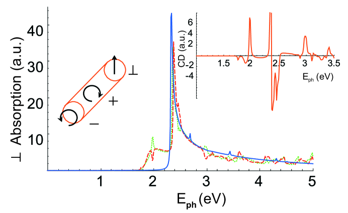
The total absorption spectrum is obtained by integration of the partial absorption coefficient Eq.(4) over the wavevector inside the first Brillouin zone and is plotted in Figure 3 for the left and right circular polarizations (dashed blue and dotted red curve, respectively). Absorption of cross-polarized light drastically differs for the hybrid and the bare tube (solid green curve in Figure 3).
The difference in absorption for two polarizations gives the circular dichroism spectrum of the DNA-SWNT hybrid: (inset of Figure 3). We stress that this circular dichroism is unrelated to the possible chirality of the DNA. Firstly, the single-stranded DNA does not make a clear helix on its own, without wrapping around the SWNT. Secondly, DNA absorption is not included in this work at all, although a recent study golovchenko showed an interesting DNA hypochromicity effect in the hybrids. Hence, the optical activity must be fully attributed to the nanotube itself and to its helical symmetry breaking. That result is further supported by the experimental data in Ref.dukovic .
Symmetry of the wrap is central to determine the circular dichroism and optical response of the hybrid. We predict that optical absorption in the perpendicular or circular polarization may be used to detect the helical wrapping. The exact geometry of the DNA wrap for an arbitrary tube is not yet precisely known. Upon variation of the parameters of the wrap and/or tube diameters, we found in our modeling that both optical effects are almost independent of the axial or equatorial displacement of the helical wrap along the SWNT surface without changing the helical angle of the DNA backbone (See Suppl.). This is because the most important factor is symmetry matching between the SWNT lattice and the DNA backbone helical angle. For various angles and for various tubes we predict different absorption spectra, though we believe that a general qualitative feature of the helical symmetry breaking, the appearance of new van Hove singularities in the optical data, must be present.
In conclusion, we obtain optical absorption spectra and circular dichroism for DNA-wrapped single-wall nanotubes. In this Communication we focus on the optical transitions for light polarized across the nanotube axis. We predict that symmetry lowering due to the Coulomb potential of the regular helical DNA wrap results in qualitative changes in the cross- or circularly polarized absorption spectrum. In particular, we predict the appearance of a new transitions in the cross-polarized absorption of the DNA-SWNT at frequencies substantially lower than that of all allowed transitions in the bare tube. Therefore, with sufficient wrapping coverage, the hybrid material is predicted to show splitting and a shift of the lowest peak, which we suggest can be used for experimental detection of the wrapping. A similar effect of the symmetry breaking is predicted to result in strong circular dichroism of the complexes.
References
- (1) C. Kocabas, S.-H. Hur, A. Gaur, M. Meitl, M. Shim, J. A. Rogers, Small 2005, 1, 1110.
- (2) J.-C. Charlier, X. Blase, S. Roche, Rev. Mod. Phys. 2007, 79, 677.
- (3) M. Zheng, A. Jagota, M. S. Strano, A. P. Santos, P. Barone, S. G. Chou, B. A. Diner, M. S. Dresselhaus, Mildred S., R. S. Mclean, G. B. Onoa, G. G. Samsonidze, E. D. Semke, M. Usrey, D. J. Walls, Science 2003, 302, 1545.
- (4) M. Zheng, A. Jagota, E. D. Semke, B. A. Diner, R. S. Mclean, S. R. Lustig, R. E. Richardson, N. G. Tassi, Nat. Mater. 2003, 2, 338.
- (5) M. Numata, M. Asai, K. Kaneko, A.-H. Bae, T. Hasegawa, K. Sakurai, S. Shinkai, J. Am. Chem. Soc. 2005, 127, 5875.
- (6) Ph. Avouris, Physics World March 2007, 40.
- (7) V. Puller, S. V. Rotkin, Europhys. Lett. 2007, 77, 27006.
- (8) S. E. Snyder, S. V. Rotkin, JETP Letters 2006, 84, 348.
- (9) S. G. Chou, H. B. Ribeiro, E. B. Barros, A. P. Santos, D. Nezich, Ge. G. Samsonidze, C. Fantini, M. A. Pimenta, A. Jorio, F. Plentz Filho, M. S. Dresselhaus, G. Dresselhaus, R. Saito, M. Zheng, G. B. Onoa, E. D. Semke, A. K. Swan, M. S. Uenlue, B. B. Goldberg, Chem. Phys. Lett. 2004, 397, 296.
- (10) Y. Miyauchi, M. Oba, S. Maruyama, Phys. Rev. B 2006, 74, 205440.
- (11) M. F. Islam, D. E. Milkie, C. L. Kane, A. G. Yodh, J. M. Kikkawa, Phys. Rev. Lett. 2004, 93, 037404.
- (12) J. Lefebvre, P. Finnie, Phys. Rev. Lett. 2007, 98, 167406.
- (13) T. Ando, T. Nakanishi, R. Saito, J. Phys. Soc. Jpn. 1998, 67, 2857.
- (14) K. Bulashevich, S. V. Rotkin, Int. Journal of Nanoscience 2003, 2, 521.
- (15) B. R. Brooks, R. E. Bruccoleri, B. D. Olafson, D. J. States, S. Swaminathan, M. Karplus, CHARMM, J. Comp. Chem. 1983, 24, 187.
- (16) A. A. Tsukanov, unpublished.
- (17) M. E. Hughes, E. Brandin, J. A. Golovchenko, Nano Lett. 2007, 7, 1191.
- (18) G. Dukovic, M. Balaz, P. Doak, N. D. Berova, M. Zheng, R. S. Mclean, L. E. Brus J. Am. Chem. Soc. 2006, 128, 9004.