The commensurate phase of multiferroic HoMn2O5
studied by X-ray magnetic scattering
Abstract
The commensurate phase of multiferroic HoMn2O5 was studied by X-ray magnetic scattering, both off resonance and in resonant conditions at the Ho- edge. Below 40 K, magnetic ordering at the Ho sites is induced by the main Mn magnetic order parameter, and its temperature dependence is well accounted for by a simple Curie-Weiss susceptibility model. A lattice distortion of periodicity twice that of the magnetic order is also evidenced. Azimuthal scans confirm the model of the magnetic structure recently refined from neutron diffraction data for both Mn and Ho sites, indicating that the two sublattices interact via magnetic superexchange.
pacs:
75.25.+z, 75.50.Ee, 77.80.-e, 78.70.CkThere has been very significant recent interest in magnetoelectric multiferroics, in particular magnetically frustrated transition-metal oxides such as TbMnO3Kimura et al. (2003), Ni3V2O8Lawes et al. (2005), RMn2O5 (R=rare-earth, Bi) Hur et al. (2004); Chapon et al. (2006, 2004); Blake et al. (2005); Vecchini et al. (2007); Muoz et al. (2002) and more recently RbFe(MoO4)2 Kenzelmann et al. (2007). In this class of materials, the onset of ferroelectricity coincides with the transition to a magnetically ordered state characterized by a complex spin configuration. While the spontaneous electric polarization is much weaker than for proper ferroelectrics, the magneto-electric coupling is strong, leading to giant effects in the physical properties upon application of an electric or a magnetic field. Some key facts about these materials have been established unambiguously: acentric magnetic ordering on the transition-metal sites is clearly the main factor in inducing ferroelectricity, and the direction of the electrical polarization can be well explained by the Landau phenomenological analysis of the magneto-electric coupling or by more general symmetry arguments Radaelli and Chapon (2007). Other aspects are much less clear: the microscopic origin of ferroelectricity is still much debated, in particular in the RMn2O5 and triangular-lattice systems, where more than one mechanism has been proposed. In addition, there is presently very little understanding of the role played by the magnetic rare earths. Rare earth magnetism not only leads to spectacular magneto-electric effects at low temperatures, but can increase greatly the value of the electrical polarization even at high temperature, as recently shown for DyMnO3 Prokhnenko et al. (2007).
Most become ferroelectric below 40 KHur et al. (2004), driven by magnetic ordering to a commensurate state with propagation vector k=(1/2,0,1/4) for R=Y,Ho,Tb Chapon et al. (2004); Blake et al. (2005) and k=(1/2,0,1/2) for Bi Muoz
et al. (2002). At around 20K, the R=Y, Ho, Tb analogues show a second magnetic transition to an incommensurate structure, with much smaller spontaneous electric polarization. Neutron diffraction studies of the ferroelectric/commensurate phase on powderChapon et al. (2006, 2004); Blake et al. (2005) and more recently single crystal specimensVecchini et al. (2007); Kimura et al. (2007), have evidenced a small moment on the magnetic rare-earth sites well above the main ordering temperature of the rare earth sublattice ( 10 K), suggesting that this moment is induced by the complex Mn magnetic ordering. For HoMn2O5, the magnitudes and directions of the Ho moments has been established independently by two groups Vecchini et al. (2007); Kimura et al. (2007), but there cannot be absolute confidence in these results, since Ho’s overall contribution to the magnetic neutron scattering intensities is very small (the Ho moments are between 0 and 1 on different lattice sites compared with an almost fully ordered Mn sublattice). More importantly, the nature of the interplay between Mn and Ho order — in particular the induced nature of the Ho moments — are impossible to address with neutron diffraction, since the Ho and Mn contributions cannot be separated at temperature close to TN
In the present Communication, we report on resonant and non-resonant X-ray magnetic diffraction experiments performed on the commensurate phase of HoMn2O5. Unlike neutron diffraction, this technique allows one to probe the Ho and Mn ordering independently. The magnetic structure of the Ho sublattice was confirmed by fitting azimuthal scans in resonant conditions at the Ho L3 edge, while off-resonance scans can be well reproduced with the overall magnetic structure determined by neutron diffraction. By studying the temperature dependence of both signals, the Ho and Mn sublattice magnetic order parameters are extracted, clearly showing that the Ho and Mn magnetic transitions are simultaneous and that the former is induced by the latter. The evolution of the Ho order parameter is well explained by a simple model accounting for the polarization by the Mn sublattice. Weak charge superlattice peaks with kL=2kM=(0,0,1/2) are also observed in the same temperature range; we find that the order parameter of the charge superstructure is proportional to the square of the Mn staggered magnetization, as predicted by Landau theory.
Synchrotron X-ray experiments were performed at Diamond Light Source, on Beamline I16 - Materials & Magnetism, dedicated to X-ray diffraction in the energy range 3-25 keV. The horizontally polarized beam delivered by a linear undulator was tuned either to the edge of Holmium (8.078 keV) or at 6.4 keV below the edge of Manganese (6.55 keV), and monochromatized by a Si double-crystal monochromator. The beam was focused to m in the horizontal and vertical directions. A single crystal of , with size approximately mm3 and faces along the , and crystallographic directions was used. The crystal was attached to a copper platform with QuickFill and mounted on the Kappa-geometry diffractometer of the beamline, in a Displex type cryofurnace. When necessary, a polarization analysis of the diffracted beam was performed with the (022) reflection of a copper crystal. Its efficiency at 6.4 keV was about 1/4 and the cross-talk between polarization channels was found to be negligible. The intensity was recorded either with an Avalanche Photo-Diode (APD) or with an energy-resolving Silicon-drift diode detector to reject the fluorescence. Further details of the beamline are available on i (16).
All measurements were performed in the commensurate phase on two strong magnetic reflections, indexed ( 3 ) and ( 4 ) and on the (4 4 0.5) ”charge” Bragg peak.The first set of measurements were conducted at 6.4 keV, i.e. at an energy far below any absorption edges of the sample. In these conditions, the magnetic scattering is not element specific and measures contributions from both Mn and Ho sites. The low fluorescence, combined with the high efficiency of the analyzer and the low noise of the APD, allowed us to measure in non-resonant conditions with good statistics (typically 1000 counts/sec) and low background (about 250 counts/sec in the channel and nearly 0 counts/sec in the channel). No resonance was found at the -edge of Manganese (not shown), confirming that its orbital moment is quenched by the crystalline electric field (at a -edge, one actually measures the orbital contribution of the specific ion to the total magnetization, which is related to the spin component by the spin-orbit coupling). A second set of measurements was conducted in resonant conditions in the vicinity of the Ho -edge. A strong dipole-dipole enhancement, due to the resonance of the electrons with the empty shell, was observed at 8.076 keV (Fig. 1). A quadrupolar contribution was also found at 8.068 keV.
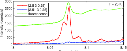
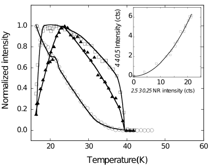
Figure 2 shows the temperature dependence of the magnetic reflection (, 3, ), off resonance and at the Ho- edge. The non-resonant measurement indicates that the transition temperature is 40K, in agreement with previous neutron diffraction Blake et al. (2005) and magnetization measurements. Around 18 K, the compounds undergoes a first order transition (not studied here) to an incommensurate magnetic order with propagation vector k=(0.48, 0, 0.28) Blake et al. (2005), splitting the (, 3, ) peak and causing the accidents observed around 18K in Fig. 2. Near the transition, the peak intensity in non-resonant conditions is essentially proportional to the square of the Mn magnetic moment, since here the Ho contribution is negligible. Its temperature dependence is consistent with the neutron data Chapon et al. (2004) and confirms that Mn ordering is the primary order parameter. In the Landau free energy expansion, the magnetic order parameter can be coupled not only to the gamma-point ferroelectric distortions, but also to an additional structural modulation () of double wave-vector, kL=(0,0,0.5). Time-reversal symmetry invariance requires at the leading term, so we also expect the charge peak intensity to be proportional to the square of the magnetic peak intensity. This is illustrated in the temperature dependence of the superlattice reflection (4 4 0.5), measured off resonance at 7.96 keV and shown in Figure 2. Its integrated intensity is proportional to the square of the integrated intensity of the (, 3, ) magnetic reflection (inset of Figure 2). This result is similar to what was recently derived from non-resonant X-ray scattering at the edge for the analog compound TbMn2O5 Koo et al. (2007). The underlying structural distortion minimizes the magnetic energy, most likely through a modulation of the -axis distances (and coupling), responding to the alternation of FM and AFM configuration across the rare earth layers.
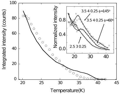
In sharp contrast to the off-resonance data, the (, 3, ) magnetic peak shows an unusual variation with temperature at the Ho- resonance. The peak appears below 40K, clearly showing that magnetic ordering of the Ho moments coincides with the magnetic ordering of the Mn sublattice but its intensity is marked by the absence of critical behavior at TN. Irrespective of the nature of the Mn-Ho coupling, discussed later, one can postulate that the Ho ordering is induced by the molecular field arising from the Mn sublattice. This field can be taken to be directly proportional to the magnetic moment on Mn sites, as estimated from the root-squared intensity of the non-resonant peak. The resonant intensity is then simply calculated using a Curie-Weiss function diverging at =10 K — the ordering temperature of the Ho sublattice — as . There is one adjustable parameter, , allowing the relative intensities of the two peaks to be scaled. Fig. 3 shows the resonant (, 3, ) integrated intensity together with the best fit to the molecular field model. While the model reproduces relatively well the overall temperature dependence, there is clearly a discrepancy between the two curves. This is likely to be due to a relative reorientation of the two sublattices as a function of temperature, as confirmed by the fact that different resonant peaks have somewhat different temperature dependencies (inset of Fig 3).
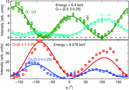
The azimuthal dependence of the magnetic cross-section in the commensurate phase has been measured in the full angular range both off-resonance at 6.4 keV and at the Ho- edge (Fig. 4). For each azimuthal scan, the intensity was integrated over a rocking curve. In the non-resonant case, magnetic scattering appears in both the and channels. At the L3 edge, magnetic scattering was observed only in the channel, as expected. The azimuthal scans are shown in Fig. 4. The scattering cross sections are proportional to the squared modulus of the scattering amplitude Blume and Gibbs (1988); Hill and McMorrow (1996), f, written as:
| (1) | |||||
| (2) | |||||
| (3) |
where the subscript nr and r refer to the non-resonant and resonant cases and the superscript refers to the scattering channel. k and k’ are unit vectors in the direction of the incident and scattered wave-vectors, respectively, and are the unit-cell magnetic structure factors. Off resonance, we considered Mn and Ho spin moments with their appropriate form factors and neglected the orbital moments. In the resonant case, only Ho sites without the usual magnetic form factor need to be taken into account. Note that we consider only the dipole-dipole scattering, which is dominant at 8.076 keV (Fig. 1).The calculated intensities have been corrected for self-absorption, multiplying them by the factor:
| (4) |
where and are respectively the incident and exit angles with respect to the crystal surface ([1 1 0]) and the linear absorption coefficient does not depend on .
In the non-resonant case, the scattering intensities have been calculated using the model of magnetic structure for HoMn2O5 recently derived by single-crystal neutron diffraction at 25K Vecchini et al. (2007). The agreement between experimental data and calculations is excellent in both the and channels as illustrated in Figure 4. In this model, the manganese ions form antiferromagnetic zig-zag chains along the -axis, with the main components of the moment in the -plane and pointing about 14∘ with respect to the -axis. A small out-of phase component along the -axis gives rise to a cycloidal modulation along the Mn4+ chains.
The azimuthal dependence at the Ho edge is also consistent with the neutron results, and confirms unambiguously that the Ho moments point mainly in the -plane.
This is consistent with the Ho single ion anisotropy: in fact, a point charge calculation of the Ho crystal field with the program McPhas Mcp , taking into account neighboring charges at distances up to 50 Å, shows that the ground state multiplet is made of two nearly degenerated singlets. Only the matrix element and have non vanishing components. This is in contrast with calculations for ErMn2O5, which show that the ground state doublet has very strong easy-axis anisotropy, as expected from the opposite sign of the Stevens equivalent operator B with respect to Ho, and also compatible with neutron diffraction study showing that the Er moments are aligned mainly along the -axis Gardner et al. (1988).
There is a residual degeneracy in the patterns of in-plane moments that can be consistent with the resonant data; however, the neutron data strongly discriminate between these alternatives. Two mechanisms, both leading to weak interactions, can account for the induced moment on the Ho site: dipole-dipole interaction on one hand and magnetic superexchange interactions between the Ho and first-neighbor Mn ions. The dipolar interaction energy, taking into account the full paramagnetic Ho moment M=10 (gJ=1.25,J=8), and the shortest Ho3+-Mn4+ distance, is 0.4K.
Calculations show that the dipolar field induced by the ordering of the Mn ions is almost orthogonal to the Ho moments, which invalidates this model.
The super-exchange interaction mediated through the Ho4(f)-O(p)-Mn4(d) orbital path is also weak given the short extension of the 4f shell. However, this mechanism is consistent with the observed structure, as already pointed out in Vecchini et al. (2007), since the Ho moment is oriented antiparallel to the resultant magnetic moment of first-neighbor Mn4+ ions.
In summary, we have studied the magnetic structure of HoMn2O5 in the commensurate/ferroelectric phase by magnetic X-ray scattering off resonance and at the Ho-L3 resonance. Our study clearly established that magnetic ordering of the Ho sublattice exists even at temperature close to TN and is induced by the magnetic ordering of the Mn sites. The azimuthal dependence of the magnetic scattering is in agreement with the overall magnetic structure derived by single-crystal neutron diffraction and provide an independent confirmation of the magnetic ordering on the Ho sites at 25 K. The magnetic configuration on the Ho sites is only consistent with coupling to the Mn4+ moments through superexchange interactions, and agrees with the Ho easy-plane anisotropy derived from a point-charge model.
References
- Kimura et al. (2003) T. Kimura, T. Goto, H. Shintani, K. Ishizaka, T. Arima, and Y. Tokura, Nature 426, 55 (2003).
- Lawes et al. (2005) G. Lawes, A. B. Harris, T. Kimura, N. Rogado, R. J. Cava, A. Aharony, O. Entin-Wohlman, T. Yildirim, M. Kenzelmann, C. Broholm, et al., Phys. Rev. Lett. 95, 087205 (2005).
- Hur et al. (2004) N. Hur, S. Park, P. A. Sharma, J. S. Ahn, S. Guha, and S.-W. Cheong, Nature 429, 392 (2004).
- Chapon et al. (2006) L. C. Chapon, P. G. Radaelli, G. R. Blake, S. Park, and S.-W. Cheong, Phys. Rev. Lett. 96, 097601 (2006).
- Chapon et al. (2004) L. C. Chapon, G. R. Blake, M. J. Gutmann, S. Park, N. Hur, P. G. Radaelli, and S.-W. Cheong, Phys. Rev. Lett. 93, 177402 (2004).
- Blake et al. (2005) G. R. Blake, L. C. Chapon, P. G. Radaelli, S. Park, N. Hur, S.-W. Cheong, and J. Rodriguez-Carvajal, Phys. Rev. B 71, 214402 (2005).
- Vecchini et al. (2007) C. Vecchini, L. C. Chapon, P. J. Brown, T. Chatterji, S. Park, S.-W. Cheong, and P. G. Radaelli, Cond-mat arXiv:0710.2065 (2007).
- Muoz et al. (2002) A. Muoz, J. A. Alonso, M. T. Casais, M. J. Martínez-Lope, J. L. Martínez, and M. T. Fernández-Díaz, Phys. Rev. B 65, 144423 (2002).
- Kenzelmann et al. (2007) M. Kenzelmann, G. Lawes, A. B. Harris, G. Gasparovic, C. Broholm, A. P. Ramirez, G. A. Jorge, M. Jaime, S. Park, Q. Huang, et al., Phys. Rev. Lett. 98, 267205 (2007).
- Radaelli and Chapon (2007) P. G. Radaelli and L. C. Chapon, Phys. Rev. B 76, 054428 (2007).
- Prokhnenko et al. (2007) O. Prokhnenko, R. Feyerherm, E. Dudzik, S. Landsgesell, N. Aliouane, L. C. Chapon, and D. N. Argyriou, Phys. Rev. Lett. 98, 057206 (2007).
- Kimura et al. (2007) H. Kimura, S. Kobayashi, Y. Fukuda, T. Osawa, Y. Kamada, Y. Noda, I. Kagomiya, and K. Kohn, J. Phys. Society Japan 76, 074706 (2007).
- i (16) http://www.diamond.ac.uk/Beamlines/Beamlineplan/I16.
- Koo et al. (2007) J. Koo, C. Song, S. Ji, J. S. Lee, J. Park, T. H. Jang, C. H. Yang, J. H. Park, Y. H. Jeong, K. B. Lee, et al., Non-resonant and resonant x-ray scattering studies on multiferroic TbMn2O5 (2007), URL http://www.citebase.org/abstract?id=oai:arXiv.org:0704.0533.
- Blume and Gibbs (1988) M. Blume and D. Gibbs, Phys. Rev. B 37, 1779 (1988).
- Hill and McMorrow (1996) J. P. Hill and D. F. McMorrow, Acta Cryst. A52, 236 (1996).
- (17) http://www.mcphas.de.
- Gardner et al. (1988) P. P. Gardner, C. Wilkinson, J. B. Forsyth, and B. M. Wanklyn, J. Phys. C: Solid State Phys. 21, 5653 (1988).