Structural anisotropy of silica hydrogels prepared
under magnetic field
Abstract
Birefringence measurements have been carried out on the Pb-doped silica hydrogels
prepared under various magnetic fields up to 5T.
The silica gels prepared at 5T were used as a medium of crystal growth of PbBr2,
whose result implied the structural anisotropy; an aligned array of crystallites
was obtained by transmission electron microscopy.
While the samples prepared at 0, 1, and 3T have no birefringence,
we found that the samples have negative birefringence on the order
of magnitude
as if the direction of the magnetic field is the optic axis of
a uniaxal crystal.
To the authors’ knowledge, the birefringent silica hydrogels were obtained
by gelation under magnetic field for the first time.
Also, scanning microscopic light scattering experiments have been performed.
The results indicate that the characteristic length distribution for birefringent
samples is narrower than that for non-birefringent ones.
Keywords: Microstructure, Optical materials and property,
Silica gels, Birefringence, Magnetic field
1 Introduction
We have found an aligned array of crystallites in gels prepared under a magnetic field of 5T, which was used as media of the crystal growth of PbBr2 [1, 2]. The crystallites were aligned with their crystallographic axis along the direction of the magnetic field, which was applied during the preparation of the gels. The magnetic field did not affect considerably if it was applied during the growth of PbBr2. It is, thus, anticipated that the magnetic field applied during the preparation of the Pb-doped silica hydorgels made the gel structure anisotropic. The first purpose is the identification of the anisotropy in the gel network.
Beside optimization of the condition of the crystal growth in silica gels such as [3, 4, 5, 6], the control of the structure of silica gels is a subject of recent studies. The control of the structure has been achieved by the selection of starting materials, pH control of the solvent, solvent exchange during polymerization stage, and aging [7, 8, 9].
There are a lot of potential uses of the silica gels with controlled structure. The transport of materials in the gels depends on the struscure in the gels. Thus, for example, the crystal growth in hydorgels can be controlled by the controlled structure in the hydrogels. If aerogels are made by drying the controlled silica hydrogels, they keep the structure in solution and then can be applied to a new type of column chromatography. If pore size is highly controlled uniformly, the filtering of the cuumn must be improved. If we realize anisotropy in mechanical and/or thermomechanical properties due to the anisotropic structure of hydrogels, those properties can be applied as a smart meterials for the sensing devices, actuators, MEMS, and so on.
Here, we focus on the structural anisotropy in silica hydrogels and structure. Effect of magnetic field applied during the gelation on the network structure of polymer gels has been elucidated. Aligned gel networks have been formed for both chemical and physical gels. Chemically cross-linked poly(N-isopropylacrylamid) forms aligned network structure perpendicular to magnetic field [10]. Physically cross-linked agarose gels also show the perpendicular alignment [11]. In case side-chain group prefers the parallel alignment, polymer chain align perpendicularly to the magnetic field because the side-chain group is basically normal to the main chain. One of such groups contains a conjugate electron. A typical example is a benzene ring, in which ring current is induced. On the other hand, in case a group has magnetic moment, the part including such a group preferes orient in parallel to the magnetic field [12].
In the case of silica gels, the interaction of polymer chain with magnetic field is not elucidated well. In this paper, after presenting the results of birefringence measurement and scanning microscopic light scattering, the discussions on the mechanism of interaction with the magnetic field are given.
2 Sample preparation
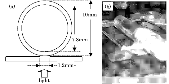
The samples were prepared in the same way as described in [4], except for the strength of the magnetic field. Sodium metasilicate was dissolved in distilled water by starting for two hours. After that, acetic acid and aqueous solution of Pb(NO3)2 were added and then stirred for two hours. This solution was settled for 7 days under various magnetic fields (0, 1, 3, and 5T) parallel to the sample tube at a 298K. The vessels used are cylindrical tube with inner being 7.8 mm. The magnetic field was applied parallel to the cylindrical axis. On the birefringence measurement a slit with the width 1.2mm to fix the geometrical condition as illustrated in Fig. 1; that is, we estimated the sample thickness for this measurement to be =7.710.09mm.
3 Method of birefringence measurement

Sénarmont method was employed for the birefringence measurement. We utilized together a spectrometer to evaluate the intensity of the light transmitted the analyzer as a function of the rotation angle of the analyzer. Usual Sénarmont method the rotation angle of the analyzer, which is a polarizer located after quarter wave plate (Sénarmont compensator), where the transmitted light disappears is evaluated by eye. If no sample, the transmitted light vanishes for the configuration of the crossed polarizers. If a birefringent sample is inserted with the axis pointing the 45∘ with respect to the transmitting axis of the polarizer, the rotation angle of the analyzer at which the transmitted light disappears changes. Instead of detection by eye, we fit the intensity as a function of the rotation angle, , of the analyzer by to determine the retardation with and being the optical path difference and the thickness of the sample. Photons were counted per 100 ms for a few minutes to obtain the intensities (averaged) and the standard deviations were also calculated. The measure of birefringence, , is obtained from , where is the wavelength of light source, commonly the mercury light, whose being 546nm. Note that here is defined as with the optic axis being parallel () to the direction of the magnetic field. Figure 2 is the typical result. In this sample the magnetic field applied was 5T and the fitting results are . Therefrom, is estimated as .
4 Structure characterization by dynamic light scattering
Scanning microscopic light scattering (SMILS) was carried out to scan and measure many different positions in the samples, in order to rigorously determine a time- and space-averaged, i.e., ensemble-averaged, (auto-) correlation function of the concentration fluctuating in the sample. All the samples were filtered through 0.1 m filters to avoid interference from dust particles. An semiconductor laser with 532 nm wavelength were used in different solutions as the incident beam. Measurements were made at four different angles, which were 40, 60, 90, and 125∘, and the typical measurement time was 90 s. The samples were maintained at a constant temperature of 30∘C throughout the experiments. Scanning measurement was performed at 31 points for each sample to determine the dynamic component of the ensemble-averaged dynamic structure factor [13]. The determined correlation function was transformed to the distribution function of relaxation time, , numerically transformed to relaxation-time distribution composed of one or two logarithmic Gaussian distribution (LGD) [14]. In the present study, we simply discuss the bahavior of a main relaxation mode of the silica hydrogels, which can be assigned to the corrective diffusion of polymer network.
5 Results and Discussions
5.1 Birefringence
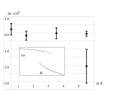
Figure 3 is the results of the birefringence measurement. In the rage of the magnetic field the samples has been classified in two classes at =5T; one has exhibited no birefringence while a negative with the order of the magnitude of . It is anticipated that there exists a coexistence region where two stable states, one of which is most stable and the other a metastable (dotted curves in the inset of Fig. 3), appear.
5.2 Scanning microscopic light scattering
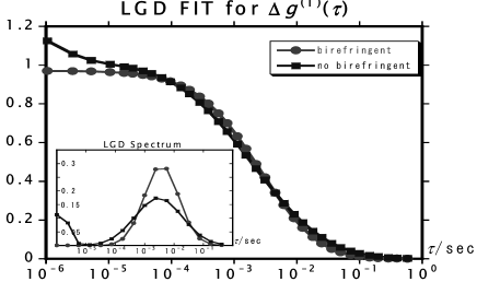
Typical results of SMILS are shown in Fig. 4. The light was input through the sample perpendicular to the sample tube. That is, the structure in the cross sectional area perpendicular to the axes along which the magnetic field was applied was investigated. We saw a tendency that the distribution of for birefringent samples is narrower that for non-birefringent ones. We conjecture that the distribution of the pore size measured by SMILS directly reflects. The optimization of the condition of measurement was difficult; the optimal combination of the laser power and the parameter of photo multiplier was different for the scattering angles depending of the samples. We wish to postpone that precise determination of the pore size.
5.3 Discussions
Here, we discuss the origin of the negative birefringence of the Pb-doped silica gels prepared under a magnetic field. Aforementioned we have interested in the structure of scale around several tens or a few hundreds nm. Pores of this scale has been reported, thought the samples have been aerogels [15]. Closed loops must exist in the hydrogels, too. Let us remain the existence of the dopants of Pb+ and that the skeletons made of silicon and oxygen atoms is of full of dangling bonds. We speculate the ring current along close loops through the mechanism similar to that of electric conduction of the conjugated polymers [16] and the force tending to direct those rings normal to the magnetic field. One can readily understand the negative birefringence from this structural anisotropy. Other possibilities cannot be, of course, ruled out. The wave functions or the molecular clouds, which govern the formation of the basic structure of the gel network, in the magnetic field have been anisotropic at the chemical reactions.
Also, narrowing of the pore size distribution can be understood consistently. Apart from the mechanism of formation, we focus here on the orientational distribution of the closed loops. In case the loops align in the plane made of incident bean and the detector, the distribution of the characteristic length become unimodal. On the other hand, if the closed loops orients directly with respect to that plane, the distribution must be broadened.
6 Concluding remarks
We have investigated the structural anisotropy of silica hydrogels prepared under the agnatic field. Some samples prepared under 5T exhibit negative birefringence with the optic axis coinciding to the direction of the magnetic field. The scanning microscopic light scattering measurments give results supporting this structural anisotropy. Such novel phenomena will be investigated in detail in the fufure.
References
- [1] T. Kaito, S-i. Yanagiya, A. Mori, M. Kurumada, C. Kaito, and T. Inoue, J. Cryst. Growth 289 (2006) 275-277.
- [2] T. Kaito, S-i. Yanagiya, A. Mori, M. Kurumada, C. Kaito, and T. Inoue, J. Cryst.Growth 289 (2006) 407-410.
- [3] S. Pandita, V. Hangloo, K. K. Bamzai, P. N. Kotru, and N. Shani, Int. J. Inorg. Mater. 3 (2001) 675-680.
- [4] H. Kusumoto, T. Kaito, S-i. Yanagiya, A. Mori, and T. Inoue, J. Cryst. Growth 277 (2005) 536-540.
- [5] P. N. S. Kumari, S. Kalainathan, and N. A. N. Raj, Mater. Res. Bull. 42 (2007) 2099-2106.
- [6] P. N. S. Kumari, S. Kalainathan, and N. A. N. Raj, Mater. Lett. 61 (2007) 4423-4425.
- [7] R. K. Ilre, The Chemistry of Silica (Wiley-Iterscience, 1979).
- [8] B. Knoblich and Th. Gerber, J. Non-Cryst. Solid 296 (2001) 81.
- [9] D. J. S. Birch and C. D. Geddes, Phys. Rev. B. 62 (200) 2977.
- [10] I. Otsuki, H. Abe and S. Ozeki, Sci. Tech. Adv. Mater. 7 (2006) 327-331.
- [11] I. Yamamoto, S. Saito, T. Makino, M. Yamaguchi, and T. Takamas, uSci. Tech. Adv. Mater. 7 (2006) 322-236
- [12] Y. Shgekura, Y. M. Chen, H. Furukawa, T. Kaneko, D. Kaneki, Y. Osada, and J. P. Gong, Adv. Mater. 17 (2005) 2695-2699.
- [13] H. Furukawa, K. Horie, R. Nozaki, M. Okada, Phys. Rev. E 68, (2003) 031406.
- [14] M. Huang, H. Furukawa, Y. Tanaka, T. Nakajima, T. Osada, and J. P. Gong, Macromolecules 40 (2007) 6658-6664.
- [15] J. Wang, J. Shen, B. Zhou, Z. Deng, L. Zhao, L. Zhu, and Y. Li, NanoStruct. Mater. 10 (1998) 909-916.
- [16] A. J. Heeger, S. Kivelson, J. R. Schrieffer, and W. P. Su, Rev. Mod. Phys. 60 (1988) 781-850.
Figure captions
Figure 1:
(a) Illustration of cross-section of test tube and
(b) the sample setting at measurement of .
Figure 2:
A typical plot of the light intensity .
Figure 3:
The birefringence V.S. the magnetic field .
The bistability is apparent at =5T.
The inset is a schematic drawing for - relation.
Figure 4:
A typical result of SMILS.
The inset is with the fit by a mode distributon of the sum
of two logarythmic Gausian distribution.
The distribution of the relaxation time is obtained therefrom.
The scattering angle for this data is 90∘.