Switchable ErSc2N rotor within a C80 fullerene cage:
An EPR and photoluminescence excitation study
Abstract
Systems exhibiting both spin and orbital degrees of freedom, of which Er3+ is one, can offer mechanisms for manipulating and measuring spin states via optical excitations. Motivated by the possibility of observing photoluminescence and electron paramagnetic resonance from the same species located within a fullerene molecule, we initiated an EPR study of Er3+ in ErSc2N@C80. Two orientations of the ErSc2N rotor within the C80 fullerene are observed in EPR, consistent with earlier studies using photoluminescence excitation (PLE) spectroscopy. For some crystal field orientations, electron spin relaxation is driven by an Orbach process via the first excited electronic state of the multiplet. We observe a change in the relative populations of the two ErSc2N configurations upon the application of 532 nm illuminations, and are thus able to switch the majority cage symmetry. This photoisomerisation, observable by both EPR and PLE, is metastable, lasting many hours at 20 K.
pacs:
76.30.-v, 81.05.TpPhotoswitchable structural phenomena are essential to many natural processes. In the laboratory, they have been observed in a wide range of organic Choi et al. (2006) and inorganic DeBoer et al. (1968) species, with switching times as short as a picosecond Nägele et al. (1997). Natural photoisomerisation (such as bleaching of rod and cone retinal pigments in sight Yoshizawa and Wald (1967)) has been exploited to engineer optical switches which can be used to store information Liu et al. (1990) and stimulate neurons Szobota et al. (2007). Fullerenes have been shown to provide remarkable nanovoids in which atoms, ions and molecules can exhibit nearly-free behaviour within a solid state environment. Examples include the quantisied rotational states in C2Sc2@C84 Krause et al. (2004) and the exceptionally narrow electron paramagnetic resonance (EPR) lines of atomic nitrogen in N@C60 AlmeidaMurphy et al. (1996). It is common for such “endohedral” fullerenes to possess several stable structural isomers, though no evidence of photoisomerisation in such systems has thus far been reported.
Although neither the fullerene C80 nor the trimetallic nitride species ErxSc3-xN (=0–3) is known to exist in isolation, the incarceration of such species within the fullerene cage results in the stable structure ErxSc3-xN@C80, where the four-atom planar unit sits in the center of the cage Stevenson et al. (1999). There are a number of configurations of the ErSc2N rotor within the cage, as determined by X-ray crystallography and photoluminescence (PL) spectroscopy Olmstead et al. (2000); Tiwari et al. (2007).
In this letter, we report the EPR signature of Er3+ within ErSc2N@C80, making this the first endohedral ion known to be directly excitable both by EPR and optical techniques Shinohara (2000); Jones et al. (2006a). The EPR spectra indicate two primary configurations with considerable axial anisotropy. The temperature dependence of the linewidths are consistent with an Orbach mechanism via a higher electronic state of the Er3+ ion, allowing us to assign the EPR peaks to configurations measured in PL. In addition, we report the photoisomerisation of the configuration of ErSc2N encapsulated within a C80 cage. We find that there are two primary configurations of the ErSc2N unit, whose populations may be manipulated by optical excitation: an excess of one configuration, obtained by thermal annealing, is converted into an excess of the other under illumination.
ErSc2N@C80 was purchased from Luna Innovations, and was further purified using high performance liquid chromatography (HPLC). The sample, containing ErSc2N@C80 molecules dissolved in toluene, was freeze-pumped in liquid nitrogen for five cycles to deoxygenate the solvent and was sealed under vacuum in a quartz EPR tube. CW EPR experiments were performed using an X-band Bruker EMX Micro EPR spectrometer equipped with a rectangular TE102 resonator (with optical access) and a low temperature helium-flow Oxford ESR900 cryostat. Measurements were performed in the temperature range 5 – 30 K. A Nd:YAG laser was used to provide 532 nm illumination with a power of about 15 mW. Photoluminescence excitation (PLE) spectra at 5 K on frozen CS2 solution were acquired using an InGaAs array detector (Princeton Instruments) mounted on a Spex 1877B triple spectrometer. A laser diode, tunable between 1420 and 1520 nm, was used for the excitation at a power of 18 mW.
Charge transfer across the molecule results in the nominal ionic configuration Er3+(ScN3-@C. The Er3+ ion is commonly used in doped glasses and crystals to generate optical amplification media Becker et al. (1999) and has been studied extensively using EPR in a variety of hosts Baker et al. (1959); Guillot-Noël et al. (2006); Hutchison et al. (1989); Bravo et al. (1999); Nolte et al. (1997); Bravo et al. (2006). It is known to fluoresce in the near-infrared regime due to the allowed optical transitions between the ground state and first excited state . Photoluminescence spectroscopy reveals that the ground state of Er3+ in this system is split into eight Kramers doublets, which can then be further split by an external magnetic field Abragam and Bleaney (1970); Jones et al. (2006b). Extensive mixing of within these states enables the observation of EPR between the doublets, which are traditionally treated as pseudo-spin 1/2 systems with a large effective factor Baker et al. (1959).
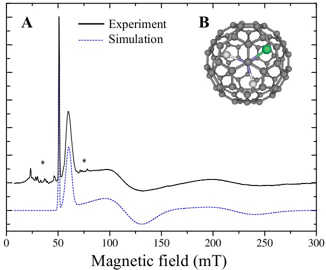
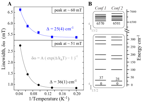
The CW EPR spectrum of ErSc2N@C80 in frozen toluene solution at 5 K shown in Figure 1. The widths of the two most intense features (at and 60 mT) are plotted as a function of temperature in Figure 2. Assuming that the features are homogeneously broadened and that the transverse relaxation time is limited only by the spin-lattice relaxation time then the width is proportional to -1. A common mechanism for relaxation in such systems with low-lying excited states is the two-phonon Orbach process Yen et al. (1965), for which
| (1) |
where represents the energy of the excited state which forms the relaxation pathway. The close correlation between (obtained from fits to Equation 1 shown as solid lines in Figure 2) and the energy of the first excited state as determined by PL Tiwari et al. (2007) suggests that these two features in the EPR spectrum are associated with the two principal configurations of ErSc2N within the fullerene cage. In order to explain the full EPR spectrum we similarly assign the broader features at higher field, though their weak intensity at 5 K makes the analogous temperature study more challenging.
Photoluminescence (PL) measurements demonstrate that the fullerene cage (and, as a result, the Er3+ ion) can be excited at 532 nm. Figure 3 shows the effect of such illumination on the CW EPR spectrum of ErSc2N@C80. The intensities of the principal features of the spectrum show a marked dependence on illumination: those centred at and 220 mT decrease in intensity, while those at about and 110 mT increase. This confirms that the high-field features have the same origin as the sharper low-field features, and indicates which pairs correspond to each of the primary configurations (which we term here Conf 1 and Conf 2). The CW spectrum in Figure 1 should therefore be modelled with two independent contributions, each generating one low-field feature and one broader high-field feature; and the relative intensities of the contributions reflect the relative populations of Conf 1 and Conf 2 configurations (see below).
Differences in the crystal field and spin-orbit coupling for the Er3+ ions in the two configurations produce two different effective spin systems. Although each is strictly a high spin system with a large crystal field splitting, only the ground state doublet is occupied at the temperatures studied here; the effective spin may be conveniently represented by an S=1/2 spin with an effective tensor. The dashed line in Figure 1 shows a simulation of the powder spectrum of a two such spin systems: , for Conf 1, and , for Conf 2. The narrow features marked with an asterisk in Figure 1 arise mostly from the hyperfine interactions with the 23% abundance of 167Er (), and some additional trace impurities in the resonator. Since the state of the Er3+ ion in this system may be measured by either photoluminesence or EPR, it may be possible to detect the magnetic resonance optically Bagraev et al. ((2006).
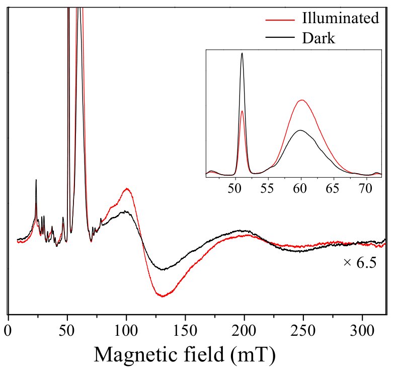
This interpretation implies that the relative populations of the two configurations of ErSc2N within the cage change under illumination at 532 nm. The change in spectra shown in Figure 3 is metastable, and persists for many hours after the illumination ceases. The original ‘dark’ spectrum can be recovered by annealing at K. Direct excitation of the Er3+ ion at 1496 and 1499 nm has no effect on the EPR spectrum, suggesting that the configuration is affected only by excitation of the fullerene cage or the molecule as a whole, and not by excitation of individual Er3+ ions. The magnitude of the effect shows a weak dependence on illumination wavelength in the region nm (the magnitude of the effect at 550 nm is that at 400 nm).
In order to deduce the change in populations between Conf 1 and Conf 2 from the change in the EPR spectrum under illumination, it is first necessary to relate the EPR intensity to the spin number. This relationship depends on many factors, including transition moment, temperature, microwave power and relaxation times. These terms can be combined into a constant of proportionality such that , where represents the intensity of any feature of the spectrum belonging to configuration , and is the number of molecules in configuration . Then, under the assumption that the total number of spin-active molecules does not change under illumination, the ratio may be extracted, and thus the fractional occupancy of the two sites before and after illumination. The results are summarised in Table 1, which examines the dependence of the two sharp features at 51 and 60 mT as a function of illumination.
![[Uncaptioned image]](/html/0802.0811/assets/x2.png)
These results lead us to the remarkable conclusion that illumination turns a slight excess of Conf 1 into a clear excess of Conf 2. It appears the Conf 1 is marginally favourable in the dark after annealing, but illumination at wavelengths of 532 nm or shorter switches the rotor preferentially to Conf 2, where it remains metastable for many hours.
Apparently, illumination at 532 nm generates an excited electronic state (either of the fullerene itself, or by driving charge transfer between the rotor and cage) in which Conf 2 is substantially favourable. The origin of such a bias is an intriguing question, and may require detailed molecular modeling in order to yield a definitive answer. X-ray diffraction studies have been performed where the ErSc2N@C80 is chemically modified to co-crystallise into host Olmstead et al. (2000). That study revealed two principal configurations of the rotor present across the whole sample which were highly correlated with the two different fullerene orientations arising from different cage functionalisations. It is plausible that the different electronic distributions in these two different cage sites influence the dominant configuration of the internal species. This is consistent with our observation that for pristine cages optical excitation can lead to a switching of the majority species. It is known that the ErSc2N rotor exhibits an electric dipole moment caused by the differences in electronegativity of erbium and scandium, as evidenced by a high degree of order observed in the XRD study of Ref Olmstead et al. (2000). Whether illumination creates an exciton on the fullerene or drives a charge transfer between the central species and the cage, the electric dipole associated with the cage may assist in producing the observed alignment.
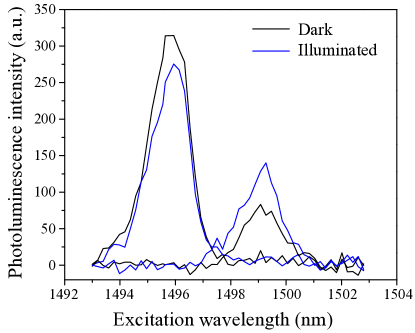
The PLE experiments described in Ref Tiwari et al. (2007) permit the selective excitation of Er3+ in the two sites, and could therefore also be used to observe the photoisomerisation described above. Alignment of the optical apparatus was performed with a 532 nm laser beam, as is common practice, at 50 K (i.e. above the configuration annealing temperature of about 30 K found from EPR experiments above). The sample was then cooled down to 5 K and excited with an infra-red laser. For the two configurations, the PLE spectra were acquired while monitoring the fluorescence at respectively 1522 nm (PL peak attributed to Conf 1) and 1519 nm (PL peak attributed to Conf 2). In the range 1490–1500 nm, PLE peaks were found at 1496 nm for Conf 1 and 1499 nm for Conf 2. 532 nm illumination was then applied for 20 minutes at 5 K and turned off, and a second set of PLE spectra were taken. The results are shown in Figure 4 and unambiguously demonstrate a a shift in site occupancy, as observed in EPR, though the noise level in this experiment precludes a quantitative comparison of occupancies.
In summary, the EPR spectrum of ErSc2N@C80 reveals two species of Er3+, each with a high degree of apparent axial anisotropy (as expected for the case where the crystal field is much larger than the Zeeman splitting), corresponding to two configurations of the ErSc2N rotor within the fullerene cage. The relative occupancies of these two orientations can be optically switched, as demonstrated in both EPR and PLE studies. The switching speed and reversibility of this photoisomerisation are yet to be determined; however, the long lifetime (over 12 hours at 20 K) of the switched orientation may find application in molecular memory elements.
We thank William Hayes and Alexei Tyryshkin for valuable discussions. This research is supported by the EPSRC through the QIP IRC www.qipirc.org (GR/S82176/01), and the Oxford Centre of Advanced Electron Spin Resonance (EP/D048559/1). JJLM is supported by St. John’s College, Oxford. AA is supported by the Royal Society. GADB is supported by the EPSRC (GR/S15808/01).
References
- Choi et al. (2006) B.-Y. Choi, S.-J. Kahng, S. Kim, H. Kim, H. W. Kim, Y. J. Song, J. Ihm, and Y. Kuk, Phys. Rev. Lett. 96, 156106 (2006).
- DeBoer et al. (1968) B. G. DeBoer, A. Zalkin, and D. H. Templeton, J. Am. Chem. Soc. 7, 1085 (1968).
- Nägele et al. (1997) T. Nägele, R. Hoche, W. Zinth, and J. Wachtveitla, Chem. Phys. Lett. 272, 489 (1997).
- Yoshizawa and Wald (1967) T. Yoshizawa and G. Wald, Nature 214, 566 (1967).
- Liu et al. (1990) Z. F. Liu, K. Hashimoto, and A. Fujishima, Nature 347, 658 (1990).
- Szobota et al. (2007) S. Szobota, P. Gorostiza, F. D. Bene, C. Wyart, D. L. Fortin, K. D. Kolstad, O. Tulyathan, M. Volgraf, R. Numano, H. L. Aaron, et al., Neuron 54, 535 (2007).
- Krause et al. (2004) M. Krause, M. Hulman, H. Kuzmany, O. Dubay, G. Kresse, K. Vietze, G. Seifert, C.Wang, and H. Shinohara, Phys. Rev. Lett. 93, 137403 (2004).
- AlmeidaMurphy et al. (1996) T. AlmeidaMurphy, T. Pawlik, A. Weidinger, M. Hohne, R. Alcala, and J. M. Spaeth, Phys. Rev. Lett. 77, 1075 (1996).
- Stevenson et al. (1999) S. Stevenson, G. Rice, T. Glass, K. Harich, F. Cromer, M. R. Jordan, J. Craft, E. Hadju, R. Bible, M. M. Olmstead, et al., Nature 401, 55 (1999).
- Olmstead et al. (2000) M. M. Olmstead, A. de Bettencourt-Dias, J. C. Duchamp, S. Stevenson, H. C. Dorn, and A. L. Balch, J. Am. Chem. Soc. 122, 12220 (2000).
- Tiwari et al. (2007) A. Tiwari, G. Dantelle, K. Porfyrakis, R. A. Taylor, A. A. R. Watt, A. Ardavan, and G. A. D. Briggs, J. Chem. Phys.in press (2007).
- Shinohara (2000) H. Shinohara, Rep. Prog. Phys. 63, 843 (2000).
- Jones et al. (2006a) M. A. G. Jones, R. A. Taylor, A. Ardavan, K. Porfyrakis, and G. A. D. Briggs, Chem. Phys. Lett. 428, 303 (2006a).
- Becker et al. (1999) P. C. Becker, N. A. Olsson, and J. R. Simpson, Erbium-Doped Fiber Amplifiers (Elsevier, 1999).
- Baker et al. (1959) J. M. Baker, W. Hayes, and D. A. Jones, Proc. Phys. Soc. 73, 942 (1959).
- Guillot-Noël et al. (2006) O. Guillot-Noël, P. Goldner, Y. L. Du, E. Baldit, P. Monnier, and K. Bencheikh, Phys. Rev. B 74, 214409 (2006).
- Hutchison et al. (1989) C. A. Hutchison, S. G. Utterback, and P. M. Martineau, Phys. Rev. B 39, 4051 (1989).
- Bravo et al. (1999) D. Bravo, A. Martín, and F. J. López, Solid State Comms 112, 541 (1999).
- Nolte et al. (1997) T. Nolte, T. Pawlik, and J. M. Spaeth, Solid State Comms 104, 535 (1997).
- Bravo et al. (2006) D. Bravo, A. Martín, J. J. Carvajal, M. Aguiló, F. Díaz, and F. J. López, J. Phys. Cond. Matt. 18, 6655 (2006).
- Abragam and Bleaney (1970) A. Abragam and B. Bleaney, Electron Paramagnetic Resonance of Transition Ions (Clarendon Press, Oxford, 1970).
- Jones et al. (2006b) M. A. G. Jones, J. J. L. Morton, R. A. Taylor, A. Ardavan, and G. A. D. Briggs, Physica Status Solidi B 243, 3037 (2006b).
- Stoll and Schweiger (2006) S. Stoll and A. Schweiger, J. Mag. Res. 178, 42 (2006).
- Yen et al. (1965) W. M. Yen, W. C. Scott, and P. L. Scott, Phys. Rev. 137, A1109 (1965).
- Bagraev et al. ((2006) N. Bagraev, W. Gehlhoff, L. Klyachkin, A. Malyarenko, V. Mashkov, V. Romanov, and T. Shelykh, arxiv.org/cond-mat/0608630 ((2006)).