Three-Dimensional Bulk Electronic Structures of Ca1.5Sr0.5RuO4 Studied by Soft X-ray Angle-Resolved Photoemission
Abstract
We report on experimental data of the three-dimensional bulk Fermi surfaces of the layered strongly correlated Ca1.5Sr0.5RuO4 system. The measurements have been performed by means of -depndent bulk-sensitive soft x-ray angle-resolved photoemission technique. Our experimental data evinces the bulk Fermi surface topology at 0 to be qualitatively different from the one observed by surface-sensitive low-energy ARPES. Furthermore, stronger dispersion of the circle-like Fermi surface sheet is observed compared with Sr2RuO4. Thus in the paramagnetic metal phase, Ca1.5Sr0.5RuO4 compound is found to have rather three-dimensional electronic structure.
pacs:
79.60.-i, 71.20.-b, 71.30.+hStrongly correlated transition metal oxides are widely studied because of a variety of such intriguing phenomena as (high-temperature) anisotropic superconductivity, Mott transition, magnetic and/or orbital ordering, and large mass enhancement. Among them, the single-layered perovskite Ca2-xSrxRuO4 is particularly interesting, since it shows various phases as functions of temperature and Maeno ; Nakatsuji . By substituting Sr2+-ions for isovalent Ca2+-ions, the unconventional superconductivity takes place below 1.5 K for . A paramagnetically metallic behavior with strong electron correlation is seen in a wide temperature region for . The system shows a paramagnetic metal to “magnetic” metal transition at 10 K, for , and eventually becomes a Mott insulator for . At the Sr concentration of , it is furthermore known that the effective mass diverges below 10 K although the system does not undergo a Mott transition Nakatsuji ; cluster . These phenomena are thought to originate not only from the enhanced electron correlation by the rotation of the RuO6 octahedra (Ru-O-Ru bonds are bent from ideal ones), but also from the reduction of orbital degree of freedom by a Jahn-Teller distortion OSMT ; oo1 ; oo2 ; oo3 and/or a Ru 4 spin-orbit interaction. Indeed, a (partial) Ru 4 orbital-ordering has been reported (proposed) for Zegkinoglou ( cluster ; OSMT ).
Angle-resolved photoemission (ARPES) is a very powerful tool to evince band dispersions and Fermi surface (FS) topology. So far, several low- ARPES experiments were performed for Ca2-xSrxRuO4 low1 ; KMShen ; surface ; low2 . However, it is known that low-energy photoemission is surface-sensitive and often provides spectral shapes which are not consistent with bulk electronic structures HE1 ; HE2 ; PRL93 ; Suga1 . Meanwhile, it has been demonstrated that high- ARPES with use of the soft x-ray can reveal detailed band dispersions of the bulk electronic states SekiyamaPRB ; Suga2 ; Yano . In addition, by virtue of the longer mean free path of higher kinetic energy photoelectrons the high-energy -dependent ARPES can probe electronic structures of three-dimensional compounds with better-resolved dispersion ( - momentum component perpendicular to the cleaved sample surface) Yano ; MFP ; LSFO .
High quality single crystal of Ca1.5Sr0.5RuO4 ( = 0.5), which was grown by the floating zone method FZM , was used for the measurement. All ARPES measurements were performed at BL25SU in SPring-8 Saitoh . The high-energy ( = 708 eV) and low-energy ( = 362 eV) soft x-ray ARPES and FS mapping were carried out for the 0 plane. In addition, dependent ( = 650-730 eV) ARPES for the =0 plane FS mapping was done with the energy step of 5 eV. The base pressure was about Pa. The (001) clean surface was obtained by cleaving the sample in situ at the measuring temperature of 20 K. The overall energy resolution was set to 200 meV. The angular resolution was 0.15∘(0.25∘) for the parallel (perpendicular) direction to the analyzer slit.
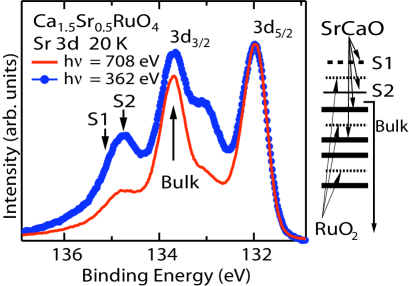
Figure 1 shows the dependence of the angle-integrated Sr 3 core-level photoemission (PES) spectra of Ca1.5Sr0.5RuO4. To our understanding these spectra contain three components corresponding to the contributions from the top-most SrCaO surface layer (S1), the second SrCaO surface layer (S2) located just below the top RuO2 layer, and the bulk layers SekiyamaPRB . The intensity of the S1 and S2 shoulders is observed to be remarkably stronger for = 362 eV (blue curve) than for = 708 eV (red curve). To estimate the bulk contribution to the spectra we did line-shape analysis in the same manner as in Ref. SekiyamaPRB . The bulk contribution is estimated to be 55% (39%) in the Sr spectra at = 708 (362) eV. These values are close to the ones evaluated for the given lattice constants and the calculated photoelectron mean free path MFP . Then, the bulk contribution in the valence-band is found to be 65 % at = 708 eV and 50 % at = 362 eV, manifesting that the 708 eV-PES mainly probes the bulk valence states.
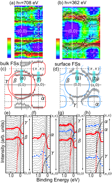
In the following, we show the FSs derived from the 708 eV- and 362 eV-ARPES spectral intensity integrated from 0.1 to 0.1 eV with respect to the Fermi level () (Figs. 2 (a) and (b)). These results are obtained from energy distribution curves (EDCs) (Figs. 2 (e)-(h)) in the paramagnetic metal phase at 20 K. Figures 2(a) and 2(b) display raw data and Figs. 2(c) and 2(d) summarize remarkable features (see captions). We have obtained (-crossing points in a reciprocal space) from both EDCs and momentum distribution curves (MDCs: not shown here) as plotted in Figs. 2 (a) and (b) with a two-dimensional cubic symmetry BZ. Although the true BZ of Ca1.5Sr0.5RuO4 is folded because of the rotation RuO6 octahedra along the -axis (12∘ oo1 ), we use the size of BZ and notations for high symmetry directions of the Sr2RuO4 crystal structure. EDCs in Figs. 2(e)-(h) correspond to shaded areas in Figs. 2(c) and (d). As shown in Fig. 2(e), two branches ( and ) located at 0.5 eV at (0,0) approach and cross between (0,0) and (0,) in the 708 eV-ARPES spectra. In the 362 eV-ARPES spectra in Fig. 2(g), we observed one additional branch between these two branches crossing . Considering that the 362 eV-ARPES is more surface sensitive, we attribute this additional branch (which spectral weight is noticeably suppressed in the 708 eV-ARPES spectra) to be resulting from the surface band different from the bulk one. Along the (,0)-(,) direction, we have likewise found that two branches cross in the 708 eV-ARPES spectra (Fig. 2(f)), whereas three branches cross in the 362 eV-ARPES spectra (Fig. 2(h)). Hence essentially dissimilar electronic structures and Fermi surface topology (at 0) between the bulk and surface states for this “quasi” two-dimensional electron system are demonstrated.
As shown in Figs. 2(a) and 2(c) by 708 eV-ARPES measurements, we have found three bulk FS sheets for Ca1.5Sr0.5RuO4, namely, one hole-like square-shaped sheet centered at (,), one electron-like square-shaped sheet and one electron-like circle-shaped sheet both centered at (0,0). Our FS topology observed for 0 plane is thus very similar to that of bulk sensetive experimental data for Sr2RuO4 and Ca0.2Sr1.8RuO4 SekiyamaPRB . If one assume that orbital character for the FS sheets remains the same as for Sr2RuO4, then and sheets should correspond mainly to the Ru orbitals whereas the sheet has mainly the Ru character. Possible effects of the BZ folding caused by the RuO6 rotation EKo are not seen (or negligibly weak) in our results in strong contrast to the low-energy ARPES result for Sr2RhO4 and Sr2RuO4 surface SRhO ; KMShen . The reason for the absence of the BZ folding effect is, however, not clear at present.
On the other hand, the 362 eV-ARPES results (Fig. 2(b)) clearly reveal three surface FS sheets (open circles) distinguished from the bulk FS sheets. While the shape of the surface sheet is rather similar to the bulk one as recognized by comparing Figs. 2(c) and (d), the electron-like surface sheet is much more circle-like compared to the bulk sheet. Furthermore, the FS topology of the sheet is qualitatively different between the bulk in Fig. 2(c) and surface in Fig. 2(d). The hole-like surface sheet observed here well traces the result of the low-energy ARPES at = 32 eV surface reproduced by the dashed curves in Fig. 2(d). The scanning tunneling microscopy (STM) measurement of Sr2RuO4 has shown a surface reconstruction Matzdorf . Except for this report no surface reconstruction has been reported for Ca2-xSrxRuO4. It has been believed that the bulk FSs can be detected even by the surface-sensitive low-energy ARPES for Ca2-xSrxRuO4 since the RuO6 rotation angle indicates similar crystal structures between the bulk (12∘) and surface (11∘) surface . However, our result evinces that the surface FS topology is noticeably different from the bulk.
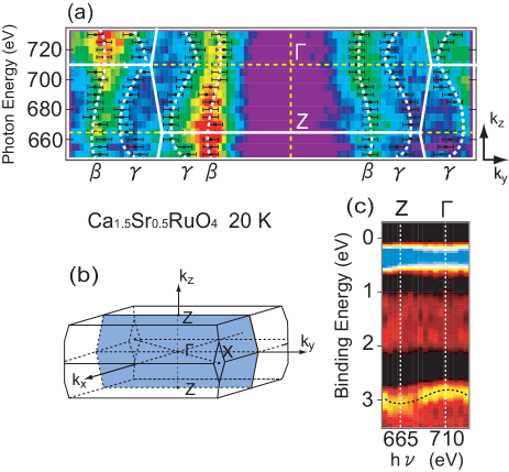
Below we discuss experimental data on interlayer coherent electron hopping or in other words dispersion of the FS observed by our bulk sensetive dependent ARPES measurements. A nearly free photoelectron model calculation with the inner potential of 9 eV identifies and Z points of a tetragonal BZ with s of 712 and 664 eV in our experiment comment .
Figure 3(a) shows the results of -dependent ARPES for Ca1.5Sr0.5RuO4, representing the dispesion of the and the FS sheets. The FS sheet is found to have significant dependence reflecting stronger three-dimensionality of the Ca1.5Sr0.5RuO4 electronic structure. This is in strong contrast to results of quantum oscillation measurements for Sr2RuO4 showing negligible dependence for the sheet dHvA ; Mackenzie . On the other hand, our result has revealed much weaker dependence of the FS sheet compared with that of the FS sheet. Indeed, a ratio of between the minimum and maximum along the -Z direction has been estimated to be 0.9 for the sheet whereas for the sheet it is 0.8. So far it has been considered that the electronic structures and FSs of such single-layered transition metal oxides as Ca2-xSrxRuO4 are highly two-dimensional. However, those for Ca1.5Sr0.5RuO4 are found to be more three-dimensional than Sr2RuO4 from our experiments.
As shown in Fig. 3(c), the spectral weight near 3 eV in the ARPES data disperses clearly as a function of . Its bottom (top) is located at Z (). This dispersion is qualitatively consistent with results of the band-structure calculation for Sr2RuO4 along the -Z () direction Oguchi ; dHvA ; SRODMFT . However there seems to be also non-dispersive contribution at 3 eV. These -independent spectral structure observed at 1-2 and 3 eV can be presumably identified as the part of the Ru 4d(t2g) lower Hubbard band similar to our recent LDA+DMFT(QMC) results for Sr2RuO4 SRODMFT . Namely, correlations in Ca1.5Sr0.5RuO4 can be enhanced due to distortion in comaprison with Sr2RuO4.
Since the bulk FS topology of Ca1.5Sr0.5RuO4 at 0 observed by us is similar to Sr2RuO4 one can expect the orbital characters of the relatively large and small sheets to be of the Ru and symmetries, respectively. In that way our results seem to contradict with theoretical expectation where strong dependence is usually expected for the Ru -derived bands rather than for the Ru band Oguchi ; dHvA ; SRODMFT . One possible explanation of our ARPES results is that the characters of the and FS sheets are interchanged with each other compared with those for Sr2RuO4. Our LDA (local density approximation) band-structure calculation LDA as well as that in Ref. EKo predict such a scenario as the smallest electron-like two-dimensional FS sheet originats from the Ru band. But this FS sheet is caused by doubling of the lattice in Ca1.5Sr0.5RuO4. As mentioned above in our ARPES results, bands coming from folding are negligibly weak. We have surveyed not only the reciprocal space shown in Figs. 2 and 3 but also the plane at = 650 eV (not shown here) covering first and second BZs by ARPES, therefore the absence of these hole-like FS sheets is thought to be intrinsic in this experiment for Ca1.5Sr0.5RuO4 system.
Anoher scenario of rather strong dependence of the sheet can be by the effect of the RuO6 rotation within the conducting plane. By virtue of the rotation, there appears small but finite -bonding between the Ru and the O orbitals. So the situation becomes similar to high-TC cuprate La2CuO4 Pavarini , where it was shown that such -bonding leads to essentially three-dimensional -symmetry Wannier function. For Ca1.5Sr0.5RuO4 this effect is weaker than for La2CuO4. Nevertheless Ru -derived band in the distorted crystal structure can form rather three-dimensional sheet. Also one should mention here that more two-dimensional square-shaped sheet has stronger nesting instability as observed in Sr2RuO4 and Ca0.2Sr1.8RuO4 SekiyamaPRB ; Sidis . Thus the Ru orbital ordering scenario within the magnetic metal phase for OSMT is possible.
To summarize, we have observed three-dimensional Fermi surfaces of Ca1.5Sr0.5RuO4 in the paramagnetic metal phase at 20 K by applying bulk sensetive soft x-ray -dependent ARPES technique. We have revealed the genuine bulk electronic structures which are different from surface sensetive data for this material. FS topology in the 0 plane is observed to be qualitatively similar with bulk FS of Sr2RuO4. However remarkable dependence of the FS sheet have proved Ca1.5Sr0.5RuO4 to have more three-dimensional electronic structure than Sr2RuO4.
We are grateful to H. Higashimichi, G. Funabashi, and T. Nakamura for supporting the experiments. This work was supported by a Grant-in-Aid for Scientific Research (15GS0213, 1814007, 18684015) and the 21st COE program (G18) of the Japan Society for the Promotion of Science and MEXT. This work was also supported by Hyogo Science and Technology Association. The ARPES was performed at SPring-8 under the approval of JASRI (2006A1169, 2007A1005). IN thanks RFFI grants 08-02-00021, 08-02-00712, 08-02-91200, 06-02-90537, travel grant of UB RAS and grant of President of Russia MK-2242.2007.2.
References
- (1) Y. Maeno et al., Nature 372, 532 (1994).
- (2) S. Nakatsuji and Y. Maeno, Phys. Rev. Lett. 84, 2666 (2000).
- (3) S. Nakatsuji et al., J. Phys. Rev. Lett. 90, 137202 (2003).
- (4) V. I. Anisimov et al., Eur. Phys. J. B 25, 191 (2002).
- (5) O. Friedt et al., Phys. Rev. B 63, 174432 (2001).
- (6) T. Mizokawa et al., Phys. Rev. Lett. 87, 077202 (2001).
- (7) T. Hotta and E. Dagotto, Phys. Rev. Lett. 88, 017201 (2001).
- (8) I. Zegkinoglou et al., Phys. Rev. Lett. 95, 136401 (2005).
- (9) A. Damascelli et al., Phys. Rev. Lett. 85, 5194 (2000).
- (10) K. M. Shen et al., Phys. Rev. B 64, 180502(R) (2001).
- (11) S.-C. Wang et al., Phys. Rev. Lett. 93, 177007 (2004).
- (12) J. Zhang et al., Phys. Rev. Lett. 96, 066401 (2006).
- (13) A. Sekiyama et al., Nature (London) 403, 396 (2000).
- (14) A. Sekiyama et al., J. Phys. Soc. Jpn. 69, 2771 (2000).
- (15) A. Sekiyama et al., Phys. Rev. Lett. 93, 156402 (2004).
- (16) S. Suga et al., J. Phys. Soc. Jpn. 74, 2880 (2005).
- (17) A. Sekiyama et al., Phys. Rev. B 70, 060506(R) (2004).
- (18) S. Suga et al., Phys. Rev. B 70, 155106 (2004).
- (19) M. Yano et al., Phys. Rev. Lett. 98, 036405 (2007).
- (20) S. Tanuma, C. J. Powell, and D. R. Penn, J. Vac. Sci. Technol. A 8, 2213 (1990).
- (21) H. Wadati et al., Phys. Rev. B 71, 035108 (2005).
- (22) S. Nakatsuji and Y. Maeno, J. Solid State Chem. 156, 26 (2001).
- (23) Y. Saitoh et al., Rev. Sci. Instrum. 71, 3254 (2000).
- (24) E. Ko, B. J. Kim, C. Kim, and H. J. Choi, Phys. Rev. Lett. 98, 226401 (2007).
- (25) B. J. Kim et al., Phys. Rev. Lett. 97, 106401 (2006).
- (26) R. Matzdorf et al., Science 289, 746 (2000).
- (27) We have measured the ARPES spectra with the polar angles near 0∘ and at the photon incident angle of 45∘.
- (28) T. Oguchi, Phys. Rev. B 51, 1385 (1995).
- (29) Y. Yoshida et al., J. Phys. Soc. Jpn. 67, 1677 (1998).
- (30) Z. V. Pchelkina et al., Phys. Rev. B 75, 035122 (2007).
- (31) A. P. Mackenzie et al., Phys. Rev. Lett. 76, 3786 (1996).
- (32) We perform TB-LMTO [O.K. Andersen and O. Jepsen Phys. Rev. Lett 53, 2571 (1984)] calculations with following NMTO [O.K. Andersen and T. Saha-Dasgupta, Phys. Rev. B 62, R16219 (2000)] analysis.
- (33) E. Pavarini et al., Phys. Rev. Lett. 87, 047003 (2001).
- (34) Y. Sidis et al., Phys. Rev. Lett. 83, 3320 (1999).