Calculations of the Exciton Coupling Elements Between the DNA Bases Using the Transition Density Cube Method
Abstract
Excited states of the of the double-stranded DNA model (A)(T)12 were calculated in the framework of the exciton theory. The off-diagonal elements of the exciton matrix were calculated using the transition densities and ideal dipole approximation associated with the lowest energy excitations of the individual nucleobases obtained from TDDFT calculations. The values of the coupling calculated with the transition density cubes (TDC) and ideal-dipole approximation (IDA) methods were found significantly different for the small inter-chromophore distances. It was shown that the IDA overestimates the coupling significantly. The effects of the structural fluctuations were incorporated by averaging the properties of the excited states over a large number of conformations obtained from the MD simulations.
I Introduction
DNA a remarkable carrier of code of life, is very stable with respect to the photochemical decay. The path chosen by Nature to protect DNA is through the very rapid decay pathways of the electronic excitation energy. Given the importance of DNA in biological systems and its emerging role as a scaffold and conduit for electronic transport in molecular electronic devices, Kelley and Barton (1999) DNA in its many forms is a well studied and well characterized system. What remains poorly understood, however, is the role that base-pairing and base-stacking plays in the transport and migration of the initial excitation along the double helix.Crespo-Hernandez et al. (2005); Markovitsi et al. (2005, 2006)
The absorption of UV radiation by DNA initiate a number of photochemical reactions that can ultimately lead to carcinogenic mutations. Besaratinia et al. (2005); Sutherland et al. (1976); Callis (1979); Sinha and Hädler (2002); Freeman et al. (1989) The UV absorption spectrum of DNA largely represents the weighted sum of the absorption spectra of it constituent bases. However, the distribution of the primary photochemical products of UV radiation, including bipyrimidine dimers, Mouret et al. (2006) is depends quite strongly upon base sequence, which implies some degree of coupling between the DNA bases. Markovitsi et al. (2005) Inasmuch as both the base stacking and base pairing are suspected to mediate the excess of electronic excitation energy, understanding of the excited-state dynamics is of primary importance for determining how the local environment affects the formation of DNA photolesions.
Recent work by various groups has underscored the different roles that base-stacking and base-pairing play in mediating the fate of an electronic excitation in DNA. Markovitsi et al. (2005); Crespo-Hernandez et al. (2005) Over 40 years ago, Löwdin discussed proton tunneling between bases as a excited state deactivation mechanism in DNALöwdin (1963) and evidence of this was recently reported by Schultz et al. Schultz et al. (2004) In contrast, ultrafast fluorescence of double helix poly(dA)·poly(dT) oligomers by Crespo-Hernandez et al.Crespo-Hernandez et al. (2005) and by Markovitsi et al. Markovitsi et al. (2005) give compelling evidence that base-stacking rather than base-pairing largely determines the fate of an excited state in DNA chains composed of adenosine and thymine bases with long-lived intrastrand states forming when ever adenosine is stacked with itself or with thymine. However, there is considerable debate regarding whether or not the dynamics can be explained via purely Frenkel exciton models Emanuele et al. (2005a, b); Markovitsi et al. (2006) or whether charge-transfer states play an intermediate role. Crespo-Hernandez et al. (2006)
Upon UV excitation, the majority of excited molecules shows a subpicosecund singlet lifetimes. Pecourt et al. (2001, 2000); Gustavsson et al. (2002); Peon and Zewail (2001) Owing to the technical difficulties in measuring the ultrashort lifetimes the study of the charge and excitation energy transfer in DNA has only recently received much of attention with the advances in the femtosecond spectroscopy. Although, so far, no clear picture of the excited -state deactivation mechanism has been offered by the experiment, two possible decay channels have been investigated. Kohler and coworkers in their recent study of the duplex poly(dA)·poly(dT) suggested that -stacking of the DNA base determines the fate of a singlet electronic excited state.Crespo-Hernandez et al. (2005) Alternative decay mechanism involves interstrand hydrogen or proton transfer. Douhal and coworkers observed excited-state proton transfer in base pair mimincs in gas-phase. Douhal et al. (1995) The experimental results suggests that these very fast decay pathways play an important role in quenching the reactive decay channels and providing DNA with intrinsic photochemical stability. However, they do not provide a clear picture which arrangement of bases, pairing or stacking, is of primary importance.
Until recently, most theoretical investigations of excitation energy transfer in DNA helices has been within the Frenkel exciton model which treats the excitation as a coherent hopping process between adjacent bases.Shapiro et al. (1975); Suhai (1984) This model has tremendous appeal since it allows one to construct the global excited states (i.e of the complete chain) in terms of linear combinations of local excited states. The key parameter in the evaluation of the electronic excitation energy transfer (EET) is the electronic coupling between the individual bases. To a first-order approximation, the base to base coupling can be estimated using a dipole-dipole approximation in which the interaction between the donor and acceptor is calculated using only the transition dipole associated with each chromophore. While this approach is certainly suitable for cases in which the distance between the donor and acceptor sites is substantially greater than the molecular length scale. In case of double stranded DNA, where the DNA bases are in relatively close contact compared to their dimensions this approach leads to the neglect of the effect of the size and spatial extent of the interacting transition densities associated with each chromophore.
By far the most precise way to calculate the coupling elements is to directly integrate the Coulomb coupling matrix element between transition densities localized on the respective basis.Krueger et al. (1998) The accuracy is then limited only by the numerical quadrature in integrating the matrix element and by the level and accuracy of the quantum chemical approach used to construct the transition densities in the first place. Futhermore, one must perform a quantum chemical evaluation of the coupling elements between each base at each snapshot along a molecular dynamics simulation in order to properly take into account the fluctuations and gyrations of the chain itself. This is a formidable task, one that has prevented an accurate benchmarking of the excited state electronic structure of realistic DNA chains.
In this paper, we present the results of simulations and calculations of accurate interbase exciton couplings for A-T strands of DNA in water in an attempt to provide such a benchmark. Starting from a molecular dynamics simulation of a model DNA sequence in water at the correct salt concentrations, we mapped out the evolution of the photochemically relevant excited states within a Frenkel exciton model in which the couplings were computed using both the ideal dipole-dipole approximation (IDA) and using the transition density cube approach (TDC).Krueger et al. (1998)
II Methodology
The calculation procedure consisted of several steps. In the first stage the molecular dynamics (MD) calculations were carried out to sample a range of conformations of (A)(T)12 model of DNA double-helix. The transition densities of the individual nucleobases obtained from time dependent density functional theory (TDDFT) calculations were subsequently superimposed with the instantaneous conformations from the MD simulations in order to calculate the coupling between the electronic transitions of the individual bases. In the final step, the excited-states of the model were calculated within the Frankel exciton model.
II.1 Exciton model
The excited states of the were calculated in the framework of the exciton theoryFrenkel (1931); Davydov (1971). In this approach the total Hamiltonian for the super system of molecules is written as the sum of Hamiltonians of isolated molecules and the intermolecular interaction potential between the molecules and .
| (1) |
The singly excited states of the system are described in term of locally excited configurations
| (2) |
where corresponds to the excited state wavefunction of the chromophore whereas all the other molecules are in their ground state . denotes the corresponding wave function of the super system. Consquently, the exciton states of the supramolecular system can be written as a linear combinations of the excited states localized on each chromophore.
| (3) |
The diagonal elements of the exciton matrix are simply excitation energies of chromophore from its ground to ith excited state, . The off-diagonal elements written as correspond to exciton coupling. It can be interpreted as the electrostatic interaction energy between the transition densities corresponding to and .
A measure of delocalization of the exciton states can be obtained from the inverse participation ratio (IPR) () which represents the number of coherently coupled chromophores in a given eigenstate . In the general case with more than one electronic transition per chromophore, is written as follows:
| (4) |
where denotes a given eigenstate and an electronic excited state of a chromophore.

For purposes of developing a model, we can cast the exciton Hamiltonian as a lattice model Bittner (2006) consisting of localized hopping interactions for exctions between adjacent base pairs along each strand () as well as cross-strand terms linking paired bases () and “diagonal” terms which account for the stacking interaction between base on one chain and base on the other chain () in which denotes coupling in the 5’-5’ direction and coupling in the 3’-3’ direction. Fig. 1 shows a schematic view of the various coupling terms between each nucleotide base.
| (5) |
where and are spinors that act on the ground-state to create and annihilate excitations on the th adenosine or thymidine base along the chain. The operators are the Pauli spin matrices with and providing the mixing between the two chains.
Taking the chain to homogeneous and infinite in extent, one can easily determine the energy spectrum of the valence and conduction bands by diagonalizing
| (8) |
where and are local excitation energies and intra-strand hopping integrals. is the coupling between Watson-Crick bases. When the interchain diagonal couplings are equal, , Eq. 8 is identical to the Hamiltonian used by Creutz and Horvath Creutz and Horvgath (1994) to describe chiral symmetry in quantum chromodynamics in which the terms proportional to are introduced to make the “doublers” at heavier than the states at since the off-diagonal coupling is now momentum dependent.
One of the serious deficiencies with this model as it stands thus far is that for DNA each of the interactions described is very sensitive to the geometric fluctuations of the DNA chain itself. Emanuele et al. (2005b, a) Hence, we need to consider each of the couplings as being parametrically dependent upon the instantaneous molecular geometry of both the individual bases and the chain itself. This is assuming there is no additional contribution from the solvent and ions surrounding the DNA chain. Assuming that the electronic time scale is fast compared to the typical time scale for geometric fluctuations of the DNA chain (s for longitudinal and s for the lateral motions of bases in DNA double helicesMcCammon and Harvey (1987)), we can consider at least the initial electronic dynamics as occurring in a fixed nuclear framework and subsequent dynamics as adiabatically following the nuclear motion. Nonadiabatic contributions can not be completely discounted; however, the dominant non-adiabatic couplings are intermolecular in origin or involve proton between adjacent bases. Clelia et al. (2005); Sobolewski and Domcke (2002); Löwdin (1963)
II.2 Transition densities and interactions
II.2.1 Exciton-exciton interactions
Each off-diagonal term in our Hamiltonian of Eq. 5 can be calculated according to
| (9) |
where the two terms in the numerator, and are the three dimensional charge distributions (transition densities) associated with the ground and electronic excited states and of molecules and , respectively, with the separation between the elements and equal to . The corresponds to the electrostatic repulsion energy between the two charge distributions and of isolated chromophores. The calculations of the Coulombic couplings using the three dimensional charge distribution takes into account the size and the spatial extent of the transition density and is valid at all molecular separations as opposed to the ideal dipole approximation (IDA). In the latter only the dipole moment of the transition density is considered for calculations of the coupling terms which makes the computations of the off-diagonal elements much more efficient. However, this approximation breaks down at the small donor-acceptor separations for which the spatial extent of the transition density becomes important.
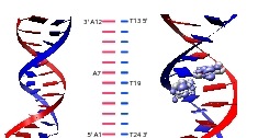
To account for the dynamics of the DNA chain itself, we performed a series of molecular simulations of the 12 base pair duplex DNA (AT) (Figure 2) with about 12,000 water molecules and counter ions. 111 Regarding the molecular dynamics simulations: the simulation was performed with the extended system (ESP) molecular dynamics program ESP . The system consisted of a 12 base pair duplex DNA (AT) with waters, sodium ions, and chloride ions in a cubic box of length Å. The atomic interactions were defined by the CHARMM (version ) force field A. D. MacKerell et al. (1998). The system was minimized and equilibrated in the NVE ensemble at K. The bonds were kept rigid using the Rattle Andersen (1983) implementation of the Shake method Ryckaert et al. (1977) and the electrostatic interactions were evaluated using the Ewald sum technique Leeuw et al. (1980). The equations of motion were integrated using the Velocity Verlet algorithm Swope et al. (1982) with a femtosecond time step. The simulation was initially run for nanoseconds. Next, the timestep was changed to femtosecond and the snapshots saved every steps. Once the system was minimized and equilibrated in the NVE ensemble at K, we integrated the dynamics for an additional 80 ps, sampling the DNA configuration every 10 fs. Even though we are dealing with a relatively small strand, it remains too large for an accurate evaluation of its electronic structure. Consequently, we make the approximation that the excited states of the molecule itself can be written as a linear combination of excited states localized on the instantaneous positions of each base along the chain. Furthermore, given the computational cost associated with evaluating the excited states of even a small molecule, it is prohibitive to perform such calculations for each base at each time-step. Our approach, then, is to perform an accurate evaluation of the local transition densities based upon the geometries of the isolated DNA bases, then map these densities onto the instantaneous positions of the bases from the molecular dynamics simulations (Figure 2). From this, we can evaluate the exciton-exciton coupling (Eq. 9) in which and are the transition densities about the instantaneous positions of bases and .
II.2.2 Excited states of individual bases
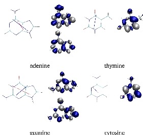
The geometries of the DNA bases, adenine, guanine, cytosine, and thymine in their most common tautomeric forms were optimized at the MP2/TZVP level of theory in chloroform using Gaussian03 suite of programs.Frisch et al. The optimized geometries were subsequently used to calculate the singlet excitation energies in gas phase at the TD-DFT level using PBE0 functional and TZVP basis set augmented with the diffusion functions on all atoms as implemented in ORCA.Neese (2006) Additionally, the excitation energies were also calculated for the standard nucleobase geometries obtained from the X3DNA.Lu and Olson (2003) In these calculations the deoxyribose and phosphate groups were replaced with hydrogens using the Chimera program.Pettersen et al. (2004) Without further optimization of the structures, the excitation energies were calculated at the same level of theory as used before for the MP2 optimized structures. Fig. 3 shows both the transition density and direction of the transition dipole moment for each base as given by TDDFT after optimization at the MP2/TZVP level in a CHCl3. Transition moments were calculated using TDDFT with PBE0 functional and aug-TZVP basis set in vacuum. The calculated excitation energies are summarized in Table 1.
| Method | Geometry | ||||||
|---|---|---|---|---|---|---|---|
| A | TDDFT | MP2 | 5.00 (0.002) | 5.29 (0.230) | 5.38 (0.069) | 5.49 (0.009) | |
| standard | 5.17 (0.001) | 5.44 (0.204) | 5.46 (0.006) | 5.52 (0.086) | |||
| MRCI | MP2 | 4.80 (0.168) | 5.01 (0.003) | 5.13 (0.446) | 5.13 (0.004) | ||
| Exp.a | 4.5–4.6 | 4.7–4.9 | 5.8–6.1 | ||||
| G | TDDFT | MP2 | 4.84 (0.024) | 5.07 (0.142) | 5.22 (0.002) | 5.24 (0.014) | 5.42 (0.304) |
| standard | 4.58 (0.001) | 5.04 (0.167) | 5.08 (0.002) | 5.25 (0.000) | 5.41 (0.313) | ||
| MRCI | MP2 | 3.68 (0.001) | 4.74 (0.286) | 5.18 (0.002) | 5.21 (0.478) | 5.57 (0.014) | |
| Exp.b | 4.4–4.6 | 4.9–5.1 | 5.5 | 6.1–6.3 | |||
| T | TDDFT | MP2 | 4.74 (0.000) | 5.22 (0.161) | 5.66 (0.000) | ||
| standard | 4.74 (0.000) | 5.21 (0.156) | 5.63 (0.000) | ||||
| MRCI | MP2 | 4.63 (0.000) | 5.35 (0.434) | 5.74 (0.004) | |||
| Exp.c | 4.6–4.7 | 5.6–6.1 | 6.4 | ||||
| C | TDDFT | MP2 | 4.78 (0.049) | 4.84 (0.000) | 5.18 (0.002) | ||
| standard | 4.70 (0.040) | 4.77 (0.000) | 5.07 (0.002) | ||||
| MRCI | MP2 | 4.69 (0.151) | 4.73 (0.002) | 5.69 (0.007) | |||
| Exp.d | 4.5–4.6 | 5.0–5.4 | 5.6–6.1 |
Average experimental excitation energies from Refs. a Clark et al. (1965); Clark (1990); Voelter et al. (1968) b Clark (1994); Voet et al. (1963); Clark (1977); Yamada and Fukutome (1968) c Voelter et al. (1968); Yamada and Fukutome (1968); Sprecher and W. Curtis Johnson (1977); Brunner and Maestre (1975) d Voet et al. (1963); Sprecher and W. Curtis Johnson (1977); Zaloudek et al. (1985); Raksanyi et al. (1978); Miles et al. (1969)
The transition densities associated with the allowed excitations of the individual nucleobases, defined by
| (10) |
where and correspond to the excited and ground states of the chromophore, were calculated using ORCA program. The densities are written in form of a charge distribution over three-dimensional grid of points, such that the integrated charge vanishes, according to
| (11) |
where is the element volume and the , , are the steps along the coordinate axes. The grid size has to be a compromise between the accuracy and the speed. Denser grids render the calculations very taxing while too small grids introduce large errors in the calculated coupling elements. A satisfactory compromise was obtained for the cube files with 40 voxels along each axis (, , and ) which corresponds to total number of 64000 elements. In case of the single nucleobase the volume of a single element is than 0.03 Å3. The changes in the magnitude of the coupling calculated for cubes with number of elements larger than 64000 was below 0.1 cm-1 . Owing to the finite size of the cube the integrated charge over space was not exactly zero. The residual charge for all the transition density cubes was below 0.01 and was compensated by adding equal amount of charge to each volume element to bring the integrated charge over the cube volume to zero.
The transition densities between the ground and excited states of the individual DNA bases were generated using TDDFT at the geometries of the bases optimized at MP2 level of theory, as described above. Before the actual calculation of the coupling elements could be carried out the transition densities and dipole moments obtained from ab initio calculations in an arbitrary coordinate system were transformed to the geometries of the bases in the studied DNA structures. This was carried out by defining the transformation superposing the plane defined by C6, N1, and N3 atoms of pyrimidine or C6, N1, and N9 atoms of purine bases in the arbitrary system with the plane defined by the corresponding three atoms of the base in the DNA structure. Subsequently, the transformation was applied to the three-dimensional grid holding the transition density and the dipole moments. The quality of the fit as measured by the root-mean squared deviation between the atom coordinates of the two overlapped structures was very good.
III Results and Discussion
III.1 Individual Nucleobases
The optimization of the standard nucleobase geometries Clowney et al. (1996) obtained from X3DNA Lu and Olson (2003) at the MP2 level has very small effect on their geometries. The only noticeable difference is the out-of-planarity of the NH2 groups of adenine, guanine, and cytosine in the optimized geometries. This is in agreement with previous theoretical studies of Shukla (Leszczynski and Skuhla (2004) and references therein) and experiment Dong and Miller (2002) where the amino groups of Ade, Gua, and Cyt were also found to be non-planar.. The root mean square deviations (RMSD) between the heavy atoms (excluding hydrogens) of the original and optimized structures is 0.045 Å for adenine (Ade), 0.040 for guanine (Gua), 0.017 Å for thymine (Thy), and 0.024 Å for cytosine (Cyt).
III.1.1 Excited-State Calculations
The MP2 optimized structures of the 9H-purines and 1H-pyrimidines were subsequently used to calculate the vertical excitation energies using time dependent density functional theory (TDDFT) at the PBE0/augTZVP level in gas phase. The results of the excited state calculations on Ade, Gua, Cyt, and Thy were also compared with the available experimental data and multireference configuration interaction (MRCI) calculations.
Adenine. The lowest TDDFT calculated vertical singlet excitation energies of adenine, 5.00, 5.29, and 5.38 eV (Table 1) correspond to the and two closely spaced transitions, respectively. While this order is in agreement with other DFT calculations (Leszczynski and Skuhla (2004)), ab initio calculations at CASPT2 level (Fulscher et al. (1997)) has shown the lowest excited state, , to be a state in agreement with the experiment. The UV-spectra of 9-methyladenine in stretched polymer poly(vinyl alcohol) films collected by Holmen (Holmen et al. (1997)) show two in-plane polarized transitions located at 4.55 and 4.81 eV. Contrary to the TDDFT results the experimental data show the low-energy transition to carry less oscillator strength. The higher level ab initio calculations performed at MRCI level also predict the lowest energy state to be the light absorbing state, calculated at lower energy, 4.81 eV, compared with TDDFT results. The second state is calculated at 5.07 eV. In accordance with the experiment and CASPT2 data the MRCI predicts the higher energy transition to be more intense than the lower energy one. The most noticeable structural change, between the MP2 optimized and standard geometry of Ade is the pyramidalization of the amine N in the former. The calculated transition energies at the TDDFT level imposed by the pyramidalization show a blue shift in the range of 0.10–0.15 eV for the flat structure. However, the separation between the two states and the character of the first excited state are not noticeable changed at this level of theory.
Guanine. For the structure with the planar geometry of the NH2 group (standard geometry) the computed lowest excitation energy at 4.59 eV is classified as the transition. For this transition the configuration with the highest percentage weight, 99%, corresponds to HOMO LUMO, with the LUMO orbital being a localized at NH2 group. The lowest energy transitions for the flat structure are calculated at 5.04 and 5.41 eV. The pyramidalization of the NH2 group in the MP2 optimized structure causes the lowest energy transition to acquires some * character. It is now calculated at higher energy 4.89 eV and defined by the configurations HOMO LUMO (91%) and HOMO LUMO+1 (5%). At this geometry the LUMO orbital is a mixture of * and * and the LUMO+1 is pure *. Similar mixing of * and * character was reported by Leszczynski and coworkers, who assigned corresponding transition for the nonplanar structure to the weak transition. In our calculations the two lowest energy transitions are calculated at 5.07 and 5.42 eV and the transition at 4.89 eV is classified as . For this assignment of the transitions the difference in their calculated energies for the planar and pyramidal geometry of NH2 group is very small, below 0.05 eV. A completely different situation was observed for the adenine, for which pyramidalization of the NH2 group caused blue shift of the transitions. The MRCI calculations yield the two lowest singlet vertical excitation energies at 4.24 and 4.34 eV which have * character. The lowest transitions at this level of theory are calculated at 4.76 and 5.24 eV. The latter transition has also larger calculated oscillator strength similarly to was was observed for adenine at this level of theory.
Thymine. The ab initio and TDDFT calculations predict the lowest energy transition in vacuo for Thy in accordance with reported experimental data. The excitation energies calculated at the optimized and standard geometries of Thy are virtually the same for both the and transitions. In an aprotic solvent thymine has the state as the lowest singlet excited state Callis (1983). Present calculations at the TDDFT level, show dark singlet excited state calculated at 4.74 eV, approximately 0.5 below bright state calculated at 5.22 eV. At the MRCI level the relative order of this two transitions is the same and the calculated energies 4.63 and 5.35 eV of the and transitions, respectively, are in good agreement with the corresponding TDDFT values.
Cytosine. The TDDFT computed vertical singlet excitation energies of cytosine, shown in (Table 1), predict the state to be the lowest energy transition calculated at 4.78 and 4.70 eV for MP2 and standard geometries, respectively. The non-planarity of the NH2 has only small effect on the energy of the transition inducing a red shift with a magnitude below 0.1 eV. The MRCI calculations predicts the same order of the two lowest transitions, with below and their energies within 0.1 eV of the corresponding TDDFT values (Table 1).
III.2 Coulombic Coupling
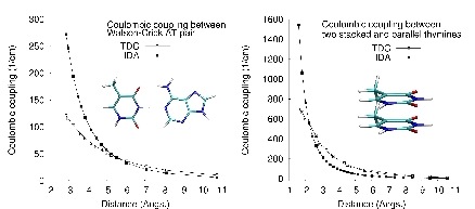
The values of the Coulombic couplings between the lowest energy transitions of the adenine and thymine and two -stacked thymines as a function of distance between the bases (Fig. 4) were calculated using the TDC and IDA methods. For the calculations the transition densities and dipoles were those obtained from the TDDFT calculations on the MP2 optimized geometry of the basese. The comparison of the coupling elements obtained with the two methods, IDA and TDC, (Fig. 4) shows a good agreement at a separation between the bases larger than 5 and 6 Å for the AT pair and two stacked thyminess, respectively. At a shorter separations, in the range of 3–4 Å, which is typical for DNA structures, the agreement between IDA and TDC is very poor with the differences between calculated couplings larger than 100% in case of AT pair. The aforementioned good agreement between IDA and TDC at a larger and poor agreement at shorter separations between nucleobases indicate that the shape and spatial extent of transition density (Fig. 4) become important and cannot be neglected at distances between the bases typical for double helices DNA. The agreement between the two methods becomes very good in the limit of very large separation, ( 8 Å).
| base 1 | base 2 | IDA | TDC | |
| Watson-Crick Base Pairs | ||||
| A | T | 229.5 (0.1) | 100.8 (0.04) | |
| -stacked bases: nearest neighbors: | ||||
| A | A | 871.7 (0.4) | 160.7 (0.2) | |
| T | T | 201.4 (0.1) | 90.2 (0.03) | |
| -stacked bases: 2nd nearest neighbors | ||||
| A | A | 57.4 (0.1) | 8.7 (0.02) | |
| T | T | 3.1 (0.005) | 1.4 (0.002) | |
| interchain diagonal cross terms | ||||
| A | T | 163.4 (0.03) | 109.1 (0.01) | |
| T | A | 90.7 (0.02) | 25.2 (0.01) |
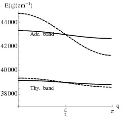
In order to compare the performance of the IDA and TDC method we calculated the coulombic couplings between the lowest transitions of adenine and thymine bases for the interstrand Watson-Crick, intrastrand -stacked, and diagonal arrangement (Fig. 1) of the base pairs for the oligomer in the idealized B-DNA geometry (Table LABEL:table2) generated using the 3DNA programLu and Olson (2003) using the IDA and TDC approximations. The corresponding band-structure as given by Eq. 8 is shown in Fig. 5.
For the stacking and pairing distances corresponding to the idealized B-DNA geometry the coupling elements calculated with the IDA approximation result with several-fold larger absolute values compared with the corresponding values calculated using the TDC method. The largest differences between the two methods are obtained for the couplings between the -stacked adenines. For the idealized B-DNA geometry the coupling between two adenines located on the same strand calculated using IDA, 872 cm-1, is more than 5-fold larger compared with the value obtained using TDC 161 cm-1. The differences in the calculated couplings using the same two methods for two stacked thymines are much smaller. For this base pair the Coulombic coupling calculated using IDA is equal to approximately 230 cm-1, more than twice the value of 101 cm-1 obtained with TDC. The absolute values of the coupling elements between the second nearest neighbors located on the same strand are much smaller. At the IDA level of approximation the coupling between the two adenines is only 57 cm-1 compared with 9 cm-1 calculated for the same base pairs using transition density cubes. The coupling between the two thymine bases on the same strand is even smaller – approximately 3 and 1 cm-1 for IDA and TDC methods, respectively.
The values of the calculated coupling elements give hint on the relative exciton delocalization along the thymine and adenine strands of the oligomer. The band structure in Fig. 5 shows that the mobility of the exciton along the thymine strand is low with both methods, IDA and TDC, giving resonable close results. The exciton mobility along the adenine strand, to the contrary, is quite different for the two methods. IDA, in this case, predicts more delocalized exciton states compared to TDA, which reflects larger magnitudes of the couplings calculated with the former method.
The magnitudes of the couplings between bases located on different strands (Table LABEL:table2), which belong to the Watson-Crick base pairs, are also larger when calculated with IDA method. The average Coulombic coupling for the Watson-Crick AT base pair calculated using IDA is 230 cm-1 compared with 101 cm-1 obtained with TDC method. The magnitudes of the couplings between bases located on different strands, which does not belong to the Watson-Crick, the diagonal terms, are generally still quite large. Especially, the magnitude of the coupling between diagonal bases in the 5’-XY-5’ direction () is comparable with the coupling for the Watson-Crick basepairs (Table 3). The values computed for the AT pair with the IDA and TDC methods are 163 and 109 cm-1, respectively. These values are almost twice and four times larger compared with the coupling between the corresponding bases in the 3’ direction ()calculated with the IDA and TDC methods, respectively. From the results of Table LABEL:table2 it is clear that the coupling elements are very sensitive with respect to the base sequence. The calculated Coulombic couplings for the -stacked arrangement of the adenine bases are by far the largest. Using IDA method the calculated coupling elements are more than two-fold larger than the corresponding values calculated with IDA for almost all arrangements.
| base 1 | base 2 | Max | Min | |||
|---|---|---|---|---|---|---|
| Interchain: Watson-Crick Base pairs | ||||||
| IDA | ||||||
| A9 | T16 | 235.7 | 29.9 | 325.8 | 105.2 | |
| A4 | T21 | 237.9 | 31.3 | 347.7 | 100.7 | |
| A7 | T18 | 235.8 | 33.3 | 363.0 | 112.0 | |
| TDC | ||||||
| A9 | T16 | 109.3 | 11.7 | 151.6 | 71.4 | |
| A4 | T21 | 101.7 | 12.8 | 146.7 | 51.1 | |
| A7 | T18 | 99.7 | 15.3 | 146.3 | 39.3 | |
| Intrachain: nearest neighbor | ||||||
| IDA | ||||||
| A3 | A4 | 769.3 | 163.5 | 1338.4 | 322.0 | |
| A6 | A7 | 858.1 | 183.9 | 1378.9 | 284.2 | |
| A9 | A10 | 935.9 | 153.6 | 1396.6 | 434.1 | |
| T21 | T22 | 473.3 | 153.7 | 1007.4 | 18.6 | |
| T18 | T19 | 435.8 | 136.7 | 1041.8 | 110.9 | |
| T15 | T16 | 315.4 | 145.1 | 874.0 | 210.4 | |
| TDC | ||||||
| A3 | A4 | 175.4 | 47.1 | 330.7 | 50.0 | |
| A6 | A7 | 197.2 | 65.4 | 363.7 | 0.0 | |
| A9 | A10 | 196.8 | 43.9 | 339.5 | 48.3 | |
| T21 | T22 | 164.9 | 45.9 | 338.9 | 38.0 | |
| T18 | T19 | 159.8 | 36.5 | 301.8 | 0.0 | |
| T15 | T16 | 124.2 | 39.7 | 277.3 | 17.4 | |
| Intrachain: 2nd nearest neighbor | ||||||
| IDA | ||||||
| A3 | A5 | 62.8 | 14.2 | 104.8 | 12.3 | |
| T20 | T22 | 25.1 | 15.5 | 74.2 | 24.8 | |
| TDC | ||||||
| A3 | A5 | 11.9 | 8.2 | 41.6 | 13.8 | |
| T20 | T22 | 9.7 | 6.7 | 30.2 | 12.2 | |
| Interchain; diagonal terms | ||||||
| IDA | ||||||
| A8 | T16 | 129.0 | 32.3 | 238.0 | 28.2 | |
| A9 | T17 | 121.8 | 38.8 | 247.0 | 1.9 | |
| TDC | ||||||
| A8 | T16 | 77.0 | 17.2 | 150.0 | 0.0 | |
| A9 | T17 | 33.0 | 9.9 | 73.5 | 0.0 |
To investigate the effect of the structural fluctuations on the calculated couplings between the adenine and thymine basepairs we analyzed 4000 conformations of selected basepairs from the 80 ps molecular dynamics trajectories of the . The extracted 4000 snapshots span the whole 80 ps simulations with each snapshot taken every 20 fs. In Table 3) the average values of the couplings calculated using IDA and TDC methods for selected pairs of the oligomer are listed. The comparison of these values with corresponding couplings calculated for basepairs in their idealized B-DNA geometry (Table LABEL:table2) shows some very interesting points. Comparing the maximum and minimum values of the coupling elements in Table 3 it can be seen that the magnitude of the couplings is very sensitive to the structural fluctuations observed in the MD simulations. The absolute value of the coupling can differ by as much as 1000 cm-1 for -stacked nucleobases. Other arrangements of the bases exhibit much smaller fluctuations of the couplings in the range of 300 cm-1 for Watson-Crick basepairs and still less for cross terms between bases on opposite strands and second nearest neighbors located on the same strand (Table 3). The fluctuations of the couplings observed for the intrastrand nearest neighbors show also more complex pattern compared to these for Watson-Crick basepairs. An example is given by the couplings calculated with TDC method for the A9-A10 step (data not shown) of oligomer. For this base pair the relatively larger and slower fluctuations on appoximately 20 ps time scale are superimposed on the rapid fluctuations. Similar slower fluctuations are observed for other -stacked adenines but not Watson-Crick pairs. For both -stacked adenines and thymines the Coulombic coupling seems to also depend on the location of a given pair along the chain. As can be inferred from data in (Table 3) the base pairs closer to the 3’ end, A9-A10 and T21-T22 show larger average values of the couplings compared with the corresponding values calculated for the base pairs closer to the 5’ end.
In spite of the large fluctuations of the couplings the values averaged over 4000 conformations are in good agreement with the corresponding couplings calculated for basepairs in standard B-DNA geometry. The best agreement is observed for the Watson-Crick base pairs while somewhat worse is seen for the -stacked thymines. Interestingly, the average magnitudes of couplings for diagonal base pairs in the 5’ and 3’ directions are very similar when calculated using IDA method. The average couplings of the A8-T16 and A9-T17 base pairs calculated with this method for the 4000 conformations from the MD simulations are equal to 129 and 122 cm-1, respectively (Table 3). The transition density cubes calculated couplings, however still show sensitivity the the direction with the average coupling in the 5’ direction for A8-T16 , 77 cm-1, compared to only 33 cm-1 in 3’ direction for A9-T17.
III.3 Exciton states
In this section we compare the properties of the excited states of the double-stranded DNA model (A)(T)12 calculated in the framework of the exciton theory where the off-diagonal elements of the exciton matrix were calculated using the ideal dipole approximation (IDA) and transition density cubes (TDC). The transition energies, oscillator strength, and the localization of the excited states were determined by diagonalization of the exciton matrix with the transition energies of the individual bases (diagonal terms) obtained from TDDFT calculations using standard geometries of the nucleobases while the coupling elements (off-diagonal terms) were determined using either IDA or TDC method. All properties were averaged over an ensamble of 240 conformations extracted form the MD simulations.

Fig. 6 shows the average values of the energies of the 24 eigenstates of the obtained with the couplings calculated using TDC and IDA method. Using the former method for calculations of the coupling elements the energies of the lowest 12 eigenstates are clearly separated from those of the remaining twelve eigenstates. The energy change between the two border eigenstates, <12> and <13>, amounts to almost 2500 cm-1 while the difference between the highest and the lowest energy eigenstates in each of the two sets is less than 1000 cm-1. The variations in the energy of a given eigenstate do not exceed 150 cm-1. The abrupt energy change between the two sets of eigenstates diminishes significantly when the IDA method is employed for calculations of the dipolar couplings (Fig. 6). The difference between the average energies of the border eigenstates <12> and <13> is less than 700 cm-1 with variations in the energies of these two eigenstates equal to 420 and 350 cm-1, respectively.
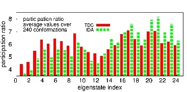
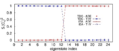
The spatial extent of the eigenstates was evaluated based on the inverse participation ratio () of a given eigenstate (Eq. 4), which indicates the number of coherently bound chromophores Bouvier et al. (2002). The plot in Fig. 7 shows the average values for each of the 24 eigenstates calculated using TDC and IDA to obtain the couplingelements of the Hamiltonian matrix. The average values of the inverse participation ratios for (A)(T)12 obtained with TDC are in the range between 4.5 and 7.1. The corresponding values obtained using TDC method are slightly smaller for the first 12 eigenstates and slightly larger for the remaining 12 eigenstates and are in the range between 3.7 and 8.2. In both cases the values are much larger than one indicating delocalization of the excitation over several bases. Markovitsi et al. Emanuele et al. (2005b); Markovitsi et al. (2006); Emanuele et al. (2005a) showed that for the columnar aggregates of identical chromophores, the maximum values of the normalized inverse participation ratio is equal to 0.7. Therefore, in case of the oligomer a completely delocalized eigenstate over one strand of the double helix would have a participation ratio equal to 8.4. Contrary to what Bouvier reported for the (A)(T)20 and (dAdT)(dAdT)10 oligomers, we found the participation ratios for all the eigenstates to be lower than 8.4. Therefore, a delocalization of the eigenstates over only adenosine or thymine chromophores but not both is expected. As can be seen from the contribution of the transitions of adenine and thymine to the eigenstates of the (A)(T)12 (Fig. 8) the lower energy eigenstates are localized almost completely on the transition on the thymine, while the higher energy eigenstates are localized on the transition of adenosine.
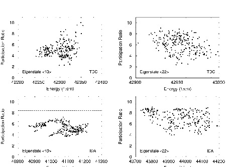
The inverse participation ratios of the eigenstates <13> and <22> as a funcion of energy are ploted in Figure 9. The IPR values for these two eigenstates of (A)12(T)12 calculated for 240 conformations taken from the MD simulations show large fluctuactions in the range of . Despite the wide range of calculated IPR values, as can be seen from the plots in Figure 9, the higher energy eigenstate, <22>, on average shows larger delocalization compared with the lower energy eigenstate, <13>, wheather the IDA or TDC method was used to calculate coupling elements. However, only for a handful conformations the value of IPR exceeds the theoretical value of 8.4 (indicated by a dashed line in Figure 9) corresponding to the completely delocalized exciton over one strand of the (A)12(T)12. This indicates that both eigenstates <13> and <22> which are localized on the transition associated with adenine remains localized on only one strand of the (A)12(T)12 composed of adenine nucleobases.
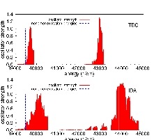
The average values of the oscillator strengths versus the average energies of the eigenstates for 240 conformations from MD simulation are plotted in Figure 10. The total oscillator strength is distributed over a small number of eigenstates clustered in two bands. The first one comprise eigenstates <9> to <12> localized on thymine strand, and the second eigenstates <21> to <24> on adenine strand. The corresponding energies are around 39700 and 43000 cm -1 for the off-diagonal couplings obtained using TDC and 40100 and 44000 cm-1 for off-diagonal terms calculated with IDA. These “bright” states correspond to the higher energy eigenstates built on the thymine and adenosine monomer transitions (Figure 10), while their “dark” counterparts, carrying negligible oscillator strength, correspond to eigenstates with lower energies. The largest oscillator strenthts are carried by the eigenstates <10> and <22> and in both cases (IDA and TDA) the energies of these eigenstates are blue shifted with respect to transition energies of the individual bases. The magnitude of the shift approximately 1000 cm -1 and less than 500 cm -1 for IDA and TDC, respectively, reflects the differences in the couplings obtained with these two methods.
IV Conlusions and Summary
We have investigated the properties of the excited states of double helix calculated in the framework of the exciton theory. In our approach we combined the quantum mechanical calculations with the molecular dynamics simulations. The TDDFT calculations were employed to calculate the energies of the singlet excited states of the individual nucleobases. The transition moments and densities of the transitions of adenine and thymine which correspond to the lowest energy transitions for these two bases were used to calculate the off-diagonal elements of the exciton matrix. The effect of the conformational changes were incorporated by averaging the calculated spectral properties of the double -stranded over large number of the conformations extracted from the molecular dynamics simulations.
The Coulombic couplings calculated using the IDA and TDC methods show a large deviations for the distances between chromophores typical for the DNA double helices. The magnitude of the couplings calculated with IDA being always larger than the corresponding values obtained with TDC. The agreement between the two methods is satisfactory only for the separations between the chromophores larger than 5 Å. The largest difference between these two methods is observed for the -stacked adenines in the standard B-DNA geometry for which the coupling calculated with IDA is over five times larger than the corresponding values calculated using TDC. The effect of the structural fluctuations on the calculated coupling elements is also very significant for both methods the values of the calculated coupling can change by an order of magnitude for different conformations of a given basepair. The difference between the smallest and largest coupling between the stacked adenines calculated using IDA for a given base pair can be as large as 1000 cm-1, smaller but still significant difference in the range of 300 cm-1 was calculated using TDC.
The properties of the excited states of the calculated in the framework of the exciton theory are affected to a different extent when the off-diagonal elements of the exciton matrix calculated using IDA and TDC methods. The eigenstates which carry the largest the oscillator strength, <10> and <22>, are slightly blue-shifted with respect to the transition energies of single nucleobases (Figure 10). The larger shift, approximately 1000 cm-1, is observed for the exciton states obtained with the off-diagonal elements of the exciton matrix calculated using the IDA approximation, compared to less than 500 cm-1 obtained with the dipolar coupling calculated using TDC method. However, the delocalization properties of these eigenstates is similar for both IDA and TDC couplings. The IPR values of the “bright” eigenstate <10> calculated with both IDA and TDC couplings are 5.5 and 6.0, respectively, while the corresponding IPR values of eigenstate <22> equal to 7.1 and 6.1 indicate only slightly large delocalization. Accordingly, comparing the IPRs obtained with TDC couplings the initial population of the bright eigenstates <10> and <22> by UV absorption will create exciton states which are delocalized over roughly 6 thymine and adenine bases, respectively. Upon relaxation the exciton states become more localized (Figures 9 and 7) as indicated by lower IPR values of the border eigenstates <1> and <13>.
Acknowledgements.
This work was funded in part by grants from the National Science Foundation and the Robert Welch Foundation. We are also grateful to Dr. Gillian C. Lynch and Prof. B. Montgomery Pettitt for providing us with the MD simulation data. We thank Dr. Stephen Bradforth for stimulating discussion motivating this work.References
- Kelley and Barton (1999) S. O. Kelley and J. K. Barton, Science 283, 375 (1999), URL http://www.sciencemag.org/cgi/content/abstract/283/5400/375.
- Crespo-Hernandez et al. (2005) C. E. Crespo-Hernandez, B. Cohen, and B. Kohler, Nature 436, 1141 (2005), URL http://www.nature.com/doifinder/10.1038/nature03933.
- Markovitsi et al. (2005) D. Markovitsi, D. Onidas, T. Gustovsson, F. Talbot, and E. Lazzarotto, J. Am. Chem. Soc. 127, 17130 (2005).
- Markovitsi et al. (2006) D. Markovitsi, F. Talbot, T. Gustavsson, D. Onidas, E. Lazzarotto, and S. Marguet, Nature 441, E7 (2006), URL http://www.nature.com/nature/journal/v441/n7094/pdf/nature049%03.pdf.
- Besaratinia et al. (2005) A. Besaratinia, T. W. Synold, H.-H. Chen, C. Chang, B. Xi, and A. Riggs, Proc Natl Acad Sci U S A 102, 10058 (2005).
- Sutherland et al. (1976) B. M. Sutherland, R. Oliver, C. O. Fuselier, and J. C. Sutherland, Biochem. 15, 402 (1976).
- Callis (1979) P. R. Callis, Chem. Phys. Lett. 61, 563 (1979).
- Sinha and Hädler (2002) R. P. Sinha and D.-P. Hädler, Photochem. Photobiol. Sci. 1, 225 (2002), URL doi:10.1039/b201230h.
- Freeman et al. (1989) S. E. Freeman, H. Hacham, R. W. Gange, D. J. Maytum, J. C. Sutherland, and B. M. Sutherland, Proc Natl Acad Sci U S A 86, 5605 (1989).
- Mouret et al. (2006) S. Mouret, C. Baudouin, M. Charveron, A. Favier, J. Cadet, and T. Douki, Proc Natl Acad Sci U S A 103, 13765 (2006).
- Löwdin (1963) P. O. Löwdin, Rev. Mod. Phys. 35, 724 (1963).
- Schultz et al. (2004) T. Schultz, E. Samoylova, W. Radloff, V. H. Ingolf, A. L. Sobolewski, and W. Domcke, Science 306, 1765 (2004), URL http://www.sciencemag.org/cgi/content/abstract/306/5702/1765.
- Emanuele et al. (2005a) E. Emanuele, D. Markovitsi, P. Millie, and K. Zakrzewska, ChemPhysChem 6, 1387 (2005a).
- Emanuele et al. (2005b) E. Emanuele, K. Zakrzewska, D. Markovitsi, R. Lavery, and P. Millie, Journal of Physical Chemistry B 109, 16109 (2005b).
- Crespo-Hernandez et al. (2006) C. E. Crespo-Hernandez, B. Cohen, and B. Kohler, Nature 441, E8 (2006), URL http://www.nature.com/nature/journal/v441/n7094/pdf/nature049%04.pdf.
- Pecourt et al. (2001) J.-M. Pecourt, J. Peon, and B. Kohler, J. Am. Chem. Soc. 123 (2001).
- Pecourt et al. (2000) J.-M. Pecourt, J. Peon, and B. Kohler, J. Am. Chem. Soc. 122 (2000).
- Gustavsson et al. (2002) T. Gustavsson, A. Sharonov, and D. Markovitsi, Chemical Physics Letters 351, 195 (2002).
- Peon and Zewail (2001) J. Peon and A. H. Zewail, Chemical Physics Letters 348, 255 (2001).
- Douhal et al. (1995) A. Douhal, S. K. Kim, and A. H. Zewail, Nature 378, 260 (1995), URL http://www.nature.com/nature/journal/v378/n6554/abs/378260a0.%html.
- Shapiro et al. (1975) S. L. Shapiro, A. J. Campillo, V. H. Kollman, and W. B. Goad, Optics Communications 15, 308 (1975).
- Suhai (1984) S. Suhai, International Journal of Quantum Chemistry, Quantum Biology Symposium 11, 223 (1984).
- Krueger et al. (1998) B. P. Krueger, G. D. Scholes, and G. R. Fleming, J. Phys. Chem. B 102, 5378 (1998).
- Frenkel (1931) J. Frenkel, Physical Review 37, 1276 (1931).
- Davydov (1971) A. S. Davydov, Theory of Molecular Excitons (McGraw-Hill, 1971).
- Bittner (2006) E. R. Bittner, The Journal of Chemical Physics 125, 094909 (pages 12) (2006), URL http://link.aip.org/link/?JCP/125/094909/1.
- Creutz and Horvgath (1994) M. Creutz and I. Horvgath, Nuclear Physics B (Proc. Supp.) 34, 583 (1994).
- McCammon and Harvey (1987) J. A. McCammon and S. C. Harvey, Dynamics of proteins and nucleic acids (Cambridge University Press, Cambridge, 1987), 2nd ed.
- Clelia et al. (2005) C. Clelia, M. Michel, P. Francois, T. Benjamin, D. Iliana, and E. Mohamed, The Journal of Chemical Physics 122, 074316 (2005).
- Sobolewski and Domcke (2002) A. L. Sobolewski and W. Domcke, European Physical Journal D: Atomic, Molecular and Optical Physics 20, 369 (2002).
- (31) M. J. Frisch, G. W. Trucks, H. B. Schlegel, G. E. Scuseria, M. A. Robb, J. R. Cheeseman, J. A. Montgomery, Jr., T. Vreven, K. N. Kudin, J. C. Burant, et al., Gaussian 03, Revision C.02, Gaussian, Inc., Wallingford, CT, 2004.
- Neese (2006) F. Neese, Orca – an ab initio, density functional and semiempirical program package, version 2.5. university of bonn (2006).
- Lu and Olson (2003) X.-J. Lu and W. K. Olson, Nucleic Acids Research 31, 5108 (2003), URL http://rutchem.rutgers.edu/~xiangjun/3DNA/index.html.
- Pettersen et al. (2004) E. Pettersen, T. Goddard, C. Huang, G. Couch, D. Greenblatt, E. Meng, and T. Ferrin, J. Comput. Chem. 25, 1605 (2004), URL http://www.cgl.ucsf.edu/chimera/docs/credits.html.
- Clowney et al. (1996) L. Clowney, S. C. Jain, A. R. Srinivasan, J. Westbrook, W. K. Olson, and H. M. Berman, Journal of the American Chemical Society 118, 509 (1996).
- Leszczynski and Skuhla (2004) J. Leszczynski and M. Skuhla, J. Comput. Chem 25, 768 (2004).
- Dong and Miller (2002) F. Dong and R. E. Miller, Science 298, 1227 (2002).
- Fulscher et al. (1997) M. P. Fulscher, L. Serrano-Andres, and B. O. Roos, J. Am. Chem. Soc. 119 (1997).
- Holmen et al. (1997) A. Holmen, A. Broo, B. Albinsson, and B. Norden, J. Am. Chem. Soc. 119, 12240 (1997).
- Callis (1983) P. R. Callis, Ann. Rev. Phys. Chem. 34, 329 (1983).
- Bouvier et al. (2002) B. Bouvier, T. Gustavsson, D. Markovitsi, and P. Millie, Chemical Physics 275, 75 (2002).
- (42) Esp: Extended systems program, copyright university of houston.
- A. D. MacKerell et al. (1998) J. A. D. MacKerell, D. Bashford, M. Bellott, J. R. L. Dunbrack, J. D. Evanseck, M. J. Field, S. Fischer, J. Gao, H. Guo, S. Ha, et al., Journal of Physical Chemistry B 102, 3586 (1998).
- Andersen (1983) H. C. Andersen, Journal Computational Physics 52, 24 (1983).
- Ryckaert et al. (1977) J.-P. Ryckaert, G. Ciccotti, and H. J. C. Berendsen, Journal Computational Physics 23, 327 (1977).
- Leeuw et al. (1980) S. W. D. Leeuw, J. W. Perram, and M. L. Klein, Proc. R. Soc. Lond. A373, 27 (1980).
- Swope et al. (1982) W. C. Swope, H. C. Andersen, P. H. Berens, and K. R. Wilson, Journal of Chemical Physics 76, 637 (1982).
- Clark et al. (1965) L. B. Clark, G. G. Peschel, and I. Tinoco, J. Phys. Chem. 116, 3615 (1965).
- Clark (1990) L. B. Clark, J. Phys. Chem. 94, 2873 (1990).
- Voelter et al. (1968) W. Voelter, R. Records, E. Bunnenberg, and C. Djerassi, J. Am. Chem. Soc. 90, 6163 (1968).
- Clark (1994) L. B. Clark, J. Am. Chem. Soc. 116, 5265 (1994).
- Voet et al. (1963) D. Voet, W. B. Gratzer, R. A. Cox, and P. Doty, Biopolymers 1, 193 (1963).
- Clark (1977) L. B. Clark, J. Am. Chem. Soc. 99, 3934 (1977).
- Yamada and Fukutome (1968) T. Yamada and H. Fukutome, Biopolymers 6, 43 (1968).
- Sprecher and W. Curtis Johnson (1977) C. A. Sprecher and J. W. Curtis Johnson, Biopolymers 16, 2243 (1977).
- Brunner and Maestre (1975) W. C. Brunner and M. F. Maestre, Biopolymers 14, 555 (1975).
- Zaloudek et al. (1985) F. Zaloudek, J. S. Navros, and L. B. Clark, J. Am. Chem. Soc. 107, 7344 (1985).
- Raksanyi et al. (1978) K. Raksanyi, I. Foldvary, and L. K. J. Fidy, Biopolymers 17, 887 (1978).
- Miles et al. (1969) D. W. Miles, M. J. Robins, R. K. Robins, M. W. Winkley, and H. Eyring, J. Am. Chem. Soc. 91, 831 (1969).