Steady-state MreB helices inside bacteria: dynamics without motors
Abstract
Within individual bacteria, we combine force-dependent polymerization dynamics of individual MreB protofilaments with an elastic model of protofilament bundles buckled into helical configurations. We use variational techniques and stochastic simulations to relate the pitch of the MreB helix, the total abundance of MreB, and the number of protofilaments. By comparing our simulations with mean-field calculations, we find that stochastic fluctuations are significant. We examine the quasi-static evolution of the helical pitch with cell growth, as well as timescales of helix turnover and denovo establishment. We find that while the body of a polarized MreB helix treadmills towards its slow-growing end, the fast-growing tips of laterally associated protofilaments move towards the opposite fast-growing end of the MreB helix. This offers a possible mechanism for targeted polar localization without cytoplasmic motor proteins.
pacs:
87.16.-b,87.16.Ac,87.16.NnI Introduction
The eukaryotic cytoskeleton organizes cell shape, cell polarity, cell division, and non-diffusive subcellular transport. F-actin, microtubules, and intermediate filaments comprise the cytoskeleton, and act with the associated proteins that provides spatial and dynamic control of cytoskeletal function (mre52, ). Prokaryotic cells have cytoskeletal analogues, such as the FtsZ-ring associated with cell division (mre86, ) together with its “divisome” of associated proteins. Bacteria also have a number of polymerizing cytoplasmic proteins, such as ParM mre55 and MinD mre86 that exhibit distinctive helical structures within the cell.
Recently, the actin homologue MreB has been shown to play a cytoskeletal role in many bacteria (mre92, ; mre87, ; mre86, ). MreB forms a continuous cytoplasmic helix that runs the length of nearly all rod-shaped prokaryotes, including Escherichia coli, Bacillus subtilis and Caulobacter crescentus (mre30, ), and it has been implicated in shape determination and polar protein localization.
In most Gram-positive bacteria MreB is present together with several paralogues, such as Mbl and MreBH in B. subtilis. The helical pitches for MreB or Mbl, separately observed by immunoflourescence microscopy, are reported to be and , respectively (mre13, ). More recent measurements of fluorescent fusions of MreB and of Mbl report pitches of , with colocalization (mre90, ). In Gram-negative species, such as E. coli and C. crescentus, only MreB is present. In E. coli, pitches of have been reported (mre73, ). In all cases, the helices are dynamic, with elements moving along the main helix at reported speeds ranging from (mre71, ) to (mre25, ). The helical structure has also been observed to condense into a ring at midcell near the time of division in E. coli (mre45, ), C. crescentus (mre23, ) and B. subtilis (where only MreBH coils) (mre88, ).
The MreB helix appears to be composed of a bundle of individual “protofilaments” (mre29, ; mre33, ; protofilament, ). Quantitative immunoblotting has been used to measure the molecular abundance of various MreBs. In B. subtilis, there are roughly 8000 MreB monomers and monomers of Mbl (mre13, ) while E. coli has roughly monomers of MreB (mre73, ). Neglecting the cytoplasmic fraction of monomeric MreB, these abundances suggest a bundle thickness of about 10 protofilaments (mre39, ).
In B. subtilis Mbl is necessary for proper insertion of new peptidoglycan, which occurs in a helical fashion (mre30, ), while MreBH is necessary for the localization and function of the cell wall hydrolase LytE that is believed to recycle the outer layers of the cell wall, also in a helical fashion (mre88, ). Cells with mutant mreB are wide, rounded and usually not viable (mre67, ). Helical bundles of MreB may contribute to the spatial localization of associated MreC, MreD, and PBP2 that, in turn, help to coordinate cell wall synthesis. It has also been suggested that helical filaments of MreB paralogues under tension can lead to spiral morphologies mre65 .
Disruption of MreB leads to loss of proper polar localization of a number of proteins such as the chemotaxis protein Tar and the Shigella flexneri virulence factor IcsA in E. coli (mre37, ), and three integral membrane proteins (PleC, DivJ, CckA) in C. crescentus (mre34, ). Polar localization in C. crescentus was disrupted by either underexpression or overexpression of MreB. When normal MreB expression was returned, polar localization was re-established (mre34, ). This suggests that MreB has a continual role in either direct polar trafficking of these proteins or in the maintenance of landmarks necessary for their proper positioning (mre92, ).
The polar proteins in C. crescentus are normally directed towards distinct (stalked and swarmer) poles in different stages (swarmer, stalked, and predivisional) of its life cycle. For example, PleC is localized to swarmer poles in swarmer and predivisional cells, DivJ is localized to stalked poles, while CckA is localized to both poles of predivisional cells (mre50, ). After MreB expression is disrupted and restored, PleC and DivJ are restored randomly to either pole (mre50, ). This suggests that MreB may be polarized within C. crescentus and that the polarity of the MreB helix is randomly restored after its disruption. However, tracking of individual YFP-MreB molecules shows unpolarized motion (mre71, ), raising questions about the mechanism of specific polar targeting. MreB-directed targeting to specific poles has not been reported in E. coli or B. subtilis.
MreB interacts with both RNA polymerase (RNAP) (mre50, ) and SetB, a chromosome defect suppressor (mre53, ), and has been implicated in the fast polar translocation of the origin-proximal regions (oriC) mre91 of newly-replicated DNA in C. crescentus mre26 and in E. coli mre50 (see however blaauwen ). Time-lapse microscopy has shown that the polar transport of oriC in B. subtilis had an average speed of and a peak speed of (mre91, ).
MreB is a homologue of the eukaryotic cytoskeletal protein actin (act04, ; act05, ). Actin filaments are used to change cell shape and to move bacteria such as Listeria monocytogenes via polymerization forces act01 , and in muscle contraction and for organelle movement via collections of myosin motor proteins (mre52, ). Actin assembly is regulated through a number of “actin-binding proteins” that variously control cross-linking, bundling, filament nucleation, end-capping, filament cutting, monomer sequestration and desequestration. MreB, in contrast, does not have any clearly identified motor proteins or associated proteins that regulate polymerization. Notably, MreB filaments spontaneously bundle in vitro without associated proteins (mre38, ).
The varied roles of MreB inside bacterial cells raise some basic questions. What is the origin of its helical configuration, and how significant are the forces that the MreB helix applies? What does the helical pitch of MreB filaments depend on? What aspects of the MreB system can be understood in terms of actin-like polymerization dynamics? Specifically, must we invoke yet-to-be discovered prokaryotic motor proteins or accessory proteins controlling MreB polymerization to explain MreB-related polar localization of proteins such as Tar, IcsA, DivJ, PleC, and CckA? Finally, does the small size of the bacterial cell affect MreB polymerization, as compared to actin polymerization in much larger eukaryotic cells?
To address these questions, we present a model of the MreB helix with stochastic polymerization dynamics of protofilaments together with global elasticity of a helical MreB bundle constrained by the bacterial cell wall. Our model provides a quantitative relationship between helical pitch, total abundance of MreB protein in a particular cell and the thickness of the protofilament bundles. The bundled MreB protofilaments are in a dynamical steady-state, and undergo constant advection as the polymerized subunits treadmill. We discuss how this advection could be harnessed for targeted polar localization without motor proteins. The steady-state dynamics also allow us to address other dynamical processes such as protein turnover in FRAP experiments (mre28, ) or recovery from A22-induced disruptions of the MreB helix (mre34, ).
II MODEL
II.1 Protofilament polymerization
Both actin and MreB polymerize into polarized filaments. Addition and dissociation of monomers occur at the ends of the asymmetric filament, both at the “barbed” (“” or fast-growing) tip and the “pointed” (“” or slow-growing) tip. The kinetics of actin polymerization are well-characterized by concentration-dependent polymerization rates and at the barbed and pointed ends, respectively, where is the cytoplasmic monomer concentration, and concentration-independent depolymerization rates and .
If a force is applied to a filament’s tip, the polymerization rate is reduced. When thermal bending fluctuations are much faster than the polymerization dynamics, the polymerization rates at either end of the filament are reduced to (act01, )
| (1) |
where is the MreB monomer length (mre72, ) and at room temperature. This force-dependent polymerization rate can also be obtained from thermodynamic arguments in the high-force limit (act20, ). We apply it to MreB polymerization dynamics within the cell.
In the absence of force, each filament grows above, and shrinks below, the critical cytoplasmic concentration
| (2) |
The asymmetry between the polymerization and depolymerization rates at barbed and pointed ends leads to treadmilling, in which the filament length remains constant while depolymerization from the pointed end is balanced by polymerization at the barbed end (mre52, ). The force-independent treadmilling rate is (act19, )
| (3) |
If the filament position is fixed, treadmilling results in a net advection of all polymerized monomers towards the pointed end.
The kinetic rate constants for MreB polymerization are yet to be determined explicitly, but appear to differ significantly from eukaryotic actin. In addition to spontaneously nucleating and bundling with much greater ease than actin, purified MreB from Thermotoga maritima polymerizes in vitro faster and exhibits a much lower critical concentration ( mre38 compared with (mre71, )). Nevertheless, in vivo observations of single molecule motion of MreB in C. crescentus suggest that the treadmilling rate is similar to actin (mre71, ). By starting with in vitro rate constants from eukaryotic actin (act05, ) and scaling the on-rates by a factor of , we recover the reported critical concentration of in vitro MreB from T. maritima. Following in vitro observations that the treadmilling rate is (mre71, ), we further scale all four rates by — preserving the MreB treadmilling rate. These scalings preserve the pointed/barbed-end asymmetries of actin and represent the least-intrusive modification of actin polymerization dynamics to make it consistent with observed MreB dynamics. The resulting barbed-end polymerization rate constant, (see Table 1), is close to the diffusion limit mre98 indicating that the pointed/barbed-end asymmetries of MreB may differ significantly from actin and/or that the appropriate for in vivo measurements may be significantly above the T. maritima value. However, our qualitative results do not depend on the precise parameter values used in this paper.
We model polymerization dynamics by the stochastic addition/dissociation of monomers at the barbed/pointed ends of individual MreB protofilaments (protofilament, ) using the scaled kinetic rate constants discussed above and listed, together with other parameters, in Table 1. Except for growing cells in Sec. III.2, a standard cell length of and cell radius of are used.
II.2 Bundle ultrastructure
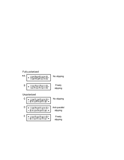
The ultrastructure of the MreB helix – the precise arrangement, orientation and length distribution of the individual protofilaments that make up the helical bundle – remains a mystery. Several hypotheses have been put forward (mre33, ) and Fig. 1 illustrates five basic possibilities. Several of these are less plausible. The slippery arrays in Fig. 1E are unlikely to be able to support the forces the cytoskeleton must withstand as it pushes against the cell wall, and recent biochemical experiments have demonstrated large lateral interactions between filaments (mre38, ). The ultrastructure in Fig. 1C leads to tapered bundles as antiparallel protofilaments, which cannot slide past each other, treadmill in opposite directions. Such tapering is not seen experimentally. In this paper we therefore consider ultrastructures composed of polarized bundle(s) of protofilaments: either one bundle (Fig. 1A) or two antiparallel bundles that freely slide with respect to each other (Fig. 1D). Since the elastic and polymerization properties of the second case follow straight-forwardly from the former, we will mostly focus on a single polarized non-slipping filament bundle (Fig. 1A) and revisit the possibility of antiparallel bundles slipping with respect to each other in the final discussion.
It is quite possible that individual MreB protofilaments do not continuously extend from one end of the bacterial cell to the other, similar to actin cables in yeast (act23, ). Indeed, in C. crescentus individual protofilaments appear to be much shorter than the cell length, only nm on average (mre71, ). For the purposes of our model, the mechanical and end-polymerization properties of discontinuous bundles of protofilaments are equivalent to bundles of continuous protofilaments. Systematic heterogeneities in the MreB helix thickness have not been reported, but our model does not depend on how the cell regulates the average number of protofilaments in a cross-section of the filament bundle. Protofilament association, dissociation and nucleation are thus implicitly included in our model. Polymerization and depolymerization away from the filament edges may occur and do not affect our steady-state results: whatever the distribution of lateral exchange, in steady state monomer incorporation and separation are balanced. Of course, significant numbers of active protofilament tips away from the cell poles can affect the dynamical timescales that we discuss.
II.3 Elastic bundle
In vitro, MreB typically polymerizes into straight filaments (mre86, ). However, MreB adopts ring-like coiled configurations in spherical mutants of normally rod-shaped organisms (mre07, ), and forms helices in rod-shaped cells (mre29, ). These observations suggest that normally straight elastic MreB protofilaments may simply buckle into helices inside the cylindrical confinement provided by the relatively hard cell wall.
A self-consistent model for the observed MreB helices consists of a particular ultrastructure of protofilaments, as in Fig. 1, buckled into a helical configuration by the cylindrical cell wall. A steady state exists where the polymerization force at the tips of the helices is balanced by the mechanical forces of the helical configuration. We treat the cell as a spherocylinder with total length and radius and total volume . The helix is assumed to extend throughout the cylindrical part of the cell, but not into the hemispherical poles, as indicated by experiment (mre13, ).
Although helical equilibria of elastic filaments have been investigated since the 1800’s, they remain a contemporary topic (ela14, ). In the elastic Cosserat model (ela01, ), a filament is parametrized by its unstretched arclength where is its total unstretched length. The Hamiltonian of such a filament is
| (4) |
where is the position of the centerline, is the local curvature, is the local twist and is the local filament thickness (measured in number of protofilaments). , and are the bending, twisting and stretching modulii of an individual protofilament, respectively. For an actin bundle, the thickness dependence of bending and twisting ranges from linear () for slippery protofilaments to quadratic () for non-sliding protofilaments, with a crosslinker dependent crossover ela16 . For MreB, lateral interactions appear strong so we assume a quadratic dependence on .
By imposing the observed helical configuration we can use variational techniques to estimate the forces working against monomer addition at the filament tips. In Appendix A, we derive the force acting at the filament tips along the filament direction in the inextensible () and freely twisting () regime:
| (5) |
where is the pitch angle of the helix and is the elastic force scale. and are the length and radius of the cylindrical portion of the cell, respectively. If the bundle thickness exhibits significant inhomogeneity, then the appropriate average thickness is the root-mean-square average thickness along the bundle length. Other than the buckling point at , is independent of for a given . The helical pitch and pitch angle are related by
| (6) |
As the pitch vanishes, , and . The force has a maximum of at .
Many of our results reflect the fact that we work in a regime where elastic forces, , and the polymerization force scale, , are similar in magnitude.
III RESULTS
III.1 Stochastic steady-state
In a typical B. subtilis cell of volume , only 4 unpolymerized monomers are necessary to achieve the critical concentration of MreB, — suggesting that stochastic effects, due to the discrete molecular nature of the polymerizing monomers, may be significant. Similarly, the number of protofilaments in a typical cross-section of the MreB helix is small (less than ), and the number of protofilaments in contact with the cell wall at the helix tips, , is even smaller. A deterministic mean-field analysis of the steady-state (see Appendix B), neglecting stochastic fluctuations, can be compared with fully stochastic simulations to explore the impact of various stochastic effects in this system.
We simulated protofilaments that grew and shrank stochastically within a common pool of monomers according to the force-dependent polymerization rates, Eq. 1, and force-independent depolymerization rates. The forces on each protofilament tip were determined by the constraint force on the bundle, Eq. 5, divided among the leading filaments at that tip, where . The filament bundle was studied once it reached a steady-state. The length of each of the protofilaments and the monomer concentration continued to fluctuate within the steady-state, as did the number of protofilament tips, , at a given end of the filament bundle.
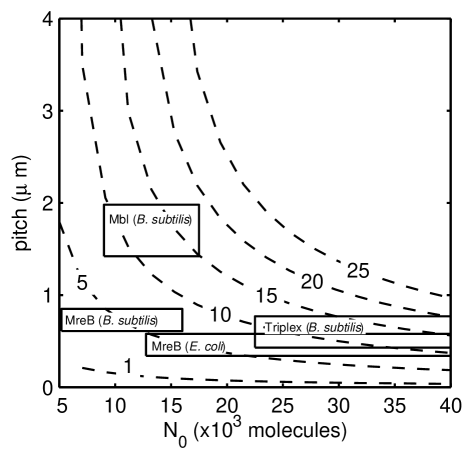
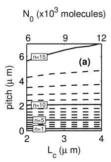
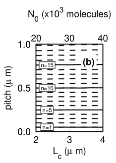
For a given cell geometry ( and ) and total number of monomers (), each bulk thickness yields a unique helical steady-state configuration with a particular pitch and average cytoplasmic monomer concentration . As seen in Fig. 2, as the abundance increases for a given number of protofilaments , the pitch decreases due to longer bundles. Conversely, at a given , thicker bundles (larger ) leads to larger pitches.
The rectangles in Fig. 2 represent independent experimental measurements of MreB abundance and cable pitch in B. subtilis (mre88, ; mre13, ) and E. coli (mre73, ). In B. subtilis, if the three MreB isoforms (MreB, Mbl, and MreBH) bundle together into a triplex structure, the total number of monomers should be the sum from each homologue, which we estimate is . We see that with current experimental pitch and abundance estimates, E. coli has protofilaments in each polarized MreB helical bundle. Under the assumption that they are bundled independently of the other isoforms in B. subtilis we estimate that Mbl has , while MreB has , with pitches from mre13 . Under the assumption that the isoforms are mutually non-slipping, we estimate in the triplex with the mutual pitch from mre90 . It is therefore possible that the distinct pitches reported by mre13 and mre90 are the result of changes in the bundling properties of the variously labeled and tagged MreB analogues.
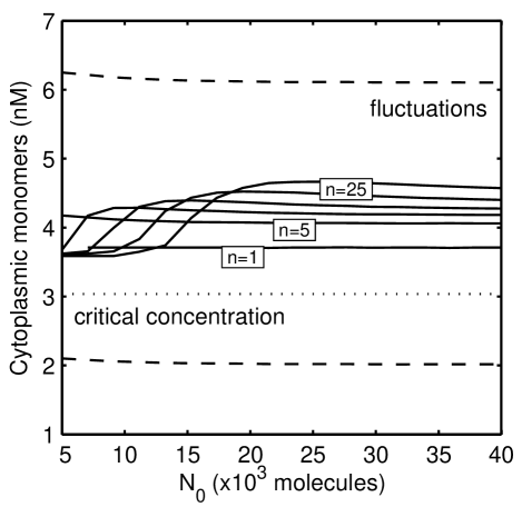
The concentration of MreB in monomeric form shown in Fig. 3 is low, representing less than of the total cellular MreB. This is in contrast to the same model using eukaryotic actin kinetics, for which is monomeric (data not shown), and in dramatic contrast with observations of FtsZ polymerization, for which 30% is associated with the Z-ring and 70% is diffuse in the cytoplasm in vivo (mre89, ). Notwithstanding, the average monomeric concentration of MreB is significantly above the critical concentration due to a reduced polymerization rate which arises from the constraining force at the bundle tips. The shape of the curves in Fig. 3 follows from the force vs. pitch relationship in Eq. 5, which grows quickly, reaches a maximum then decreases again as the pitch angle approaches . As we shall see, force and cytoplasmic concentration increase together, while, as we have seen, pitch and are inversely related. As a result, for each the cytoplasmic concentration exhibits a similar maximum vs. as force does vs. pitch. This maximum, corresponding to , is at larger for larger . We also see in Fig. 3 that the fluctuations in the monomeric concentration are very large and approximately independent of and . The cell is often instantaneously below the critical concentration despite the upward bias due to the constraint forces.
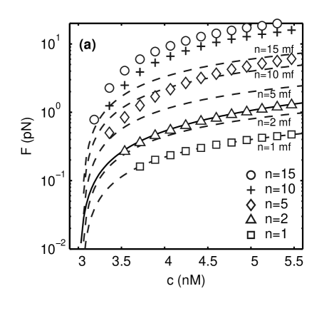
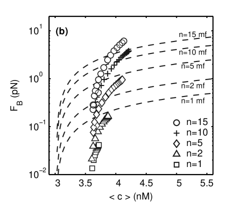
As seen in Fig. 4a, the maximal force sustainable by the bundle in steady state increases with the cytoplasmic concentration . For this part of the figure was held fixed, to facilitate comparison with both the mean-field calculations in Appendix B and the stochastic calculations in Appendix C. The increase of with comes from Eq. 1, the force-induced reduction of the polymerization-rate, and reflects the excess of the monomer fraction over the critical concentration line seen in Fig. 3. increases with the number of protofilaments, , at a fixed cytoplasmic concentration, since it is distributed over the leading protofilaments. The excess of over the maximal mean-field prediction (dashed lines), where , is recovered analytically for (solid line) in Appendix C. The excess arises because any of the protofilaments may grow and extend the bundle length, after which the remaining near-tip protofilaments will quickly catch up without force retardation. The result is enhanced growth at a given , or, equivalently, a larger at which steady-state is reached. The stochastically-enhanced force is more significant as increases, and also increases with due to the increased polymerization rate.
In Fig. 4b, we examine the additional effects on due to the large fluctuations in that were apparent in Fig. 3. Here we plot vs. the average for fully fluctuating filament bundles. Various were explored by varying . We include the mean-field results (dashed lines) for reference. Two differences with Fig. 4a are apparent. The first is that the fully fluctuating results have a maximal force sustainable by the elastic bundle, . With fixed we could use an arbitrarily stiff bundle to explore a wider range of forces, but here we need to choose a particular stiffness (by equating ) to accurately couple fluctuations in with those of . The second difference is that for the fully-fluctuating system is below the mean-field results for , and is lower than the results in Fig. 4a for a fluctuating . This systematic decrease can be seen as arising from averaging the concave-down curves from Fig. 4 over the very large fluctuations, which leads to a stronger decrease for smaller average .
There are significant effects on the forces sustained by the filament bundle both due to fluctuations in the small number of protofilaments supporting the force at the bundle tip, , and due to the large fluctuations in the monomer concentration, . Because of the curvature of the vs curve, these fluctuation effects modify the mean-field steady-state bundle force in opposite directions. While for small numbers of protofilaments () the overall effect is to reduce for a given , the net effect for the physiological range of bundle thicknesses (Fig. 2) and cytoplasmic concentrations (Fig. 3) is an increased force.
III.2 Cell growth
The MreB helix grows as the cell grows, doubling its lateral length before dividing. Our quasi-static approach can accommodate cell growth. We assume that the total number of monomers is proportional to the cell length and that the number of protofilaments is length-independent. We simulated cell growth in a regime towards the low- end of Fig. 2, with so that an average cell contains MreB monomers, and towards the high- end, with . Fig. 5 shows how the steady-state helical pitch varies as the cell length, , ranges between .
For most bundle thicknesses, , the pitch is nearly constant as the cell elongates. However, for thicker bundles in the low- regime, the helical pitch increases significantly as the cell doubles in size. This increase is due to fluctuations at small cell lengths, . While it appears strange that at large , the stochastic effects are larger, the pitch is determined by the filament length , which only depends on the maximum protofilament length. While the mean protofilament length fluctuates less with increasing , the maximum protofilament length is an extremal property of the bundle — and increases with increasing . At small , the relative fluctuations in the cytoplasmic fraction also increases. For the larger regime of Fig. 5b, the pitches are much smaller, larger, and relative length fluctuations correspondingly smaller.
In the physiological regimes shown by boxes in Fig. 2, pitch should not change significantly during cell growth if the overall concentration of MreB monomers remains constant. Mbl in B. subtilis may have longer pitches in longer cells (mre25, ; mre29, ), though this is not a strong effect (mre13, ). Experimental observations of considerable variability of the number of helical turns per cell within cells of the same strain, size and growth conditions (mre25, ) may imply corresponding variability of MreB expression or of the cross-sectional number of protofilaments — which complicates analysis.
III.3 Macromolecule trafficking
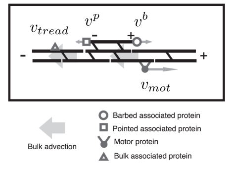
A vital role of MreB is the polar localization of proteins such as Tar in E. coli (mre37, ), the cell polarity markers DivJ and PleC in C. crescentus (mre34, ), and the origin-proximal regions of the newly-replicated chromosome in E. coli, B. subtilis and C. crescentus (mre50, ). One possibility is that these passengers associate with yet-to-be-discovered motor proteins that use MreB as a track to the poles (mre37, ). A second possibility is that these proteins simply bind to the helix and advect with the continuous treadmilling, eventually ending up at one of the polar tips. A third possibility is that they associate with leading tips of MreB protofilaments (mre33, ), perhaps via intermediary proteins analogous to formin for actin filaments. Here we quantitatively analyze these possibilities, which are depicted in Fig. 6, together with associated translocation speeds with respect to the fixed bacterial axis.
For any transport mechanism, a characteristic speed along the filament bundle, , translates into a speed relative to the cell’s axis. Assuming the protein initially binds at a uniformly random location along the bundle, the average time to reach a pole is then
| (7) |
This time can be compared to the cell division time to see whether it provides a plausible mechanism for polar localization.
The myosins that transport organelles along actin tracks in eukaryotes travel at speed (act25, ). Attached to putative myosin homologues, macromolecules could be translocated to the poles in seconds. This is well within cell division times, so this would be a viable polarization mechanism. However, no cytoplasmic motor homologue has been identified in prokaryotic cells. Furthermore, almost all myosins travel towards the pointed (“”) tip along actin filaments (act28, ), so a single polarized MreB bundle would probably only support motor-driven localization to one pole while unpolarized bundles would not support selective targeting to one pole and not the other.
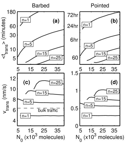
Static association of proteins to the side of MreB bundles would, through treadmilling, lead to a translocation speed equal to the rate of advection times the monomer spacing, , in the direction of the pointed end of the bundle. In steady state, the advection rate is independent of the buckling force, the bundle thickness, or the cytoplasmic concentration and leads to , shown by the dashed line in Fig. 7c. Applying Eq. 7 to a typical configuration with and , this yields minutes, which is plausible compared to cell-division timescales and comparable to oriC translocation times mre91 .
Proteins that could bind either directly, or via putative tip-binding proteins (analogous to formin in eukaryotes (mre33, ; act27, )) to the barbed end of free protofilaments could be translocated in the opposite direction to the treadmilling advection. In an unbuckled () filament, lateral protofilaments treadmill at the same rate of the net backwards advection of the bundle, accomplishing no net movement relative to the cell’s axis. In a buckled filament, however, the monomer concentration is considerably above the critical concentration of a free protofilament, as illustrated in Fig. 3. As a result, unconstrained laterally associated barbed ends grow faster than the bulk of the bundle, with
| (8) | |||||
For each bundle thickness and pitch simulated, the translocation times are shown in Fig. 7a and speeds in Fig. 7c. Typical bundles with molecules of MreB and protofilaments thick would transport passengers in minutes, considerably faster than laterally-associated proteins and in the opposite direction.
Proteins associated with the slow-growing pointed end have a net velocity given by
| (9) | |||||
Note that free pointed ends are disassembling on average, though not as rapidly as the treadmilling, so that . These translocation times are shown in Fig. 7b and speeds in Fig. 7d. According to Eqs. 8 and 9, they are slower by a factor of compared to free barbed ends, taking several hours. These times are probably too slow to be biologically relevant.
These protofilament-associated translocation modes offer a non-motor based mechanism for specific targeting of proteins to either pole in cells with a single polarized bundle of MreB. The cell could specify the specific pole destination of a particular protein by specifying which part of an MreB protofilament it binds to: barbed-associated proteins would end up at the pole at the barbed-tip of the MreB bundle with a speed of , while laterally-associated proteins would end up at the pointed-tip pole with speed . Of course, these translocation mechanisms may also supplement a (hitherto undiscovered) motor-based mechanism to provide targeting to either pole with polarized MreB bundles. We do not see any way of specific targeting of proteins to a given pole if the MreB bundle is not polarized or if there are anti-parallel bundles, either with or without motor proteins.
If protofilaments dissociate at a significant rate from the main bundles then these translocation times represent lower bounds. Additionally, any putative MreB-binding proteins could strongly affect the polymerization kinetics. For example, ADF/cofilin in eukaryotes increase for actin by times. A bacterial homologue of such a protein would decrease the delivery time of pointed-associated proteins by . Similarly, in the presence of profilin, formin increases the barbed growth rate of actin by - to -fold (mre27, ), and such a modification would decrease the delivery time of barbed-associated proteins to within a minute.
Proteins associated with pointed or barbed ends of protofilaments could be translocated towards those poles at and , respectively. However the proteins could also be directly recruited to distinct poles of the cell, due to the free ends of the MreB bundles. The relative magnitude of translocation vs. direct recruitment is dependent on the number of barbed or pointed protofilament ends. Tip-associated translocation requires a significant number of laterally associated protofilaments tips to maximize the translocation flux, while tip-directed polar recruitment requires unbroken protofilaments to minimize non-polar binding sites. Observations in C. crescentus (mre71, ) indicate that protofilaments are short, supporting tip-associated translocation as a viable mechanism in vivo.
III.4 Recovery dynamics of helices
In experiments on B. subtilis involving fluorescence recovery after photobleaching (FRAP) of fluorescence-tagged Mbl, four helical turns on one side of the cell’s longitudinal axis were bleached while the other half continued to fluoresce. It took approximately minutes for the bleached halves to recover fluorescence at the same level as the unbleached halves (mre29, ). We obtain an upper bound for this time by calculating the average time unbleached parts of the MreB protofilaments take to treadmill into the bleached regions; one half-turn in the polarized model and one quarter-turn in the non-polarized models. Thus, in silico,
| (10) |
for polarized array structures and half of that for non-polarized array structures. For this yields between 3 and 6 minutes. The agreement of this timescale with experiment suggest that monomer renewal by exchange with the cytoplasm may not dominate the FRAP recovery time.
Several experiments have applied the MreB-specific small molecule A22 to quickly and reversibly break down the MreB cytoskeleton by blocking polymerization (mre26, ; mre50, ). Cells remain viable after recovery from A22-induced disruption of MreB and reform their helical patterns in less than 1 minute for C. crescentus (mre26, ). In the context of our model, recovery from A22-treatment corresponds to re-establishment of the steady-state MreB helix from a pool of cytoplasmic monomers. We simulated our stochastic model from a nucleus of protofilaments, each of length 3 (as suggested for actin (act21, )), under the assumption that the nucleation time is short (mre38, ). Thicker bundles (larger ) reach their final steady-state length much faster than thin bundles, due to the presence of more free filament ends. The equilibration times vary between seconds. Fewer bundled protofilaments took longer to reach a steady state, the longest being , with , in which the final length is reached in seconds. These are consistent with A22 recovery timescales.
We can also address the timescale of breakdown. Assuming that A22 simply blocks polymerization, but does not change depolymerization dynamics, then it will take minutes for each protofilament to disassemble with no internal free ends. If each protofilament has length with free pointed (or barbed) ends, then we would expect the disassembly to be correspondingly faster by . Experiments constrain the actual disassembly time in C. crescentus (mre26, ) to be min, implying and . Analysis of single-molecule experiments in C. crescentus estimates (mre71, ) by assuming that protofilaments treadmill in place. If protofilaments also advect along the bulk MreB cable, as we assume in our model, this experimental estimate represents an upper bound and is consistent with our result.
These estimates of assembly and disassembly timescales of the MreB helix can also be applied to the reported midcell condensation of MreB in E. coli (mre45, ) and C. crescentus (mre23, ), and of MreBH in B. subtilis (mre89, ). Two possible mechanisms are an elastic compression of the intact MreB helix to midcell driven by some (posited) motor protein, or disassociation of the MreB helix and (transient) association with some midcell binding partners. These mechanisms may occur in tandem. The timescales of condensation and recovery are minutes in C. crescentus which is considerably longer than A22-induced breakdown. The maximum filament-end force needed to compress a typical MreB helix in our model from to a midcell spiral is , and is comparable to the force generated by the RNA polymerase (mre50, ) and the anchoring forces of integral membrane proteins (act29, ). Thus, both motor driven compression and depolymerization are plausible mechanisms for the observed midcell condensation.
IV DISCUSSION
Our model explains how polymerization forces in a bundle of MreB protofilaments can maintain a helical configuration in mechanical equilibrium. Helices are observed increasingly often within bacteria (mre92, ) and our model suggests an elastic mechanism for maintaining such helicity when filament bundles extend the length of the cell. For MreB, our model predicts that the observed pitches and protein abundance are consistent with bundles of protofilaments thick.
Helical pitch is directly observable through fluorescence microscopy, and the total abundance of MreB can be controlled by an inducible promoter (mre29, ). Increasing the MreB expression level should systematically decrease the pitch according to the curves in Fig. 2. We have assumed a cytoplasmic pool of monomers, while there is evidence of an oligomeric pool of Mbl in B. subtilis (mre29, ). Since only monomers will contribute to force-generation through polymerization, an oligomeric pool that was not part of the MreB bundle would simply lead to a shift along the axis in Fig. 2 and 3.
Filament treadmilling is ongoing in the steady-state. A significant result is that parts of the filament may travel faster than the bulk treadmilling rate, even in the opposite direction, while other parts may travel much slower. Since the cytoplasmic concentration is held above the critical concentration of free protofilaments by the constraining forces at the filament tips, the barbed end of protofilaments that associate laterally with the MreB bundles can travel at speeds greater than the bulk advection speed. These speeds are fast enough to be biologically relevant in the transport of polar-targeted proteins and the origin-proximal part of the chromosome. These transport modes may explain how MreB cables transport passengers without motor proteins, and may also reconcile experimental observations of very different speeds of MreB protofilaments: fast speeds of (mre25, ) may correspond to the lateral protofilaments described above, while slow speeds comparable to actin treadmilling speeds (mre71, ) may correspond to protofilaments involved in the bulk of the cables. It also suggests that observations of bidirectional transport along MreB cables mre71 does not necessarily rule out a single polarized MreB bundle.
It has been suggested that cell polarity arises from the inherent polarity of MreB polymers (mre33, ; mre34, ). The different modes of transport in our model provide a mechanism for translating the polarity of a cable to the entire cell. We propose that proteins destined for one pole associate laterally with the cable, and are naturally translocated towards the “pointed” pole. Proteins destined for the other pole bind to the barbed ends of free protofilaments and are naturally translocated towards the “barbed” pole. We have demonstrated that such translocation happens within a physiologically relevant timescale. Such selective targeting requires polarized MreB filaments. Proper polarization of the MreB cables themselves may require an upstream nucleator, for which TipN in C. crescentus is a candidate (mre48, ). While antiparallel MreB cables can support transport to both poles, we do not see how it can support targeted transport to specific poles.
Evidence for polarity of MreB in C. crescentus is mixed (mre34, ; mre71, ), however both PleC and DivJ appear to have targeted localization to specific poles during the cell cycle (mre50, ). This is consistent with a single polarized MreB cable in C. crescentus. We are not aware of evidence about MreB cable polarization in E. coli. While FRAP recovery of Mbl cables in B. subtilis is symmetric (mre29, ), this could be due to lateral exchange and/or the motion of laterally associated protofilaments.
Any transport along treadmilling MreB bundles would require either direct association of targeted proteins with MreB, or association through an intermediary. Interactions of only a few cytoplasmic proteins with MreB have been identified. These include SetB, implicated in chromosome segregation (mre53, ), RNA polymerase (mre50, ), and the chaperonin GroEL (mre50, ). Of these, GroEL is known to interact with a large number of cytoplasmic proteins (mre93, ), while RNA polymerase is needed for their transcription. GroEL is localized to polar and septal regions in E. coli (mre96, ) while RNAP is associated with nucleoids in punctate patterns similar to those seen for helically distributed proteins mre94 ; mre95 . It would be interesting to see if any of these MreB associated cytoplasmic proteins exhibit pole directed transport within bacterial cells.
Transport speeds may be decreased if short protofilaments necessitate multiple binding and release events to translocate from one pole to the other. Tip-directed transport also allows for direct recruitment to free protofilament tips, such as at the bundle ends. The details of bundle ultrastructure and of protein translocation will be required to sort out these competing effects.
We have shown that many of the dynamical phenomenon associated with MreB, including polar protein localization, MreB helix dissolution and reformation, and FRAP studies, could result from polymerization and treadmilling of bundled MreB protofilaments without motor proteins. In order to make precise quantitative predictions, direct measurements of the kinetic rate constants for MreB polymerization would be invaluable. The Thermotoga-scaled MreB parameters that we used are consistent with experiment and require the least amount of ad hoc parameter adjustment. Pitch-abundance and concentration-abundance relationships did not change in character between eukaryotic actin and our Thermotoga-scaled MreB kinetics. Conversely, translocation times and timescales for dissolution and reformation of MreB helices are strongly parameter-dependent. However, our qualitative result of bidirectional transport is general since, from Eq. 8, is always in the opposite direction from the bulk treadmilling.
There are two significant sources of stochastic effects in our model. Stochastic fluctuations due to the number of force-bearing protofilaments, , significantly increases the net force applied to the bundle (Fig. 4A). There are also large fluctuations in the cytoplasmic monomer concentration in the steady-state, (Fig. 3), and these systematically decrease the net forces (Fig. 4B). We have shown that the stochastic effects of , due to the small cell volume combined with the low critical concentration, and the stochastic effects of , due to the relatively small number of protofilaments, are both significant.
We have assumed that MreB polymers are naturally straight, without intrinsic curvature or twist. There are suggestions that MreB helices are always right-handed (mre13, ). Handedness could result from nucleation conditions or constraints at the helix ends, which would not affect our results. Handedness could also arise from a non-zero intrinsic twist in Eq. 4. For small , the elastic force in Eq. 5 would remain unaffected. It has been hypothesized that MreB exhibits an anisotropic membrane affinity requiring one face of the monomers to always orient towards the membrane together with significant intrinsic curvatures (mre104, ). If so, our polymerization dynamics could still be applied though with a more complex energy functional mre104 . However, we would expect qualitatively similar results if helical extension remains force-limited.
We have demonstrated that many of the dynamical phenomena associated with MreB including polar protein localization, MreB helix dissolution and reformation, and FRAP studies, can be addressed by polymerization and treadmilling of bundled MreB protofilaments without the need to invoke motor proteins. The study of the MreB helix ultrastructure, as well as the detailed study of the translocation of MreB-associated pole-directed proteins in the various bacterial species with various MreB analogues, will shed further light on the viability of our results in vivo.
Acknowledgements.
We thank NSERC for financial support, and Rut Carballido-Lopez for discussions.Appendix A Global elasticity model
Following Antman (ela01, ), an elastic filament is described by the position of its centerline and an orthonormal basis of directors specifying the orientation of its cross-section. This approach has been used extensively for DNA (ela14, ), but is seldom used for eukaryotic actin (though see (ela15, )) since actin does not systematically form structures in vivo that are much smaller than its persistence length. In contrast, MreB forms a helix with a radius of , which is smaller than the persistence length of a single protofilament () and much smaller than for a bundle of several protofilaments.
The stretching modulus of a filament bundle of actin is where is the number of protofilaments in a typical cross-section of the bundle. For a typical filament with , the energy scale to stretch the filament by monomer addition is , whereas a comparable energy scale for bending the helix is . We therefore assume that the MreB filament bundle is inextensible and parametrize the filament by its arclength . The angular strains in the filament are then simply , where is the curvature and is the local twist. We set , following the standard shear-free assumption of biopolymers.
The centerline of a helix with pitch angle (measured from the cell’s axis, ) and radius is
| (11) |
Using the tangent, normal and binormal unit vectors, , and the Frenet-Serret Theorem (ela01, ) we obtain the curvature of the helix
| (12) |
and the torsion of the filament’s centerline,
| (13) |
The directors are, in general, a rotation of through an angle . The difference between the physically relevant total twist and the centerline torsion is (ela13, ).
Using the linearly elastic Hamiltonian from Eq. 4 and and assuming that is free to rotate to eliminate , we find
| (14) | |||||
| (15) | |||||
| (16) |
Regardless of any mechanisms holding the MreB bundle in a helical configuration, one additional monomer must provide an energy of to polymerize itself to the tip of the longest protofilament(s). This provides an estimate for the force acting upon the tip of the filament bundle,
| (17) |
where . This implies that for . This is comparable to pull-out forces of integral membrane proteins (act29, ).
Eq. 14 provides an estimate for the force generated by the MreB cytoskeleton against the cell’s end caps. An estimate for the difference in longitudinal spring constant of the cell with and without a properly formed MreB helix is,
| (18) |
For and , this yields . For E. coli, the spring constant of the entire cell is (mre49, ) and is much larger for B. subtilis, thus the spring constant differential provided by the MreB helix is insignificant. Similarly, Eq. 15 provides an estimate for the radial line pressure the helix exerts on the lateral walls:
| (19) |
For and , this yields . Eq. 19 represents a force contrast between pushing the cell wall directly above the helix and elsewhere. A bundle with has approximate thickness and the local excess pressure over the cables is . In comparison, turgor pressure inside E. coli has been measured at (mre49, ), indicating that the rigidity of the MreB bundles does not directly provide significant structural support for the cell well.
We may also use the elastic model to check the self-consistency of our quasi-static helical configuration, Eq. 11. At physiological temperatures, all cellular components undergo thermal fluctuations of the order . However, a typical helical bundle with and has an elastic energy of . We therefore assume that the helical pitch does not significantly fluctuate due to thermal effects. An estimate for the timescale for elastic reorganization of the MreB helix can also be obtained from the relaxation time of the lowest hydrodynamic mode in the filament (act30, ),
| (20) |
where is the cytoplasmic viscosity. In comparison, the characteristic time for polymer elongation is given by the steady-state treadmilling rate,
| (21) |
Since we can assume that elastic relaxation is fast compared to the polymerization dynamics of interest.
Appendix B Mean-field steady-state
By neglecting stochastic fluctuations, we can analytically relate pitch, total monomer number and cytoplasmic concentration. The two essential ingredients are stationary filament lengths due to treadmilling in the presence of elastic constraint forces, and conservation of the monomer number. If protofilaments reach each tip, then from Eqs. 1, 2, and 5,
| (22) |
We also have conservation of total number of monomers,
| (23) |
where is the total number of monomers, is the number of molecules per for a given cell volume, and is averaged along the length of the helix. Together these equations give a relationship between concentration and pitch angle,
| (24) |
and an equivalent relationship between bulk thickness and pitch angle,
| (25) |
Appendix C Stochastic bundle tip,
If a force is applied to both ends of a protofilament, both and will be reduced by a factor of (act01, ; act20, ). Though the treadmilling rate is unaffected by the forcing, the treadmilling concentration becomes
| (26) |
In the absence of forces, every protofilament of a bundle treadmills with equivalent rates and critical concentrations. However, force generation by a bundle in which the load is applied only on the most advanced protofilaments behaves differently. Lagging filament tips grow without force retardation and with a reduced treadmilling concentration, thereby catching up to the tip more often than otherwise — increasing and reducing the load per loaded filament. This leads to a larger total load for the bundle for the same monomer concentration . A similar effective increase in force generation due to stochastic fluctuations has been noted in passing (act03, ), although not analyzed in detail. Here we explicitly work out the details for . For , the effects of force generation by a stochastic are expected to be even more significant, as is seen in Fig. 4.
We parametrize the system of two filaments by , the number of monomer spacings between the two tips. For any , two competing Poisson events could increase the spacing: addition at the leading tip and dissociation at the lagging tip. Reduction of occurs by the complementary two events. The master equations lead to the following equations for stationary probabilities :
| (27) |
| (28) |
| (29) |
The factors of arise when and there are two leading tips sharing the load, halving the force per filament. These equations lead to recurrence relations for . Although cannot exceed the length of the filament, the vanish exponentially as increases so we approximate . Imposing normalization, , we find
| (30) |
where . An equivalent expression for applies for the pointed end. The average polymerization velocity for these stationary probabilities is
and is a nontrivial function of both and (through ). To solve for the treadmilling concentration at a given force, we insert the barbed and pointed equivalent versions of Eq. C into , which is a cubic polynomial in , and we extract the one real, stable root.
The relation between the total applied force, , and the cytoplasmic monomer concentration is shown by the solid line in Fig. 4. Note that this derivation (and the simulations in Fig. 4) imposes a static , though fluctuates. The disagreement with the mean-field result (dashed line), indicates that the fluctuations are important for the total force.
References
- (1) B. Alberts, D. Bray, J. Lewis, M. Raff and K. Roberts, Molecular biology of the cell, 4th ed. (Garland Science, New York, 2002).
- (2) K. A. Michie and J. Lőwe, Annu. Rev. Biochem. 75, 467 (2006).
- (3) J. Møller-Jensen, J. Borch, M. Dam, R. Jensen, P. Roepstorff and K. Gerdes, Mol. Cell 12, 1477 (2003).
- (4) R. Carballido-Lopez, Microbiol. Mol. Biol. Rev. 70, 888 (2006).
- (5) Y.-L. Shih and L. Rothfield, Microbiol. Mol. Biol. Rev. 70, 729 (2006).
- (6) R. A. Daniel and J. Errington, Cell 113, 767 (2003).
- (7) L. J. F. Jones, R. Carballido-Lopez and J. Errington, Cell 104, 913 (2001).
- (8) H. J. Defeu Soufo and P. L. Graumann, Mol. Micro. 62, 1340 (2006).
- (9) T. Kruse, J. Møller-Jensen, A. Løbner-Olesen and K. Gerdes, EMBO 22, 5283 (2003).
- (10) S. Y. Kim, Z. Gitai, A. Kinkhabwala, L. Shapiro and W. E. Moerner, Proc. Natl Acad. Sci. USA 103, 10929 (2006).
- (11) H. J. D. Soufo and P. L. Graumann, EMBO 5, 789 (2004).
- (12) Y.-L. Shih, T. Le and L. Rothfield, Proc. Natl Acad. Sci. USA 100, 7865 (2003).
- (13) R. M. Figge, A. V. Divakaruni and J. W. Gober, Mol. Micro. 51, 1321 (2004).
- (14) R. Carballido-López, A. Formstone, Y. Li, S. D. Ehrlich, P. Noirot and J. Errington, Dev. Cell 11, 399 (2006).
- (15) R. Carballido-Lopez and J. Errington, Dev. Cell 4, 19 (2003).
- (16) A. Fiebig and J. A. Theriot, Proc. Natl Acad. Sci. USA 101, 8510 (2004).
- (17) We use “protofilament” as a single polymerized strand of MreB monomers, “bundle” as one or more laterally associated protofilaments, while “filament” and “cable” are generic terms for similar structures.
- (18) H. P. Erickson, Nature 413, 30 (2001).
- (19) A. Formstone and J. Errington, Mol. Micro. 55, 1646 (2005).
- (20) C. W. Wolgemuth, Y. F. Inclan, J. Quan, S. Mukherjee, G. Oster and M. A. R. Koehl, Phys. Biol. 2, 189 (2002).
- (21) Y.-L. Shih, I. Kawagishi and L. Rothfield, Mol. Micro. 58, 917 (2005).
- (22) Z. Gitai, N. Dye and L. Shapiro, Proc. Natl Acad. Sci. USA 101, 8643 (2004).
- (23) T. Kruse, B. Blagoev, A. Løbner-Olesen, M. Wachi, K. Sasaki, N. Iwai, M. Mann and K. Gerdes, Genes Dev. 20, 113 (2006).
- (24) O. Espeli, P. Nurse, C. Levine, C. Lee and K. Marians, Mol. Micro. 50, 495 (2003).
- (25) Z. Gitai, N. A. Dye, A. Reisenauer, M. Wachi and L. Shapiro, Cell 120, 329 (2005).
- (26) A. Karczmarek, R. M.-A. Baselga, S. Alexeeva, F. G. Hansen, M. Vicente, N. Nanninga, T. den Blaauwen, Mol. Micro. 65, 51 (2007).
- (27) C. D. Webb, P. L. Graumann, J. A. Kahana, A. A. Teleman, P. A. Silver and R. Losick, Mol. Micro. 28, 883 (1998).
- (28) A. Mogilner and G. Oster, Curr. Biol. 13, 721 (2003).
- (29) T. D. Pollard and G. G. Borisy, Cell 112, 453 (2003).
- (30) A. Mogilner and G. Oster, Biophys. J. 71, 3030 (1996).
- (31) O. Esue, D. Wirtz and Y. Tseng, J. Bacteriol. 188, 968 (2006).
- (32) D.-J. Scheffers, L. J. F. Jones,and J. Errington, Mol. Micro. 51, 749 (2004).
- (33) F. van den Ent, L. Amos and J. Löwe, Nature 413, 39 (2001).
- (34) A. Mogilner and L. Edelstein-Keshet, Biophys. J. 83, 1237 (2002).
- (35) L. Edelstein-Keshet and G. Ermentrout, J. Math. Biol. 40, 64 (2000).
- (36) G. I. Bell, Science 200, 618 (1978).
- (37) T. S. Karpova, J. G. McNally, S. L. Moltz and J. A. Cooper, J. Cell Biol. 142, 1501 (1998).
- (38) T. Kruse, J. Bork-Jensen and K. Gerdes, Mol. Micro. 1, 78 (2005).
- (39) N. Chouaieb, A. Goriely and J. H. Maddocks, Proc. Natl Acad. Sci. USA 103, 9398 (2006).
- (40) S. S. Antman, Nonlinear Problems of Elasticity, 2nd ed. (Springer, New York, 2004).
- (41) C. Heussinger, M. Bathe and E. Frey, cond-mat/0702097.
- (42) J. Stricker, P. Maddox, E. D. Salmon and H. P. Erickson, Proc. Natl Acad. Sci. USA 99, 3171 (2002).
- (43) M. Rief, R. S. Rock, A. D. Mehta, M. S. Mooseker, R. E. Cheney and J. A. Spudich, Proc. Natl Acad. Sci. 97, 9482 (2000).
- (44) A. L. Wells, A. W. Lin, L.-Q. Chen, D. Safer, S. M. Cain, T. Hasson, B. O. Carragher, R. A. Milligan and H. L. Sweeney, Nature 401, 505 (1999).
- (45) D. Kovar and T. Pollard, Nature Cell Biology 6, 1158 (2004).
- (46) E. C. Garner, C. S. Campbell and R. D. Mullins, Science 306, 1021 (2004).
- (47) L. Edelstein-Keshet and G. Ermentrout, Bull. Math. Biol. 60, 449 (1998).
- (48) E. Evans, D. Berk and A. Leung, Biophys. J. 59, 838 (1991).
- (49) H. Lam, W. B. Schofield and C. Jacobs-Wagner, Cell 124, 1011 (2006).
- (50) W. Houry, D. Frishman, C. Eckerskorn and F. Lottspeich, Nature 402, 147 (1999).
- (51) H. Ogino, M. Wachi, A. Ishii, N. Iwai, T. Nishida, S. Yamada, K. Nagai and M. Sugai, Genes to Cells 9, 765 (2004).
- (52) J. E. Cabrera and D. J. Jin, Mol. Micro. 50, 1493 (2003).
- (53) J. E. Cabrera and D. J. Jin, J. Bacteriol. 188, 4007 (2006).
- (54) S. S. Andrews and A. P. Arkin, Biophys. J. BioFAST 106.102343 (2007).
- (55) A. Levine, T. Liverpool and F. MacKintosh, Phys. Rev. Lett. 93, 38102 (2004).
- (56) G. van der Heijden and J. Thompson, Nonlinear Dynamics 21, 71 (2000).
- (57) X. Yao, M. Jericho, D. Pink and T. Beveridge, J. Bacteriol. 181, 6865 (1999).
- (58) J. Howard, Mechanics of Motor Proteins and the Cytoskeleton (Sinauer Associates, Sunderland, MA, 2001)
- (59) A. Mogilner and G. Oster, Biophys. J. 84, 1591 (2003).
- (60) X. Liu and G. H. Pollack, Biophys. J. 83, 2705 (2002).
- (61) K. Luby-Phelps, D. Taylor and F. Lanni, J. Cell Biol. 102, 2015 (1986).
| Symbol | Value | Meaning | Reference |
| Eukaryotic actin kinetic rates for polymerization | |||
| Barbed-end addition rate constant | (act05, ) | ||
| Pointed-end addition rate constant | (act05, ) | ||
| Barbed-end dissociation rate | (act05, ) | ||
| Pointed-end dissociation rate | (act05, ) | ||
| critical concentration | (Eq. 2 and (act05, )) | ||
| free treadmilling rate | (Eq. 3 and (act05, )) | ||
| Assumed MreB kinetic rates for polymerization | |||
| Barbed-end addition rate constant | (scaled to fit and ) | ||
| Pointed-end addition rate constant | (scaled to fit and ) | ||
| Barbed-end dissociation rate | (scaled to fit and ) | ||
| Pointed-end dissociation rate | (scaled to fit and ) | ||
| critical concentration | (mre38, ) | ||
| free treadmilling rate | (mre71, ) | ||
| Bending modulus of MreB protofilament | (from actin, (act07, )) | ||
| Stretching modulus of MreB protofilament | (from actin, (act07, )) | ||
| Monomer spacing of MreB | (mre38, ) | ||
| Radius of B. subtilis cell | (mre88, ) | ||
| Length of cylindrical portion of B. subtilis cell | (mre88, ) | ||
| Cytoplasmic viscosity | (act26, ) | ||