Present address: ]Helmholtz-Zentrum Berlin für Materialien und Energie GmbH Present address: ]Helmholtz-Zentrum Berlin für Materialien und Energie GmbH
Magnetic domain fluctuations in an antiferromagnetic film observed with coherent resonant soft x-ray scattering
Abstract
We report the direct observation of slow fluctuations of helical antiferromagnetic domains in an ultra-thin holmium film using coherent resonant magnetic x-ray scattering. We observe a gradual increase of the fluctuations in the speckle pattern with increasing temperature, while at the same time a static contribution to the speckle pattern remains. This finding indicates that domain-wall fluctuations occur over a large range of time scales. We ascribe this non-ergodic behavior to the strong dependence of the fluctuation rate on the local thickness of the film.
pacs:
75.10.Nr, 75.70.Rf, 78.70.CkSlow dynamics of magnetic domains on time scales of nanoseconds and longer are of high practical importance. Domain wall dynamics play a crucial role in magnetization reversal processes; thermally activated domain wall motions determine the lifetime of magnetically stored information. Slow dynamics on nanometer length scales is best probed by x-ray photon correlation spectroscopy (XPCS) Dierker et al. (1995); Thurn-Albrecht et al. (1996); Mochrie et al. (1997); Malik et al. (1998); Seydel et al. (2001); Sikharulidze et al. (2002, 2003); Madsen et al. (2004, 2005); Shpyrko et al. (2007); Turner et al. (2008) using the coherent diffraction or speckle pattern, which is generated when a coherent light beam scatters from a disordered structure. Any fluctuations in the disorder lead to a change in the speckle pattern; the dynamics in the sample can be obtained by measuring the time averaged intensity correlation function (ICF) Berne and Pecora (2000); Pusey and van Megen (1989); Kroon et al. (1996) of the speckle intensities on time scales ranging from 50 ns Sikharulidze et al. (2002) to hours Malik et al. (1998); Shpyrko et al. (2007). Importantly, with PCS one can obtain directly the fluctuating and static parts of the sample Pusey and van Megen (1989); Kroon et al. (1996), which makes PCS highly attractive for the investigation of systems where pinning or jamming effects occur Malik et al. (1998).
In order to study magnetism the speckle experiment has to be sensitive to spin degrees of freedom. X-ray scattering in the conventional x-ray range, even at electronic resonances, has a low magnetic scattering cross section Vettier (2001). The only exception are systems where a magnetic phase transition in UAs has indeed been observed by the loss of speckle contrast Yakhou et al. (2001). But actinide systems are only of limited practical relevance, whereas in most interesting magnetic systems the magnetism is carried by or electrons. magnetism can be probed at the resonances in the conventional x-ray range, but such experiments usually require polarization analysis of the scattered photons, which is hard to combine with the high spatial resolution required to resolve the speckle pattern. The scattering cross section for magnetism on and off the resonance is very low and only few magnetic scattering studies of transition-metal systems in the conventional x-ray range exist.
In some cases one may probe magnetism indirectly, via its coupling to structural degrees of freedom Shpyrko et al. (2007). But while a coupling of spin order to a charge density wave or to charge order is found for many systems, both orders will generally form on different temperature scales. Examples are layered nickelates Yoshizawa et al. (2000) or cobaltates Zaliznyak et al. (2000), for which the temperature scales for charge and for spin order are clearly different. An extreme case is La1.5Sr0.5CoO4, where charge order sets in below 750 K, while static spin order is not observed above 35 K Zaliznyak et al. (2000). It is therefore not to be expected that the charge dynamics are generally representative for the dynamics of magnetic order. This means that the indirect approach cannot be generally applied and one needs to probe the magnetic signal directly.
This is possible in soft x-ray range where resonant scattering provide a high magnetic contrast. PCS at the Co resonance was used to study the influence of disorder on the static domain pattern of Co/Pt multilayers for different magnetic fields Pierce et al. (2005). In this Letter we show that using soft x-ray PCS it is actually possible to directly probe fluctuating magnetic domains near a second-order phase transition and to address the question, how this transition is affected by static disorder in the system.
We studied an 11 monolayers (ML) thin epitaxially grown Ho-metal film sandwiched between Y-metal layers Ott et al. (2006); Leiner et al. (2000). The structural film properties were characterized with x-ray reflectivity measurements in the conventional x-ray range. We found that the roughness at the two Ho/Y interfaces causes thickness variations over the film of 2 monolayers. The Ho film grew pseudomorphically on Y Bentall et al. (2003) with a structural in-plane correlation length of about 80 nm, which is only slightly shorter than that of the Y layers (100 nm).
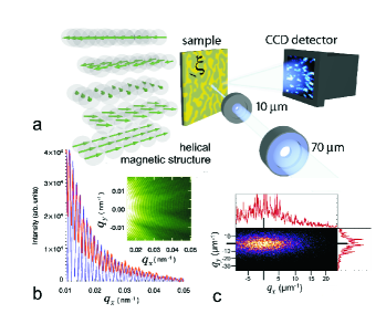
() The diffraction pattern of the 10 m pinhole. denotes the momentum transfer. A line cut through the center is shown in red, together with a least squares fit of an Airy pattern in blue. From the contrast we determine the coherent fraction of the light to be 40 %.
() A typical speckle pattern of the magnetic satellite peak at 30 K. The two intensity line traces plotted in red go along and through the center of the peak. The tick marks for refer to and vice versa.
Holmium metal displays a helical magnetic phase (sketched in Fig. 1(a)) over a wide temperature range leading to superstructure peaks in the magnetic diffraction signal separated by a wavevector from the structural peaks Helgesen et al. (1994). The ordering temperature depends on the film thickness such that films below 10 ML, which is about one helix period length in bulk Ho, do not show any helical order Weschke et al. (2004); Cinti et al. (2008, 2009, 2009). The 11-ML film studied here is hence near the stability limit for helical order and thus close to two-dimensionality. On the other hand the stability of the helical phase in such a film should be very susceptible to slight thickness variations of the Ho layer because is a steep function of the thickness.
The experiments were carried out at the U49/2-PGM1 and UE46 beam lines of BESSY (Helmholtz Zentrum Berlin) using the soft x-ray diffractometer built at the FU Berlin. In order to observe the magnetic signal from the film, we used the strong magnetic contrast that is found at the () excitation in the soft x-ray range at a photon energy around 1344 eV corresponding to a photon wavelength =9.2 Å Ott et al. (2006); Konings (2007).
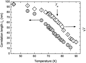
We intercepted the first-order satellite with a direct-exposure soft x-ray CCD camera as displayed in Fig. 1(a). With incoherent light, we observe a smooth diffraction peak. As is shown in Fig. 2 (diamond symbols), the scattered intensity in this peak decreases when the nominal transition temperature =76 K is approached, but does not vanish up to 90 K Ott (2004). The peak profile is well described by a single Lorentzian, with a half-width that equals the inverse of the in-plane correlation length of the magnetically ordered regions. As shown by the circular symbols in Fig. 2, we find to decrease from 90 nm (similar to the structural correlation length) at 45 K to 10 nm at 85 K. The loss of correlation already sets in around 50 K and keeps on changing over an unusually wide temperature interval of more than 40 K. This may either be an intrinsic effect caused by the proximity of the film to two-dimensionality or an effect of static disorder induced by the interface roughness.
In order to disentangle the roles of static and dynamic effects at this second-order type phase transition, we selected the transversely-coherent fraction of the BESSY II undulator radiation using a 10 m pinhole in front of the sample coherence . This causes the smooth magnetic diffraction peak to break up into a myriad of speckles [Fig. 1(c)], which form the diffraction pattern of the magnetic domain structure of the particular illuminated spot. Speckle fluctuations at different distances from the peak center are related to real-space fluctuations on different length scales [Fig. 1(c)]. We obtain the most intense signal from magnetic disorder on length scales of more than 100 nm, which we assign to helicity or phase domains.
We followed the time evolution of the speckle pattern by recording movies over a period of several hours with exposure times of 4 or 10 seconds. Snapshots from these movies for 52 K and 70 K are presented in the small frames of Fig. 3. The complete movies for various temperatures are available online web . At 52 K the speckle pattern is static on a time scale of one hour. At intermediate temperatures the speckle pattern starts to change with time and already at 70 K the movement of the speckles is very vivid.
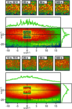
In order to quantify how much of the speckle pattern is moving, we took the time average of all the frames in one movie, shown in the large panels of Fig. 3. At 52 K the average pattern is equal to that of a single frame. Closer to the phase transition subsequent speckle patterns differ strongly, and the time-averaged speckle pattern is much smoother than the individual frames thus showing that domain-wall fluctuations have started. But even the time averages of films at higher temperatures over hours show some graininess due to the existence of static speckles connected to non-fluctuating parts of the domain pattern. These static speckles are found on all length scales, i.e. at all distances from the peak center, which is very obvious in the line cuts in Fig. 3(b). Our finding thus implies that some regions of the sample are fluctuating, while others remain fixed over the measurement period: the system behaves non-ergodic.
We argue that the most likely cause for the observed wide range of fluctuation times are variations in the Ho film thickness. As noted above, the ordering temperature of Ho films is critically dependent on the film thickness in the range of 10 to 12 monolayers Weschke et al. (2004); Cinti et al. (2008, 2009, 2009). This leads to a picture in which at low temperatures the magnetization has settled down in an irregular static domain structure with the magnetic domain size determined by the structural in-plane correlation length of the film. Domain walls exist between regions of opposite helicity or helical order phase slips As the temperature increases, the thinnest regions approach their local Néel temperature and start to fluctuate. At higher temperatures, gradually the thicker regions join in, explaining the observed reduction of the magnetic correlation length and the increasing fluctuation rates in the speckle movies.
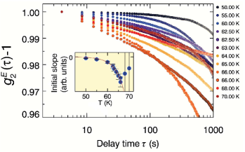
The increase of the fluctuation rates with temperature are also reflected in the time-averaged intensity correlation function (ICF). We performed the ensemble averaging required for non-ergodic systems Pusey and van Megen (1989); Kroon et al. (1996) by averaging over all the pixels with a distance from the peak center of less than 0.004 nm-1 corresponding to correlation lengths of 250 nm and larger. The normalized ensemble averaged ICF is defined as , where gives the delay time between two data samples and the brackets indicate time and ensemble averaging. In Fig. 4 we show the results for for the different temperatures, with the curves normalized to the first data point. For long correlation times the curves show an unsystematic behavior, which we ascribe to slow drifts of the beam line and setup. For short correlation times, however, the signal changes faster and faster with upon heating. This we quantified by determining the slope of in the first 50 seconds as depicted in the inset. The change of slope reflects an increase of the domain wall dynamics that speeds up with increasing temperature. The last two points in the inset indicate that for temperatures above 66 K the scattered intensity becomes too low for a reliable analysis. Lumma et al. (2000); Falus et al. (2004)
In conclusion, we show that already with the limited coherent flux available at present 3rd-generation light sources, x-ray photon correlation spectroscopy at soft x-ray resonances provides unique information on the influence of disorder on magnetic phase transitions. In general, such transitions are analysed in terms of scaling theory and critical exponents, which is only relevant for strictly ergodic systems. The non-ergodic behavior observed here in an ultrathin Holmium film is prototypical for systems in which domain walls are pinned in the potential landscape formed by disorder, in this case due to local thickness variations that cause local variations in . This image also provides a natural explanation for the smearing out of the magnetic phase transition of the film over a wide temperature range found for this sample.
Finally, we wish to point out that resonant soft x-ray scattering is also sensitive to charge and orbital order. Soft x-ray resonant PCS therefore provides a new experimental approach for the study of the phase transitions that are found abundantly in complex correlated electron compounds. Many of these systems have chemical disorder due to doping, which provides an intrinsic pinning landscape. For non-ergodic systems a characterization of the fluctuation rate on a wide range of time and length scales is necessary. Clearly, with the new x-ray free-electron laser sources, which deliver a fully coherent photon beam Grübel et al. (2007), the study of more highly interesting systems with soft x-ray PCS will become possible.
Acknowledgements.
The authors thank the BESSY and HZB staff of U49/2-PGM1 and UE46-PGM, notably D. Schmitz for expert assistance during the measurements. We thank G. Wegdam and D. Bonn for discussions on PCS. The authors gratefully acknowledge financial support by the Stichting voor Fundamenteel Onderzoek der Materie (FOM), the Deutsche Forschungsgemeinschaft (DFG) through SFB 608, the BMBF through projects 05ES3XBA/5, 05KS7PC1, 05KS7PK1, and BESSY as a partner of the EU’s I3 IA-SFS project (BESSY-ID.06.2.198; RII 3CT-2004-506008).References
- Dierker et al. (1995) S. B. Dierker, R. Pindak, R. M. Fleming, I. K. Robinson, and L. Berman, Phys. Rev. Lett. 75, 449 (1995).
- Thurn-Albrecht et al. (1996) T. Thurn-Albrecht, W. Steffen, A. Patkowski, G. Meier, E. W. Fischer, G. Grübel, and D. L. Abernathy, Phys. Rev. Lett. 77, 5437 (1996).
- Mochrie et al. (1997) S. G. J. Mochrie, A. M. Mayes, A. R. Sandy, M. Sutton, S. Brauer, G. B. Stephenson, D. L. Abernathy, and G. Grübel, Phys. Rev. Lett. 78, 1275 (1997).
- Malik et al. (1998) A. Malik, A. R. Sandy, L. B. Lurio, G. B. Stephenson, S. G. J. Mochrie, I. McNulty, and M. Sutton, Phys. Rev. Lett. 81, 5832 (1998).
- Seydel et al. (2001) T. Seydel, A. Madsen, M. Tolan, G. Grübel, and W. Press, Phys. Rev. B 63 073409 (2001).
- Sikharulidze et al. (2002) I. Sikharulidze, I. P. Dolbnya, A. Fera, A. Madsen, B. I. Ostrovskii, and W. H. de Jeu, Phys. Rev. Lett. 88 115503 (2002).
- Sikharulidze et al. (2003) I. Sikharulidze, B. Farago, I. P. Dolbnya, A. Madsen, and W. H. de Jeu, Phys. Rev. Lett. 91, 165504 (2003).
- Madsen et al. (2004) A. Madsen, T. Seydel, M. Sprung, C. Gutt, M. Tolan, and G. Grübel, Phys. Rev. Lett. 92 096104(2004).
- Madsen et al. (2005) A. Madsen, T. Seydel, M. Tolan, and G. Grübel, J. Synchr. Rad. 12, 786 (2005).
- Shpyrko et al. (2007) O. G. Shpyrko, E. D. Isaacs, J. M. Logan, Y. Feng, G. Aeppli, R. Jaramillo, H. C. Kim, T. F. Rosenbaum, P. Zschack, M. Sprung, S. Narayanan, and A. R. Sandy, Nature 447, 68 (2007).
- Turner et al. (2008) J. J. Turner, K. J. Thomas, J. P. Hill, M. A. Pfeiffer, K. Chesnel, Y. Tomioka, Y. Tokura, and S. D. Kevan, New. J. Phys. 10, 053023 (2008).
- Berne and Pecora (2000) B. J. Berne and R. Pecora, Dynamic light scattering (Dover publications Inc., Mineola, New York, 2000).
- Pusey and van Megen (1989) P. N. Pusey and W. van Megen, Physica A 157, 705 (1989).
- Kroon et al. (1996) M. Kroon, G. H. Wegdam, and R. Sprik, Phys. Rev. E 54, 6541 (1996).
- Vettier (2001) C. Vettier, Journ. Electron. Spectrosc Relat. Phenom 117-118, 113 (2001).
- Yakhou et al. (2001) F. Yakhou, A. Letoublon, F. Livet, M. de Boissieu, and F. Bley, J. Magn. Magn. Mater. 233, 119 (2001).
- Yoshizawa et al. (2000) H. Yoshizawa, T. Kakeshita, R. Kajimoto, T. Tanabe, T. Katsufuji, and Y. Tokura, Phys. Rev. B 61, R854 (2000).
- Zaliznyak et al. (2000) I. A. Zaliznyak, J. P. Hill, J. M. Tranquada, R. Erwin, and Y. Moritomo, Phys. Rev. Lett. 85, 4353 (2000).
- Pierce et al. (2005) M. S. Pierce, C. R. Buechler, L. B. Sorensen, J. J. Turner, S. D. Kevan, E. A. Jagla, J. M. Deutsch, T. Mai, O. Narayan, J. E. Davies, K. Liu, J. H. Dunn, K. M. Chesnel, J. B. Kortright, O. Hellwig, and E. E. Fullerton, Phys. Rev. Lett. 94, 017202 (2005).
- Ott et al. (2006) H. Ott, C. Schüßler-Langeheine, E. Schierle, A. Y. Grigoriev, V. Leiner, H. Zabel, G. Kaindl, and E. Weschke, Phys. Rev. B 74, 094412 (2006).
- Leiner et al. (2000) V. Leiner, D. Labergerie, R. Siebrecht, C. Sutter, and H. Zabel, Physica B 283, 167 (2000).
- Bentall et al. (2003) M. J. Bentall, R. A. Cowley, R. C. C. Ward, M. R. Wells, and A. Stunault, J. Phys.: Condens. Matter 15, 7155 (2003).
- Helgesen et al. (1994) G. Helgesen, J. P. Hill, T. R. Thurston, D. Gibbs, J. Kwo, and M. Hong, Phys. Rev. B 50, 2990 (1994).
- Weschke et al. (2004) E. Weschke, H. Ott, E. Schierle, C. Schüßler-Langeheine, D. V. Vyalikh, G. Kaindl, V. Leiner, M. Ay, T. Schmitte, H. Zabel, and P. J. Jensen, Phys. Rev. Lett. 93, 157204 (2004).
- Cinti et al. (2008) F. Cinti, A. Cuccoli,and A. Rettori, Phys. Rev. B 78, 020402(R) (2008).
- Cinti et al. (2009) F. Cinti, A. Cuccoli,and A. Rettori, Phys. Rev. B 79, 134420 (2009).
- Cinti et al. (2009) F. Cinti, A. Cuccoli,and A. Rettori, J. Appl. Phys. 105, 07E117 (2009).
- Konings (2007) S. Konings, PhD thesis, University of Amsterdam (2007).
- Ott (2004) H. Ott, PhD thesis, Freie Universität Berlin (2004).
- (30) The coherent flux was ph/s, the bandwidth , the longitudinal coherence length was about 10 microns. The coherent fraction of the radiation as determined from the contrast in the Airy pattern was 0.4.
- (31) http://www.science.uva.nl/research/cmp/konings/speckle.
- Lumma et al. (2000) D. Lumma, L. B. Lurio, S. G. J. Mochrie, and M. Sutton, Rev. Sci. Instr. 71, 3274 (2000).
- Falus et al. (2004) P. Falus, M. A. Borthwick, and S. G. J. Mochrie, Review of Scientific Instruments 75, 4383 (2004).
- Grübel et al. (2007) G. Grübel, G. B. Stephenson, C. Gutt, H. Sinn, and T. Tschentscher, Nucl. Instr. Meth.B 262, 357 (2007).