P.O. Box 10348, Ruston, LA 71272 11email: {johnjack, apaun}@latech.edu
Modeling the effects of HIV-1 virions and proteins on Fas-induced apoptosis of infected cells
Abstract
We report a first in modeling and simulation of the effects of the HIV proteins on the (caspase dependent) apoptotic pathway in infected cells. This work is novel and is an extension on the recent reports and clarifications on the FAS apoptotic pathway from the literature. We have gathered most of the reaction rates and initial conditions from the literature, the rest of the constants have been computed by fitting our model to the experimental results reported. Using the model obtained we have then run the simulations for the infected memory T cells, called also latent T cells, which, at the moment, represent the major obstacle to finding a cure for HIV. We can now report that the infected latent T cells have an estimated lifetime of about 42 hours from the moment they are re-activated. As far as we know this is the first result of this type obtained for the infected memory T cells.
1 Introduction
Apoptosis (or programmed cell death) is the process through which a multi-cellular organism removes the cells that pose a threat locally (at the tissue level) or globally to the organism. During the development of organisms, apoptosis is initiated in order to remove unneeded cells at various developmental stages. Apoptosis is a form of cellular suicide, an attempt by an organism to cleanly rid itself of cells that are unwanted, improperly formed, injured, infected, etc. It is a vital process of multicellular organisms, because of this, the mechanisms of apoptosis are hard-coded in all the cells of an organism and it can be executed in several ways; we will focus on one particular signaling pathway, which utilizes members of the Caspase protein family. The apoptotic pathway has been recently under a high degree of research focus [19, 35, 36] due to the fact that it is estimated that more than 50% of the cancers result due to the inhibition or damage in apoptotic signal transduction. Using the model from [19] we have recently simulated the FAS-mediated apoptosis in [9] by using a deterministic waiting time algorithm from [26] which was then modified and improved into a nondeterministic waiting time algorithm in [21]. The main benefit for using the nondeterministic algorithm proposed by us is the fact that it is a discrete simulation method (accounting for the discrete nature of the signal transduction pathways) as opposed to differential methods and is also fast as opposed to the stochastic simulations such as Gillespie’s algorithm (see [15, 16]), without losing the nondeterministic behavior.
We are extending in the current paper the model from [21] to include HIV-1 proteins in the nondeterministic discrete simulation of the effects of the HIV on the Fas-induced apoptosis. In the following we will give a brief reason for the extension.
The HIV virus is currently causing a global epidemic according to the report published in Geneva on November 21, 2006 by the World Health Organization. The virus infected roughly 1% of the global population with about 7.3% of the infected people dying in 2006.
The HIV-1 virus, which is responsible for 99% of the HIV infections overall, has some remarkable features. It infects cells from the immune system of the body: mainly CD4+ T cells, but also monocytes and macrophages. The initial infection seems to be through the R5 virus type that uses the co-receptor CCR5 for cell entry as reported in [33]. Reports have shown that the R5 evolves through mutations into the X4 variant of HIV-1 (using co-receptor CXCR4) in the infected individuals late during the course of the infection (see for example [33, 11, 27]). There is a significant difference between the R5 and X4 variants of the virus, R5 is inducing apoptosis in the bystander T cells while the X4 is responsible for the synctium formation (a multi nucleic cell) which proceeds to the killing of even more immune cells. The fact that the X4 is usually not present in the first stages of the HIV infection, but is more prevalent in the last stages of AIDS suggests that a normal immune system could counteract the X4 strand effectively. So one could derive that the immune system is first weakened by the R5 strand through apoptosis before the X4 strand can “survive” and collapse it.
In this paper, we focus on simulating the effects on the Fas-induced apoptosis caused by the HIV-1 strand R5 proteins. We report a simulation for the Fas-mediated pathway in HIV-1 infected cells. There are no other simulations of this type in existence. The HIV uses cellular machinery to produce its proteins, in the creation of new virions. Therefore, when a T cell is activated by the immune system, it can become infected by HIV, produce HIV proteins (and new virus’) for a couple of days, then die by apoptosis. Yet, sometimes the CD4+ T cells will become inactive after the HIV genome is incorporated into the cellular DNA. This results in a so-called latent infection, which has been of major focus in recent research (reviewed in [17]). In [17] the authors explain that latently infected cells are persistent in patients undergoing active HIV treatment (such as HAART). It is clear that a better understanding of the latent cells will assist biologists in the development of new treatments. In Section 2 we will highlight the key aspects of our Fas-induced apoptosis model, Section 3 will describe the new simulation technique. In Section 4 we review the role of HIV proteins in cell death and in Section 5 we present the results our from model’s simulations and we finish with discussions in Section 6.
2 Fas-mediated signal cascade
There are several distinct death receptors, which, when activated, can lead to cellular apoptosis through a tightly regulated molecular signaling cascade [3]. We have focused our previous efforts on simulating the well-understood Fas-mediated pathway [21]. According to the literature [28] and [20], understanding the complex signaling cascade of Fas-mediated apoptosis can assist in the creation of remedies for cancer and autoimmune disorders. In this paper we provide an expansion of our previous model, described in [21], in order to decipher and predict the behavior of HIV-infected cells. But first, we discuss the Fas-mediated signaling pathway.
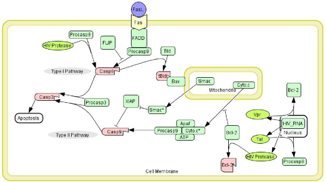
There are two versions of the Fas-mediated pathway [32], they are generally referred to as the type I and type II pathways. Both pathways begin with the Fas ligand binding to the Fas receptor (CD95) on the cell membrane. The ligand forces a conformational change of the Fas receptor, which then goes on to recruit the Fas-associated death domain (FADD). Once FADD is bound (up to three FADD per Fasl-Fas site), Procaspase 8 and FLIP can be competitively recruited to the death domain. FLIP is an inhibitor of the pathway, in that its binding to the death domain removes binding sites for Procaspase 8. The recruitment of at least two Procaspase 8 molecules can lead to cleavage into active Caspase 8 form. The divergence in the two pathways occurs after active Caspase 8 is in the cytoplasm; the end result of both pathways is the cleavage of Procaspase 3 into active Caspase 3. If the initial concentration of Caspase 8 is large enough, Caspase 3 is directly phosphorylated by Caspase 8 (type I pathway). Otherwise, Caspase 8 truncates Bid, which binds with two Bax molecules enabling the release of cytochrome c from the mitochondria (type II pathway). Bcl-2 can hinder the release of cytochrome c (thus, inhibiting apoptosis) by binding to Bid, tBid, Bax or the tBid:Bax molecular complexes. Once released, cytochrome c binds to Apaf and ATP before recruiting and activating Caspase 9. The active Caspase 9 can then go on to activate Procaspase 3. XIAP can prohibit apoptosis by binding to Caspase 9, thus preventing the phosphorylation of Caspase 3.
Though the molecules of the Fas-mediated pathways (type I and type II) have been analyzed experimentally (pro- and anti-apoptotic), we believe the only way to understand the dynamics of the complex apoptotic system is through computer simulations. In [19] and [21], three different modeling methods are employed that yield results similar to experimental observations. We use our ideas from [21] and modify the model to incorporate the functions of HIV proteins.
3 The membrane system model
There are three schools of thought on modeling molecular interactions. Microscopic chemistry - that is, the consideration of molecular dynamics for every single molecule in the system (position, momenta of atoms, etc.) - is computationally intractable; the time scale involved in the simulations, uncertainty about cellular components, and the number of atoms involved are factors that make this approach impossible to implement in a computational system. Differential equations are the macroscopic approach. This approach has been applied to the Fas pathway previously in [19].
We believe that differential equations are not the best approach for modeling signal cascades involving low numbers of molecules. We discussed this in general for the Fas-mediated pathway in [21] and now we expand our ideas into the realm of HIV proteins, where proteins like Vpr are at a level of only 700 molecules upon viral infection [6]. Our approach, referred to as mesoscopic chemistry, is similar to the microscopic approach. However, we ignore insignificant molecules, like water and non-regulated parts of the cellular machinery, and we do not directly model the position and momenta of the molecules, but we model molecular interactions using statistical equations governing how often and which molecules interact. Our approach allows finer modeling of a molecular system than the macroscopic approach, and yet it is more tractable than the microscopic approach.
The membrane system paradigm is based on the compartmental structure found in cells. Each compartment has different rules and objects, and the system evolves as the rules are carried out based on the objects in the compartments. As we are modeling molecules in a cell, it is a natural approach to use a membrane system.
We provide now all of the different types of rules (reactions) our membrane system can simulate:
-
Transformation, complex formation and dissociation rules:
These rules are used to specify chemical reactions taking place inside a compartment of type , more specifically they represent the transformation of into ; the formation of a complex from the interaction of and ; and the dissociation of a complex into and . The rules of type (4) model the formation of a complex from the interaction of 3 molecules: , and and the rules of type model the dissociation of the complex into 3 molecules and .
-
Diffusing in and out:
When chemical substances move or diffuse freely from one compartment to another we use these types of rules, where moves from or to a compartment of type .
-
Binding and unbinding rules:
Using rules of the first type we can specify reactions consisting in the binding of a ligand swimming in one compartment to a receptor placed on the membrane surface of another compartment. The reverse reaction, unbinding of substance from a receptor, can be described as well using the second rule.
-
Recruitment and releasing rules:
With these rules we represent the interaction between two chemicals in different compartments whereby one of them is recruited from its compartment by a chemical on the other compartment, and then the new complex remains in the latter compartment. In a releasing rules a complex, , located in one compartment can dissociate into and , remaining in the same compartment as , and being released into the other compartment.
Our interest in the simulator lies in the molecular multiplicity evolution over time. Our rules are carried out at varying lengths of time, obeying the Law of Mass Action - that is, the reaction rate depends proportionally on the multiplicities of the reactants. Therefore, the number of reactants for a given reaction logically governs the speed at which that reaction will occur. In our model, the time for each reaction to occur is calculated (Waiting Time or WT), and after sorting all waiting times, the reaction with the least waiting time is applied once (reactants decreased/products increased by one molecule). The simulation clock is aggregated (WT), and the WT of each the reaction affected by the applied reaction (i.e., number of reactants of reaction changed) are recalculated. The process continues until the desired simulation time has been reached.
To calculate the waiting time for each reaction we have an associated reaction rate constant111Some of the reaction rate constants are from [19] and the HIV protein associate rates are derived from various sources. For each reaction we pre-compute a kinetic constant : where is the volume of the system, is Avogadro’s constant () and is the number of reactants involved in the reaction. The time needed for the reaction to finish will then be computed in the following way: where and represent the multiplicities of the two reactants of the reaction . On the other hand, if the reaction simulated is a dissociation or other first order reaction involving one reactant, , then the time is computed as . Now we shall discuss how the HIV proteins interact with the Fas-mediated pathways (type I and type II), and the method with which we set up a simulation of these effects.
4 The roles of HIV proteins in apoptosis
The onset of AIDS is associated with the loss of immune function, due to the death of CD4 T cells (among others). As part of our simulation, we have focused on the mechanisms through which an infected cell is kept alive in order to produce new provirus’, aiding in the spread of the infection.
The method through which an HIV virus infects a cell is well understood. A spike on the virus (the gp120 molecule) binds to the CD4 receptor of primary T cells, and in conjunction with subsequent binding to a coreceptor (CCR5 or CXCR4), a path is opened for the virus to inject its contents into the cell [8] and [39]. Reverse transcriptase translates the RNA into DNA and the code is implanted into the cells own DNA for future production, once the cell is activated. During this time, the immune system fails to detect and destroy the infected cell. HIV proteins that were packaged in the virion with the RNA interact with various signaling cascades (e.g., Fas pathway) within the cell preparing it for viral reproduction while avoiding annihilation by the immune system. It is not until the immune system activates the T cell that viral reproduction occurs.
In this study, we focus on the specific functions of the HIV proteins related to the apoptosis of CD4 T cells; reviews are found in [31], [29] and [2]. The functions of some of these proteins are still up for debate, however, we have pooled the collective knowledge of the biological community in order to categorize and model the described functions of various HIV proteins. For an illustration of the FAS-mediated apoptosis pathway and the involvement of the HIV proteins, we refer the reader to Fig. 1.
The HIV protein, Tat, has shown both pro-apoptotic and anti-apoptotic behavior. In [4], the authors were able to show that Tat expression in the cell leads to up-regulation of Procaspase 8 (proapoptotic molecule). Tat has been associated with upregulation of Bcl-2 (antiapoptotic molecule) [14]. Conversely, [31] argues that Tat downregulates Bcl-2, resulting in increased sensitivity to the Fas-mediated type II pathway. Tat has also been implicated in the upregulation of the Fas ligand in infected cells [4] and [40]. However, the upregulation is assumed to be a mechnism to destroy CTLs (thus, keeping the cell alive) and bystander cells through Fas-mediated apoptosis, the increase of Fas ligand does not appear to affect apoptosis of the infected cell [31].
Another HIV protein, Vpr, has been implicated in both prohibiting and assisting apoptosis. Since Vpr is located in the virions, a low concentration is avaible early in the infected cell life-cycle (assumed to be 700 molecules according to [6]). At low multiplicity, Vpr has been shown to prohibit apoptosis by up-regulating Bcl-2 and down-regulating Bax (proapoptotic molecule) [12].
HIV protease, which is responsible for cleaving the proteins created from the viral genetic code, has also been shown to affect the signaling cascade in apoptosis. The HIV protease has been shown to cleave Bcl-2 into a deactive state [34], while it is also responsible for cleaving Procaspase 8 into active Caspase 8 form [24], both events lead to an increased susceptibility for Fas-mediated apoptosis.
The HIV RNA travels to the nucleus and becomes incorporated into the cellular genetic code. While the T cell is activated by the immune system, the HIV proteins are manufactured through the cellular machinery (transcription into HIV RNA and translation through the ribosomes in the cytoplasm). The simulator makes stochastic choices on where to send the Tat, Vpr, and HIV Protease (cytoplasm, mitochondria, or extracellular). Using the information on the three HIV proteins, we were able to create 11 new rules for our simulator. Our results are discussed below.
5 Results of the Infected Model
Based on the rules in Appendix A we simulated two different version of T cells. What we refer to as the ”Non-latent” is a simulation of the T cell just after the contents of a virion have infected it, so there is Vpr active in the cytoplasm and the RNA is on its way to being incorporated into the DNA of the active CD4 cell. We call the second simulation method ”Post-latent”, which is a simulation of the reactivation of an infected, but latent CD4 cell. We seek to achieve greater understanding on the nature of latently infected cells.
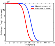
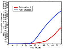
As we consider activation of Procaspase 3 to be an indication of the onset of apoptosis, we see in Fig. 2 that both the Post-latent and Non-latent cells die in approximately two days. Our simulations do indicate that reactivated latent cells will die more quickly than freshly infected cells. The lack of Vpr molecules in the latent cells is most likely the cause, as the anti-apoptotic effect of the protein would not be available. Also in Fig. 2 we show the truncation of Bid, a necessary step in the induction of the type II pathway. Active Caspase 8 is responsible for the truncation of Bid, so we again see the affects of the increased rate of Caspase 8 activation. The amplification of the signal through the mitochondria occurs earlier in the Post-latent than in the Non-latent simulation.
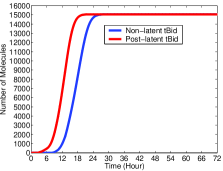
In an HIV infected cell, Procaspase 8 can be activated by two ways, interactions with FADD or HIV Protease. The upregulation of Procaspase 8 by Tat increases the activation rate of Procaspase 8 (because it increases the chance of Procaspase 8 interacting with the existing FADD or HIV Protease molecules). As the number of molecules of Fas ligand is the same between both simulations, the increased rate of apoptosis is due to the creation and activity of HIV Protease and Tat. In Fig. 3, we show the activation of Procaspase 8 and Procaspase 9 for each of the simulations.
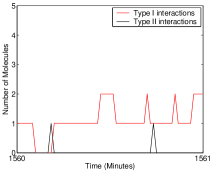
We next seek to determine whether the Procaspase 3 is being activated through the type I or the type II pathway. According to our rule set, an interaction between Procaspase 3 and active Caspase 8 or Caspase 9 can have two outcomes, resulting in activation of the Procaspase 3 molecule or not. Interestingly, it appears that the two pathways work together to annihilate the cell. The Non-latent simulation shows the first interaction of a molecule of Procaspase 3 occurs with a Caspase 8 molecule just after 10 hours into the run. It isn’t until a 14 hours later (24.5 hours into the run, allowing for signal transduction through the mitochondria) that we begin to see Procaspase 3 interactions with active Caspase 9. As we have shown in [21], given a sufficiently high concentration of procaspase 8 in the T cell, there is no need for signal amplification. However, the initial level of Procaspase 8 in our model reflects Jurkat T Cells [19], and we are seeing cooperation between Caspase 8 and Caspase 9 leading to cellular apoptosis. In Fig. 4 we get an overall image of the effects of both pathways on Procaspase 3 activation.
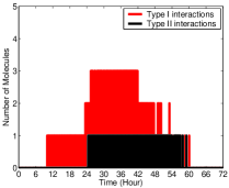
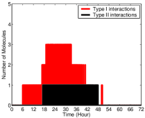
We find similarities in the Procaspase 3 activation between the Non-latent and Post-latent simulation runs. In the Post-latent run, we see type I interactions occur about 6 hours into the simulation, while type II molecular binding occurs after 17 hours (11 hours after the type I interactions). Fig. 5 show us an the overall interactions between Procaspase 3 with active Caspase 8 or Caspase 9.
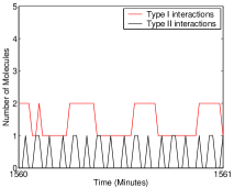
The graphs in Fig. 4 and 5 imply that there are more interactions in the type I pathway. This does not necessarily mean that the type I pathway has a greater responsibility in activation of Caspase 3 than the type II pathway. Caspase 3 activation is a two step process. First, Caspase 8 or 9 binds to the Procaspase 3 molecule, then after a period of time the binding ends with either an active Caspase 3 or the original Procaspase 3 molecule. Technically, based on the kinetic rates and the concentration of molecules involved, Caspase 8 can stay binded to Procaspase 3 for longer than Caspase 9. Therefore, though it appears that Caspase 8 binds to Procaspase 3 more frequently, it might not be the case that this results in more activation of Caspase 3 via Caspase 8 than Caspase 9. The graphs illustrate this possibility by the fact that you see constant jumps for the type II interactions (Caspase 9 and Procaspase 3), which implies less waiting time during the binding period.
6 Discussion
Based on the biological evidence from the literature, we were able to construct a simulation of the effects of HIV proteins on the Fas-mediated apoptosis pathway. We feel that our unique approach provides insight into the molecular dynamics of the Fas pathway. Our simulator leads to apoptosis in approximately two days, which is in agreement with observed results. But, we have shown that cooperation between the type I and type II pathway seems to be the method of the cell death.
In this preliminary simulator, we have focused primarily on the difference between reactivated latent cells and actively infected cells, but we are looking to refine and expand our technique. We will apply new information on the nature of latent HIV infected cells as it becomes available. However we will also explore alternative HIV models. For instance, it would be interesting to model the effects HIV proteins on bystander cell apoptosis. The type 1 HIV has been implicated in reducing T cell levels by not only destroying the cell it invades, but through the killing of so-called bystander (uninfected) cells [14]. Various mechanisms have been debated as to the means through which the virus causes the destruction of the bystander (uninfected) cells. Along with Fas-induced apoptosis, other possible mechanisms for bystander cell death are reviewed in [31], [29] and [2]. Upon being exocytised by an infected cell, several distinct types of HIV proteins can affect neighboring bystander cells.
Soluble and membrane-bound Env can bind to the CD4 receptor of bystander cells (as is the case during infection). Along with subsequent binding to a co-receptor, it can induce apoptosis. In [10] and [5], it has been shown that ligation of the CD4 receptor by Env, is sufficient to increase apoptosis in bystander cells. The reasons for increased apoptosis following Env-CD4 binding can be attributed to Bcl-2 down-regulation [18], increased Caspase 8 activation [1], and upregulation of Fas [25], FasL and Bax [31].
Extracellular Tat can be endocytised by a bystander cell, resulting in pro-apoptotic behavior. The addition of Tat to a culture of uninfected cells has been shown to increase apoptosis [23]. Endocytised Tat can up-regulate levels of Caspase 8 [4] and increase expression of the Fas ligand [31], similar to its effects in infected cells. Extracellular Vpr can disrupt the mitochondrial membrane, leading to increased translocation of cytochrome c [31].
Lastly we would like to note the fact that the experimental information about the latent HIV infected T cells is scarce, the main reason being the fact that these cells are found in small numbers in vivo, thus it is rather hard to obtain sufficient numbers for experiments in vitro. We hope that our results will help guide the researchers in the lab and we are looking forward to experimental results about these enigmatic cells that will suggest improvements and refinements of our model.
References
- [1] Algeciras-Schimnich, A. et al. (2002) CCR5 mediates Fas- and Caspase 8 dependent apoptosis of both uninfected and HIV infected primary human CD4 T cells.
- [2] Alimonti, J. et al. (2003) Mechanisms of CD4 T Lymphocyte cell death in human immunodeficiency virus infection and AIDS. Jour of Gen Vir, 84, 1649-1661.
- [3] Ashkenazi, A., & Dixit, V. (1998) Death receptors: signaling and modulation. Science 281. 1305-1308.
- [4] Bartz, S. & Emerman, M. (1999) Human immunodeficiency virus type 1 Tat induces apoptosis and increases sensitivity to apoptotic signals by up-regulating FLICE/Caspase 8. J Virol 73, 1956 1963.
- [5] Biard-Piechaczyk M., et al. (2000) Caspase-dependent apoptosis of cells expressing the chemokine receptor CXCR4 is induced by cell membrane-associated human immunodeficiency virus type 1 envelope glycoprotein (gp120). Virology 268, 329-344.
- [6] Briggs, J. et. al. (2004). The stoichiometry of Gag protein in HIV-1. Nature Structural & Molecular Biology 11, 672-675.
- [7] Brumme Z.L., Goodrich J., Mayer H.B., et al. (2005) Molecular and clinical epidemiology of CXCR4-using HIV-1 in a large population of antiretroviral-naive individuals. J Infect Dis 192, 466 74.
- [8] Chan, D. & Kim, P. (1998) HIV entry and its inhibition. Cell, 93, 5 681-684.
- [9] Cheruku, S., Paun, A. et. al. (2007) Simulating FAS-induced apoptosis by using P systems. Progress in Natural Science, to appear 2007.
- [10] Cicala C., et al. (2000) HIV-1 envelope induces activation of caspase-3 and cleavage of focal adhesion kinase in primary human CD4(+) T cells. Proc. Natl.Acad. Sci. U.S.A. 97, 1178-1183.
- [11] Connor, R. I., Sheridan, K. E. et. al. (1997) Change in coreceptor use correlates with disease progression in HIV-1- infected individuals. J. Exp. Med. 185, 621 628.
- [12] Conti, L. et al. (1998) The HIV-1 vpr protein acts as a negative regulator of apoptosis in a human lymphoblastoid T cell line possible implications for the pathogenesis of aids.
- [13] Crise, B, et al. (1990). CD4 is retained in the ER by the human immunodeficiency virus type 1 glycoprotein precursor. J Vir, 64, 11 5585-5593.
- [14] Finkel, T. (1995) Apoptosis occurs predominantly in bystander cells and not in productively infected cells of HIV- and SIV-infected lymph nodes. Nature Medicine 1. 129 - 134.
- [15] Gillespie, D.T. (1976). A General Method for Numerically Simulating the Stochastic Time Evolution of Coupled Chemical Reactions. Journal of Computational Physics, 22, 403-434.
- [16] Gillespie, D.T. (1977). Exact Stochastic Simulation of Coupled Chemical Reactions. The Journal of Physical Chemistry, 81, 25, 2340-2361.
- [17] Han, T., et al. (2007) Experimental approaches to the study of HIV-1 latency. Microbiology. 5, 95-106.
- [18] Hashimoto, F., et al. (1997) Modulation of Bcl-2 Protein by CD4 Cross-Linking a possible mechanism for lympphocyte apoptosis in human immunodeficiency virus infection. Blood, 90, 2, 745-753.
- [19] Hua, F., Cornejo, M., Cardone, M., Stokes, C., Lauffenburger, D. (2005). Effects of Bcl-2 Levels on FAS Signaling-Induced Caspase-3 Activation: Molecular Genetic Tests of Computational Model Predictions. The Journal of Immunology, 175, 2, 985–995 and correction 175, 9, 6235-6237.
- [20] Igney, F. & Krammer, P. (2002) Death and anti-death: tumour resistance to apoptosis. Nat Rev Cancer 2. 277-288.
- [21] Jack, J., et al. (2007) Simulating apoptosis using discrete methods: a membrane system and a stochastic approach. Theoretical Computer Science (submitted)
- [22] Jacotet E., et al. (2000) The HIV-1 viral protein R induces apoptosis via a direct effect on the mitochondrial permeability transition pore.
- [23] McCloskey, T., et al. (1997) Dual role of HIV Tat in regulation of apoptosis in T cells. J. Immunol. 158. 1014-1019.
- [24] Nie, Z. et al. (2002) HIV-1 protease processes procaspase 8 to cause mitochondrial release of cytochrome c, caspase cleavage and nuclear fragmentation.
- [25] Oyaizu, N., et al. (1994) Cross-linking of CD4 molecules upregulates Fas antigen expression in lymphocytes by inducing interferon-gamma and tumor necrosis factor- alpha secretion. Blood. 84. 2622-2631.
- [26] Pérez-Jiménez, M.J., Romero-Campero, F.J. (2007) P Systems, a New Computational Modeling Tool for Systems Biology, Transactions on Computational Systems Biology, to appear.
- [27] Richman, D. D.,Bozzette, S. A. (1994) The impact of the syncytiuminducing phenotype of human immunodeficiency virus on disease progression. J. Infect. Dis. 169, 968 974.
- [28] Rieux-Laucat, F., et al. (2003) Autoimmune lymphoproliferative syndromes: genetic defects of apoptosis pathways. Cell Death Differ. 10. 124-133.
- [29] Ross, T. (2001) Using death to one’s advantage: HIV modulation and apoptosis. Leukemia, 15, 332-341
- [30] Salghetti, S, et al. (1995) Human immunodeficiency virus type 1 Nef and p56Ick protein-tyrosine kinase interact with a common element in CD4 cytoplasmic tail. Proc. Natl. Acad. Sci. U.S.A. 92 349-353
- [31] Selliah, N. & Finkel, T. (2001) Biochemical mechanisms of HIV induced T cell apoptosis. Cell Death and Differentiation, 8, 127-136.
- [32] Scaffidi, C., et al. (1998) Twp CD95 (APO-1/Fas) signaling pathways. EMBO J 17. 1675-1687.
- [33] Schuitemaker, H., et. al. (1992) Biological phenotype of human immunodeficiency virus type 1 clones at different stages of infection: progression of disease is associated with a shift from monocytotropic to T-cell-tropic virus populations. J. Virol. 66, 1354 1360.
- [34] Strack, et al. (1996) Apoptosis mediated by HIV protease is preceded by cleavage of bcl-2.
- [35] Susin, S.A. et al. (2000) Two Distinct Pathways Leading to Nuclear Apoptosis. Journal of Experimental Medicine 192 (4).
- [36] Takaoka, A., et al. (2003) Integration of interferon-alpha/beta signalling to p53 responses in tumour suppression and antiviral defence. Nature 424 (6948).
- [37] Wildum, S. et al. (2006) Contribution of Vpu, Env, and Nef to CD4 Down-Modulation and Resistance of Human Immunodeficiency Virus Type 1-Infected T Cells to Superinfection. J Vir, 80, 16 8047-8059.
- [38] Willey, R. et al. (1992) - Human Immunodeficiency Virus Type 1 Vpu Protein Induces Rapid Degradation of CD4 J Vir, 66, 7193-7200.
- [39] Wyatt, R. & Sodroski, J. (1998) The HIV-1 envelope glycoproteins: fusogens, antigens, and immunogens. Science, 280, 5371 1884-1888.
- [40] Yang, Y. et al. (2002) HIV Tat Binds Egr Proteins and Enhances Egr-dependent Transactivation of the Fas Ligand Promoter. Journ of Bio Chem, 277, 22, 19482-19487.
Appendix 0.A The Model
Our model consist of the following P system:
where:
-
•
In the alphabet we represent the proteins and complexes of proteins involved in the signalling transduction:
HIVRNA, Tat, Vpr, HIV Protease, FASL, FAS, FASC, FADD, FASC-FADD, FASC-FADD2, FASC-FADD3, FASC-FADD2-CASP8, FASC-FADD3-CASP8, FASC-FADD2-FLIP, FASC-FADD3-FLIP, FASC-FADD2-CASP82, FASC-FADD3-CASP82, FASC-FADD2-CASP8-FLIP, FASC-FADD3-CASP8-FLIP, FASC-FADD2-FLIP2, FASC-FADD3-FLIP2, FASC-FADD-CASP8, FASC-FADD-FLIP, CASP8, FLIP, FASC-FADD3-CASP83, FASC-FADD3-CASP82-FLIP, FASC-FADD3-CASP8-FLIP2, FASC-FADD3-FLIP3, CASP8, CASP8, CASP3, CASP-CASP3, CASP, CASP8-Bid, tBid, Bid, Bax, tBid-Bax, tBid-Bax2, Smac, Smac∗, Cyto.c, Cyto.c∗, XIAP, Smac∗-XIAP, Apaf, Cyto.c∗-Apaf-ATP, CASP9, Cyto.c∗-Apaf-ATP-CASP9, Cyto.c∗-Apaf-ATP-CASP92, CASP9∗, CASP9∗-CASP3, CASP9-XIAP, CASP3∗-XIAP, Bcl, Bcl-Bax, Bcl-tBid222Smac is an abbreviation for second mithocondria-derived activator of caspase.
-
•
The set of labels, , can also be used to identify the different compartments where the signalling transduction takes place, namely, the environment (), the cell surface (), the cytoplasm (), the mitochondria (), and the nucleus ().
-
•
In the membrane structure , the compartments are represented. The compartments 1, 2, 3, and 4 can be labeled respectively with , , , , and .
-
•
where the multiset represents the initial amount of molecules in the compartment and is the label associated with the compartment. For Example:
-
–
and FASL
-
–
and FAS
-
–
and FADD10038, CASP820071, FLIP48779, CASP3120442, Bid15055,
Bax50182, XIAP18066, Apaf60221, ATP6022141 CASP912044, Vpr -
–
and Smac60221, Cyto.c60221 Bcl
-
–
and HIVRNA
-
–
-
•
, , and are the sets of rules associated with the environment, cell surface, cytoplasm, mitochondria, and the nucleus. These rules represent the chemical reactions that take place in each compartment or region of the cell.
-
–
-
–
-
–
-
–
-
–
-
–
We list the four sets of rules we simulated, which are based upon the apoptosis pathway. Each set incorporates a mechanism for Bcl2 involvement in the inhibition of apoptosis.
The deterministic kinetic constants (reaction rates) mentioned in the previous table are given in the following; we refer the interested reader to [19] for more details about the rates and references for their estimation.