Non-equilibrium mechanics and dynamics of motor-activated gels
Abstract
The mechanics of cells is strongly affected by molecular motors that generate forces in the cellular cytoskeleton. We develop a model for cytoskeletal networks driven out of equilibrium by molecular motors exerting transient contractile stresses. Using this model we show how motor activity can dramatically increase the network’s bulk elastic moduli. We also show how motor binding kinetics naturally leads to enhanced low-frequency stress fluctuations that result in non-equilibrium diffusive motion within an elastic network, as seen in recent in vitro and in vivo experiments.
pacs:
87.16.Ka, 87.15.La, 62.20.DcThe mechanics of living cells are largely governed by the cytoskeleton, a complex network of filamentous protein aggregates and various specialized proteins and enzymes that couple the filaments together and generate forcesAlberts-1994-26 . As materials, in vitro networks of cytoskeletal filaments have been shown to have unusual mechanical properties, including a highly non-linear elastic responseMackintosh-1997-20 ; Gardel-2004-2 ; Storm-2005-5 ; Bausch-2006-6 ; Chaudhuri-2007-27 and negative normal stressesJanmey-2007-21 . Cytoskeletal networks in vivo, however, are far from equilibrium materials, due in large part to molecular motors that exert internal forces within the networks. This presents a challenge for quantitative statistical/thermodynamic modeling. Recent studies of in vitro networks that include molecular motors have shown nearly a 100-fold stiffening of the networks due to motor activity, as well as pronounced low-frequency, non-equilibrium fluctuationsMizuno-2007-1 . Here, we develop a model for such active gels that can explain both the strong stiffening of networks with motor activity, as well as the large non-equilibrium fluctuations at low frequencies. We also show how motor (un)binding kinetics naturally leads to a very simple and general form of stress fluctuations and diffusive-like motion, which are consistent with observed non-equilibrium dynamics in living cellsLau-2003-10 ; Brangwynne-2007 . This model can form the basis for quantitative design principles for creating synthetic polymeric materials with tunable elastic properties and muscle-like activation.
Active solutions consisting of polymers and motors motors constitute a strikingly new kind of material that can actively change/adapt its macroscopic mechanical properties due to small-scale motor activity that drives relative sliding of polymers past each otherLiverpool-2001-38 ; Humphrey-2002-9 ; LeGoff-2002-37 ; Kruse-2005-39 ; Liverpool-2006-28 . In permanently cross-linked networks, however, such motor activity can produce tensile stressesMizuno-2007-1 . This muscle-like contraction is sketched in Fig. 1A. It is well known that single semi-flexible polymers stiffen under extensionBustamante-1994-3 , and that this can result in macroscopic stiffening of networks under external strainMackintosh-1995-4 ; Gardel-2004-2 ; Storm-2005-5 . This effect can also account for the observed dramatic stiffening of active networksMizuno-2007-1 ; Koenderink-2007 . Assuming an average state of tension in the network strands due to motor activity, we can calculate the expected degree of network stiffening as follows. The tension in a single filament is calculated as a function of longitudinal extension as in Ref. Mackintosh-1995-4 , from which an effective spring constant is calculated. In the nonlinear regime, this increases as Gardel-2004-2 . The network modulus is given by , where is the density (length per volume) of polymer, and is the distance between cross-linksGittes-1998-8 ; Morse-1998-32 . The predicted stiffening is shown in Fig. 1B, where the filament tension has been normalized by the characteristic tension required to pull out the fluctuations on a filament of length in the network. Here, is the persistence length. For a network of actin filaments, such as in Mizuno et al.Mizuno-2007-1 , where m and m, this characteristic average tension is of order 0.1pN, meaning that a tension of just a few pN, which is easily reached by myosin motors, can lead to the observed -fold stiffening of active networks.
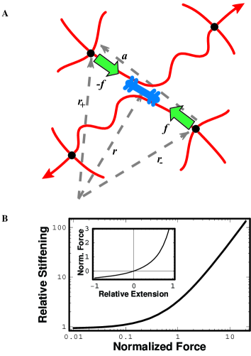
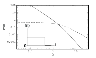
The quasistatic picture sketched in Fig. 1A shows a motor (myosin minifilament) generating a pair of equal and opposite forces applied at points , separated by . We expect to be a few microns in an in vitro network. Since actin filaments are not able to support compressive loads over this distance, the resulting force dipole is contractile: the points are pulled together by a sort of muscle-like activity. While individual myosin motors are non-processive and are incapable of persistent, directed motion, they self-assemble into minifilaments, which are processive. These minifilaments still have a finite duty ratio. When they unbind the tension is instantaneously released, as sketched in the inset of Fig. 2Mizuno-2007-1 . Such a step-like force corresponds to a power spectrum of force fluctuations that varies as , proportional to the square Fourier transform of .
As we show, this physical picture of step-like contractile forces naturally leads to non-equilibrium fluctuations that dominate only at low frequencies, as sketched in Fig. 2. Surprisingly, this generates motion that appears to be diffusive: , but occurring in an elastic material. The effective diffusion constant is controlled by motor activity and not temperature. Using well-established viscoelastic properties of cross-linked F-actin networksGittes-1998-8 ; Morse-1998-32 , we find distinct regimes of both thermal and athermal (motor-induced) fluctuations sketched in Fig. 2, which are consistent with the observations both in vivo Lau-2003-10 and in vitroMizuno-2007-1 .
To model the active gel we use a continuum description for a viscoelastic homogeneous and isotropic medium, but in which the motor activity couples to this medium as illustrated in Fig. 1A.
For in vitro networks such as in Ref. Mizuno-2007-1 , the distance between cross-links, and thus is expected to be of order 3-10m. On this scale, we can model the action of a motor as the introduction of a pair for equal and opposite applied forces in the (visco-)elastic continuum. The resulting displacement field at position of the network we describe by a linear response function depending on position and frequency as
| (1) |
using the fact that the motor-generated forces are equal and opposite. Stability also requires that and be parallel. The response function to a point force can be written in terms of and , where .
We calculate these two response components within a two-fluid approximation, in which the cytoskeletal filaments are treated as a porous elastic network immersed in a viscous solventBrochard-1977-35 ; Milner-1993-34 ; Gittes-1997-19 ; Levine-2000-16 . Here, the network displacement and solvent velocity satisfy the coupled equations
| (2) |
| (3) |
where and are Lame coefficients, is the solvent viscosity, and the forces represent the forces on the network and solvent, respectively. Given a meshwork with a pore size , the coupling is expected to be of order . These are solved for the response of the combined system to an applied point force. The resulting response functions are given by
| (4) |
and
| (5) |
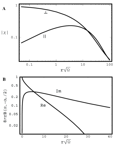
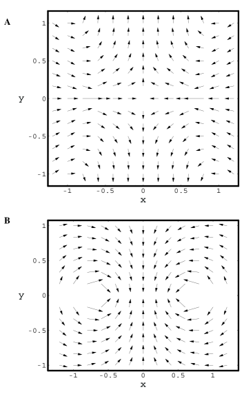
where and . Here, is the shear modulus and is the longitudinal modulus, where is the Poisson ratio, and . This coupling can be understood in terms of the solvent flow through the highly porous gel: rapid solvent flow through the filament mesh gives rise to large shear stresses, effectively dragging the network with the solvent. This drag prevents the large-scale relative motion of the network and solvent beyond a range of order . On larger length scales or at higher frequencies , the drag effectively inhibits the relative motion of solvent and network so that for , the combined network and solvent act as a single incompressible materialGittes-1997-19 ; Levine-2000-16 , and both vanish (Fig. 3A). Here, the response of the medium is purely transverse (the displacement vector field is divergenceless) and is given by the generalized Oseen tensor, given by leading terms in square brackets aboveLevine-2000-16 . The corresponding volume-preserving flow response of an incompressible gel when subject to a symmetric pair of point forces is shown in Fig. 4A.
In this incompressible case, the displacement field of the network resulting from motor activity varies with an overall frequency dependence proportional to the ratio of the force to the shear modulus , according to Eqs. (1-3). Thus, we find for the model illustrated in Fig. 1A that . Cross-linked biopolymer networks typically exhibit a constant or weakly frequency-dependent elastic regime as a function of frequency. Here, we expect to see , which is consistent with recent displacement fluctuations observed in cellsLau-2003-10 , and which corresponds to diffusive motion. At higher frequencies, such networks typically exhibit a power-law increase in the shear modulus with frequencyGittes-1997-19 ; Gittes-1998-8 ; Morse-1998-32 , in which . In this frequency regime stain fluctuations in the active gel take the form , as shown in Fig. 2. For comparison, the equilibrium thermal fluctuations for such a network are shown as the dashed line. At low frequencies the motor-driven fluctuations will dominate over the ever-present thermal fluctuations, consistent with the results of both Lau et al.Lau-2003-10 and Mizuno et al.Mizuno-2007-1 .
Since biopolymer and cytoskeletal networks are generically porous with pore sizes of order m, they can deform compressibly. This density mode, however, is strongly suppressed by drag at high enough frequencies. The loss of the density mode at high frequencies is illustrated in Fig. 3A, where the effects of finite compressibility, represented by , vanish at high frequency. Although the basic physics of these effects have been discussed before for both flexible polymer systemsBrochard-1977-35 ; Milner-1993-34 and semi-flexible biopolymer systemsGittes-1997-19 ; Levine-2000-16 , there has been no direct experimental observation of these compressibility effects in porous biopolymer systems.
We can isolate the effects of the network compressibility by the examining the combination
| (6) |
which is plotted in Fig. 3B. This measurable combination of response functions strictly vanishes in the incompressible limit. This, along with the specific combined and dependence, may permit the first direct measurement of compressibility effects that are expected to be characteristic of biopolymer/cytoskeletal networks. Furthermore, the flow/displacement field corresponding to this compressible mode (shown in Fig. 4B in the limit ) strongly differ from the case of an incompressible system (Fig. 4A). Here, the longitudinal (irrotational displacement field) contributions to the response function are and . The difference in spatial structure of these strain fields may also be used to experimentally identify the effects of compression.
To consider the effect of multiple contractile events within the medium, we can represent the resulting displacement field at the origin at by a sum
| (7) |
where is the response to a contractile force pair. We suppress the frequency dependence. This sum represents the combined effect of temporally uncorrelated contractile events occurring homogeneously throughout the medium. This assumption remains valid provided that the events rarely occur with a separation of order during the typical processivity time . Such a sum or average has been performed in calculating the PSD in Fig. 2 for the case of an incompressible network. In this case the scaling described above is a good approximation.
This model shows how motor activity within a semi-flexible gel, together with the well-established non-linear response of such networks leads to a strong stiffening of the network, and that this stiffening increases more than linearly with the motor force. This can account for the recently observed nearly 100-fold network stiffening with motor forces of order 1-10 pNMizuno-2007-1 . Furthermore, the (un)binding kinetics of the motors naturally leads to a specific characteristic time dependence of the force fluctuations in active gels. Given a finite processivity time over which minifilaments remain bound and generate force, the unbinding results in force fluctuations for frequencies . This spectrum is a direct result of the expected sharp time dependence of motor unbinding, and is insensitive to slow variations of force during motor motion. For frequencies , the divergence of the force spectrum will be suppressed. Our model, is for uncorrelated motor activity, in that the total fluctuations can be represented as a sum of independent fluctuations due to individual motor force generation and unbinding. At sufficiently high motor densities, one might expect cooperativity of motor activity, whose consequences can be studied in extension of the present model.
This work was supported in part by the (Netherlands) Foundation for Fundamental Research on Matter (FOM), NSF Materials World Networks (grant no. DMR-0354113), and the NSF through the Kavli Institute for Theoretical Physics. The authors thank J. Crocker, A. Grosberg, A. Lau, T. Lubensky, D. Mizuno, M. Rubinstein, and C. Schmidt for helpful discussions.
References
- (1) B. Alberts, et al., Molecular Biology of the Cell, 3rd edition (Garland, New York, 1994).
- (2) F.C. MacKintosh and P.A. Janmey, Current Opinion in Solid State & Materials Science 2: 350 (1997).
- (3) M.L. Gardel, et al., Science 304: 1301 (2004).
- (4) C. Storm, et al., Nature 435: 191 (2005).
- (5) A.R. Bausch and K. Kroy, Nature Physics 2: 231 (2006).
- (6) O. Chaudhuri, S.H. Parekh and D.A. Fletcher, Nature 445: 295 (2007).
- (7) P.A. Janmey, et al., Nature Materials 6: 48 (2007).
- (8) D. Mizuno, C. Tardin, C.F. Schmidt and F.C. MacKintosh, Science 315: 370 (2007).
- (9) A.W.C. Lau, et al., Phys. Rev. Lett. 91: 198101 (2003).
- (10) C. Brangwynne, F.C. MacKintosh, and D.A. Weitz, PNAS, in press; C. Brangwynne, et al., arXiv:0709.2952.
- (11) T.B. Liverpool, A.C. Maggs and A. Ajdari, Phys. Rev. Lett. 86: 4171 (2001).
- (12) L. Le Goff, F. Amblard and E.M. Furst, Phys. Rev. Lett. 88: 018101 (2001).
- (13) D. Humphrey, et al., Nature 416: 413 (2002).
- (14) T.B. Liverpool, Phil. Trans. A 364: 3335 (2006).
- (15) K. Kruse, et al., Eur. Phys. J. E 16: 5 (2005).
- (16) C. Bustamante, et al., Science 265: 1599 (1994).
- (17) F.C. MacKintosh, J. Käs and P.A. Janmey, Phys. Rev. Lett. 75: 4425 (1995).
- (18) G.H. Koenderink, et al., unpublished.
- (19) F. Gittes and F.C. MacKintosh, Phys. Rev. E 58: R1241 (1998).
- (20) D.C. Morse, Macromolecules 31: 7044 (1998).
- (21) F. Brochard and P.G. Degennes, Macromolecules 10: 1157 (1977).
- (22) S.T. Milner, Phys. Rev. E 48: 3674 (1993).
- (23) F. Gittes, et al., Phys. Rev. Lett. 79: 3286 (1997); B. Schnurr, F. Gittes, F.C. MacKintosh and C.F. Schmidt, Macromolecules 30: 7781 (1997).
- (24) A.J. Levine and T.C. Lubensky, Phys. Rev. Lett. 85: 1774 (2000).